


Application of T2* weighted angiography and 3D ASL in evaluation of cerebral perfusion after unilateral internal carotid artery chronic occlusion
-
摘要:
目的 分析T2*加权血管成像及三维动脉自旋标记技术在单侧颈内动脉(ICA)慢性闭塞后脑灌注状态评估方面的临床应用价值 方法 回顾性收集2018年1月~2022年3月经三维时间飞跃法磁共振血管成像诊断为单侧ICA闭塞患者44例,并均行T2*加权血管成像及三维动脉自旋标记序列检查,分析闭塞侧ICA供血区突出血管征(PVS)阴性(阴性组,n=25)与阳性(阳性组,n=19)的脑血流量(CBF)值差异,同时比较PVS阳性组以及阴性组ICA闭塞侧与镜像区CBF值差异 结果 PVS阴性组ICA闭塞侧额叶、顶叶、颞叶及侧脑室旁白质区CBF值均高于PVS阳性组,差异有统计学意义(P < 0.05);PVS阴性组ICA闭塞侧额叶、顶叶、颞叶及侧脑室旁白质区CBF值与镜像区的差异无统计学意义(P>0.05),PVS阳性组ICA闭塞侧额叶、顶叶、颞叶及侧脑室旁白质区CBF值低于镜像区,差异有统计学意义(P < 0.05) 结论 单侧ICA慢性闭塞后,T2*加权血管成像及三维动脉自旋标记能够客观反映侧支循环建立及脑灌注状态,可为临床治疗方案选择提供影像依据 Abstract:Objective To analyze the clinical value of T2* weighted angiography and three-dimensional arterial spin labeling in the evaluation of cerebral perfusion after unilateral internal carotid artery (ICA) chronic occlusion. Methods Forty-four patients with unilateral ICA occlusion diagnosed by three-dimensional time leap magnetic resonance angiography from January 2018 to March 2022 were retrospectively collected, and all underwent T2* weighted angiography and threedimensional arterial spin labeling sequence examination. The differences of cerebral blood flow (CBF) between the negative and positive groups of ICA supply area prominent vascular sign (PVS) on the occluded side were analyzed, and the CBF values of ICA occluded side and mirror area in the positive and negative groups were compared. Results The CBF values of frontal lobe, parietal lobe, temporal lobe and paraventricular white matter of ICA occlusion in PVS negative group were significantly higher than those in PVS positive group (P < 0.05). There was no significant difference between the CBF values of frontal lobe, parietal lobe, temporal lobe and paraventricular white matter area in ICA occlusion side in PVS negative group and those in mirror area (P>0.05). The CBF values of frontal lobe, parietal lobe, temporal lobe and paraventricular white matter area in ICA occlusion side in PVS positive group were significantly lower than those in mirror area (P < 0.05). Conclusion After chronic occlusion of unilateral ICA, T2* weighted angiography and three- dimensional arterial spin labeling can objectively reflect the establishment of collateral circulation and cerebral perfusion, which can provide image basis for the selection of clinical treatment. -

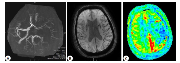
图 1 患者3D-TOF MRA(A)、SWAN(B)及3DASL(C)图像
患者男, 69岁, 头晕头痛5 h入院. A: 3D-TOF MRA提示左侧ICA闭塞; B: SWAN提示左侧额颞叶PVS阳性; C: 3DASL提示左侧额颞叶及侧脑室旁低灌注.
Figure 1. Images of 3D-TOF MRA(A)、SWAN(B) and 3DASL(C) of a patient.
表 1 PVS阳性组及阴性组ICA闭塞侧脑灌注量比较
Table 1. Comparison of cerebral perfusion volume of ICA occlusion side between PVS positive group and negative group [Mean±SD, (mL/min)×100 g]
分组 额叶CBF值 顶叶CBF值 颞叶CBF值 侧脑室旁CBF值 PVS阳性组(n=19) 33.68±7.18 32.82±7.21 33.49±6.92 29.89±6.11 PVS阴性组(n=25) 41.68±6.82 40.63±6.87 42.66±6.94 35.14±7.21 t 3.7676 3.6566 4.3468 2.5515 P < 0.001 < 0.001 < 0.001 0.015 PVS: 突出血管征; CBF: 脑血流量.  下载: 导出CSV
表 2 PVS阴性组ICA闭塞侧与镜像区CBF值比较
Table 2. Comparison of CBF values between ICA occluded side and mirror area in PVS negative group [n=25, Mean±SD, (mL/min)×100 g]
分组 额叶CBF值 顶叶CBF值 颞叶CBF值 侧脑室旁CBF值 ICA闭塞侧 41.68±6.82 40.63±6.87 42.66±6.94 35.14±7.21 镜像区 43.17±7.35 42.02±7.14 42.98±7.47 36.86±8.03 t 0.7430 0.7014 0.1569 0.7969 P 0.461 0.486 0.876 0.429 ICA: 颈内动脉.
下载: 导出CSV
表 3 PVS阳性组ICA闭塞侧与镜像区CBF值比较
Table 3. Comparison of CBF values between ICA occluded side and mirror area in PVS positive group [n=19, Mean±SD, (mL/min)×100 g]
分组 额叶CBF值 顶叶CBF值 颞叶CBF值 侧脑室旁CBF值 ICA闭塞侧 33.68±7.18 32.82±7.21 33.49±6.92 29.89±6.11 镜像区 40.26±6.41 40.85±6.93 42.74±7.25 36.06±6.84 t 2.9799 3.5000 4.0230 2.9324 P 0.005 0.001 <0.001 0.006
下载: 导出CSV
-
[1] 周建国, 符大勇, 马先军, 等. ASL对大脑中动脉M1段闭塞后侧支循环建立显示的临床应用[J]. 实用放射学杂志, 2018, 34(8): 1164-6, 1171. [2] 马永青, 尹喜, 王成伟. 磁敏感加权血管成像在指导急性脑梗死溶栓治疗及评估预后中的临床价值[J]. 实用放射学杂志, 2019, 35(9): 1389-94. doi: 10.3969/j.issn.1002-1671.2019.09.003 [3] 黄双凤, 崔伟, 梁志刚. 磁敏感加权成像在急性缺血性卒中患者中的应用[J]. 国际脑血管病杂志, 2020, 28(6): 457-62. [4] 林天烨, 有慧, 冯逢, 等. 动脉自旋标记MR技术进展及应用[J]. 中华放射学杂志, 2019, 53(5): 431-4. [5] 周建国, 卢明聪, 孟云, 等. 单侧大脑中动脉M1段闭塞后脑动脉硬化与侧支循环建立相关性研究[J]. 临床放射学杂志, 2021, 40(2): 212-6. https://www.cnki.com.cn/Article/CJFDTOTAL-LCFS202102004.htm [6] 孟云, 符大勇, 周建国, 等. 磁敏感加权成像在急性缺血性脑卒中方面的临床应用价值[J]. 中风与神经疾病杂志, 2019, 36(2): 143-6. https://www.cnki.com.cn/Article/CJFDTOTAL-ZFSJ201902012.htm [7] 王光耀, 荆京, 孟霞, 等. 缺血性脑血管病梗死模式的分类及病因、发病机制研究进展[J]. 中国卒中杂志, 2019, 14(9): 950-4. https://www.cnki.com.cn/Article/CJFDTOTAL-ZUZH201909023.htm [8] 张萍淑, 吴小英, 孔祥慧, 等. 慢性单侧颈内动脉颅外段闭塞患者脑动脉血流分流观察[J]. 山东医药, 2017, 57(43): 68-70. https://www.cnki.com.cn/Article/CJFDTOTAL-SDYY201743021.htm [9] 赵云凯, 薛国芳. 侧支循环对缺血性脑卒中的影响及其影像评估[J]. 中华老年心脑血管病杂志, 2021, 23(1): 105-7. https://www.cnki.com.cn/Article/CJFDTOTAL-LNXG202101031.htm [10] 国家卫生健康委员会脑卒中防治工程委员会神经影像专业委员会, 中华医学会放射学分会神经学组. 脑血管病影像规范化应用中国指南[J]. 中华放射学杂志, 2019, 53(11): 916-40. [11] 周建国, 符大勇, 卢明聪, 等. 3D ASL在不同程度颈动脉狭窄患者脑分水岭灌注评估中的临床应用[J]. 分子影像学杂志, 2021, 44(1): 59- 62. doi: 10.12122/j.issn.1674-4500.2021.01.11 [12] 葛永桂, 郭婷婷, 王玉洁. 影响大脑中动脉狭窄后侧支循环建立的因素及其研究进展[J]. 中华老年心脑血管病杂志, 2019, 21(2): 211-3. https://www.cnki.com.cn/Article/CJFDTOTAL-LNXG201902027.htm [13] 郭茜, 曹树刚, 葛婷婷, 等. 同侧大脑后动脉偏侧优势与大脑中动脉供血区缺血性卒中患者转归的相关性[J]. 国际脑血管病杂志, 2018, 26(6): 418-21. [14] 贾亚南, 刘翠翠, 刘俊艳. 磁敏感加权成像不对称皮层静脉征与急性缺血性卒中预后的关系研究[J]. 中国卒中杂志, 2019, 14(7): 639-44. https://www.cnki.com.cn/Article/CJFDTOTAL-ZUZH201907004.htm [15] Yu JC, Wang LM, Li ZZ, et al. Related factors of asymmetrical vein sign in acute middle cerebral artery stroke and correlation with clinical outcome[J]. J Stroke Cerebrovasc Dis, 2017, 26(10): 2346- 53. [16] 穆丽颖, 张乐, 王维平, 等. SWI对急性缺血性脑卒中梗死增长及早期预后的预测价值[J]. 中国临床医学影像杂志, 2021, 32(8): 569-73. https://www.cnki.com.cn/Article/CJFDTOTAL-LYYX202108010.htm [17] Yuan T, Ren GL, Quan GM, et al. Fewer peripheral asymmetrical cortical veins is a predictor of favorable outcome in MCA infarctions with SWI-DWI mismatch[J]. J Magn Reson Imaging, 2018, 48(4): 964-70. [18] 倪晓星, 曹树刚, 王建, 等. 磁敏感加权成像显著低信号血管征在急性脑梗死诊疗中的应用价值[J]. 中华神经科杂志, 2020, 53(1): 64- 71. [19] 周建国, 符大勇, 孟云, 等. 双时相ASL在急性缺血性脑卒中灌注评估中的临床应用价值[J]. 实用放射学杂志, 2018, 34(2): 315-6, 318. [20] 陈峰, 王洁, 甘解华, 等. 侧支循环评分及磁共振3D-ASL技术对急性前循环大血管闭塞患者再灌注治疗预后的评估价值[J]. 浙江医学, 2021, 43(10): 1091-5. https://www.cnki.com.cn/Article/CJFDTOTAL-ZJYE202110015.htm [21] 耿立娜, 袁涛, 全冠民, 等. 皮髓质静脉征评估急性缺血性脑卒中的研究进展[J]. 国际医学放射学杂志, 2019, 42(5): 539-42. https://www.cnki.com.cn/Article/CJFDTOTAL-GWLC201905008.htm [22] 游润发, 周海军. 动脉自旋标记技术在常见脑部疾病中的应用进展[J]. 中华神经医学杂志, 2019, 18(1): 93-7. https://www.cnki.com.cn/Article/CJFDTOTAL-LYSJ201502023.htm [23] 周建国, 符大勇, 卢明聪, 等. 三维动脉自旋标记在单侧颈内动脉闭塞后侧支循环评估中的价值[J]. 分子影像学杂志, 2020, 43(1): 130-3. 记在单侧颈内动脉闭塞后侧支循环 [24] 王宪雯, 吴芳, 刘玥宏, 等. 伪连续动脉自旋标记成像评估出血型烟雾病患者脑血流动力学研究[J]. 磁共振成像, 2022, 13(1): 6-10. https://www.cnki.com.cn/Article/CJFDTOTAL-CGZC202201002.htm [25] 周建国, 符大勇, 卢明聪, 等. 大脑中动脉M1段闭塞后动脉偏侧优势和动脉内高信号对比研究[J]. 国际医药卫生导报, 2022, 28(3): 307-10. -

 点击查看大图
计量
- 文章访问数: 196
- HTML全文浏览量: 146
- PDF下载量: 12
- 被引次数: 0