


Application of 3.0 T MRI combined with serum DR-70 detection in early diagnosis of colorectal cancer
-
摘要:
目的 探讨3.0 T MRI与血清纤维蛋白降解复合物(DR-70)联合检测在早期结直肠癌诊断中的价值。 方法 选取本院2018年8月~2020年8月收治的早期结直肠癌患者96例,另选取同期40例结直肠良性肿瘤患者,实施3.0 T MRI行动态增强和扩散加权成像等序列扫描和血清DR-70检测,利用Spearman相关性分析3.0 T MRI相关参数和血清DR-70水平与早期结直肠癌的关系,并采用ROC曲线评估这两种检测指标对早期结直肠癌的诊断效能。 结果 96例患者中本次共检出结直肠癌94例,漏诊2例,检出率为97.92%;3.0T MRI能较好地观察到肿瘤的病灶部位、形态、侵袭等情况,肿瘤病灶在T1图像呈中等偏低信号,T2图像呈略高信号,动态增强扫描早期呈不均匀强化,延迟期表现为稍低信号,在扩散加权成像中呈明显高信号,在表观扩散系数(ADC)图上呈低信号;观察组MRI参数ADC值低于对照组(P < 0.05),而DR-70水平高于对照组(P < 0.05);Spearman相关性分析显示ADC值与结直肠癌呈负相关(r=-0.383,P < 0.05),而DR-70水平则与结直肠癌呈正相关(r=0.460,P < 0.05);ROC曲线显示:ADC、DR-70联合诊断结直肠癌曲线下面积为0.850,明显高于各单项指标曲线下面积(P < 0.05)。 结论 结直肠癌患者利用3.0T MRI技术和DR-70联合检测能显著提高结直肠癌的诊断鉴别效能。 Abstract:Objective To investigate the value of 3.0 T MRI combined with serum fibrin- degrading complex (DR-70) in the diagnosis of early colorectal cancer. Methods Ninety-six patients with early colorectal cancer admitted to our hospital from August 2018 to August 2020 were selected as observation group, and forty patients with colorectal benign tumor were selected during the same period as control group. Implementation of 3.0T MRI dynamic enhancement and diffusion weighted imaging sequence scan and serum DR-70 test, using the Spearman correlation analysis the relationship between early colorectal cancer of 3.0T MRI parameters and serum DR-70 level. ROC was used to evaluate the diagnostic efficacy of the two detection indexes for early colorectal cancer. Results Among 96 patients, 94 cases of colorectal cancer were detected, 2 cases were missed, the detection rate was 97.92%. 3.0T MRI could better observe the location, morphology, invasion and other conditions of the tumor lesion. The tumor lesion showed medium low signal in T1 image, slightly high signal in T2 image, uneven enhancement in the early stage of dynamic enhancement scan, slightly low signal in the delay stage, significantly high signal in diffusion weighted imaging, and low signal in ADC image. The ADC value of MRI parameters in observation group was significantly lower than that in control group (P < 0.05), while the DR-70 level was significantly higher than that in control group (P < 0.05). Spearman correlation analysis showed that ADC value was significantly negatively correlated with colorectal cancer (r=-0.383, P < 0.05), while DR-70 level was significantly positively correlated with colorectal cancer (r=0.460, P < 0.05). ROC curve showed that the area under the curve of ADC and DR-70 combined diagnosis of colorectal cancer was 0.850, which was significantly higher than the area under the curve of each individual index (P < 0.05). Conclusion 3.0 T MRI and DR-70 combined detection can significantly improve the diagnostic and differential performance of colorectal cancer patients. -

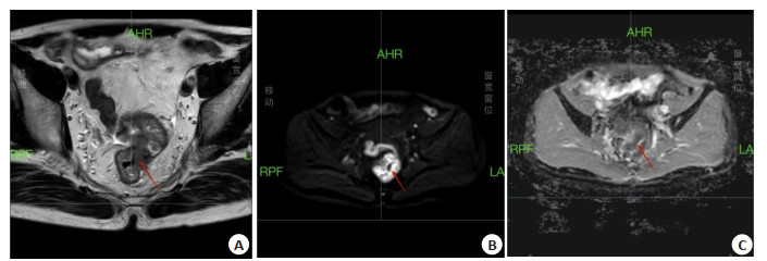
图 1 59岁男性患者,病理诊断为直肠中分化腺癌
A: T2WI图; B: DWI图; C: ADC图.
Figure 1. A59-year-old male patient, pathological diagnosis was moderately differentiated adenocarcinoma of rectum.

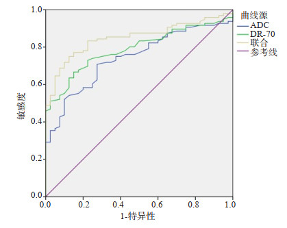
图 2 ADC、DR-70对结直肠癌的ROC曲线分析
Figure 2. ROC curve analysis of ADC and DR-70 on colorectal cancer.
表 1 两组ADC、DR-70比较
Table 1. Comparison of ADC and DR-70 between the two groups(Mean±SD)
组别 ADC(×10-3mm2/s) DR-70(μg/mL) 观察组(n=96) 1.30±0.41 1.15±0.31 对照组(n=40) 1.61±0.23 0.88±0.12 t 4.472 5.471 P < 0.001 < 0.001 *P < 0.05 vs联合指标.  下载: 导出CSV
表 2 ADC、DR-70对结直肠癌的ROC曲线分析
Table 2. ROC curve analysis ofADC and DR-70 for colorectal cancer
指标 曲线下面积 最佳截断值 敏感度 特异性 95% CI 约登指数 ADC 0.743* 1.51×10-3mm2/s 0.708 0.725 0.660~0.825 0.433 DR-70 0.791* 0.99 μg/mL 0.667 0.850 0.717~0.865 0.517 联合 0.850 - 0.750 0.875 0.786~0.913 0.625 *P < 0.05 vs联合指标.
下载: 导出CSV
-
[1] Sung H, Ferlay J, Siegel RL, et al. Global cancer statistics 2020: GLOBOCAN estimates of incidence and mortality worldwide for 36 cancers in 185 countries[J]. CAACancer J Clin, 2021, 71(3): 209-49. doi: 10.3322/caac.21660 [2] Munro MJ, Wickremesekera SK, Peng LF, et al. Cancer stem cells in colorectal cancer: a review[J]. J Clin Pathol, 2018, 71(2): 110-6. doi: 10.1136/jclinpath-2017-204739 [3] Dekker E, Tanis PJ, Vleugels JLA, et al. Colorectal cancer[J]. Lancet, 2019, 394(10207): 1467-80. doi: 10.1016/S0140-6736(19)32319-0 [4] 国家癌症中心中国结直肠癌筛查与早诊早治指南制定专家组, 赫捷, 陈万青, 等. 中国结直肠癌筛查与早诊早治指南(2020, 北京) [J]. 中国肿瘤, 2021, 30(1): 1-28. https://www.cnki.com.cn/Article/CJFDTOTAL-ZHLU202101001.htm [5] 赵胜兵, 王树玲, 方军, 等. 国内外结直肠癌早诊早治现状[J]. 中华消化内镜杂志, 2019, 36(2): 143-7. doi: 10.3760/cma.j.issn.1007-5232.2019.02.016 [6] Nasseri Y, Langenfeld SJ. Imaging for colorectal cancer[J]. Surg Clin NorthAm, 2017, 97(3): 503-13. doi: 10.1016/j.suc.2017.01.002 [7] Saridemir S, Güven HE, Aksel B, et al. Serum AMDL DR-70 levels: a new concept in the diagnosis and follow-up of colorectal carcinoma [J]. Biomark Med, 2020, 14(8): 621-8. doi: 10.2217/bmm-2020-0004 [8] 中华人民共和国国家卫生和计划生育委员会医政医管局, 中华医学会肿瘤学分会. 中国结直肠癌诊疗规范(2017年版)[J]. 中国实用外科杂志, 2018, 38(10): 1089-103. https://www.cnki.com.cn/Article/CJFDTOTAL-ZGWK201810001.htm [9] Baidoun F, Elshiwy K, Elkeraie Y, et al. Colorectal cancer epidemiology: recent trends and impact on outcomes[J]. Curr Drug Targets, 2021, 22(9): 998-1009. doi: 10.2174/1389450121999201117115717 [10] Díaz-Tasende J. Colorectal cancer screening and survival[J]. Rev Esp Enferm Dig, 2018, 110: 681-3. [11] Hall NC, Ruutiainen AT. Colorectal cancer: imaging conundrums[J]. Surg Oncol Clin NAm, 2018, 27(2): 289-302. doi: 10.1016/j.soc.2017.11.004 [12] de Souza GD, Souza LRQ, Cuenca RM, et al. Pre-and postoperative imaging methods in colorectal cancer[J]. ABCD, Arq Bras Cir Dig, 2018, 31(2): e1371-2. [13] Li YN, Xin JQ, Sun YB, et al. Magnetic resonance imaging-guided and targeted theranostics of colorectal cancer[J]. Cancer Biol Med, 2020, 17(2): 307-27. doi: 10.20892/j.issn.2095-3941.2020.0072 [14] Wang Y, Chen Y, Lyu X, et al. Value and related factors of preoperative diagnosis of extramural vascular invasion of rectal cancer by 3.0T magnetic resonance imaging[J]. Zhonghua Zhong Liu Za Zhi Chin J Oncol, 2019, 41(8): 610-4. [15] Goiffon RJ, O'Shea A, Harisinghani MG. Advances in radiological staging of colorectal cancer[J]. Clin Radiol, 2021, 76(12): 879-88. doi: 10.1016/j.crad.2021.06.005 [16] 黎叶芳, 马淑华, 袁珠, 等. 3.0T MRI和多层螺旋CT在直肠癌术前TN分期中的临床应用对比[J]. 实用癌症杂志, 2018, 33(3): 481-4. doi: 10.3969/j.issn.1001-5930.2018.03.039 [17] 吉盛超, 耿承军, 杨晓亮, 等. 3.0 T MRI在直肠癌术前分期和评估中的应用价值[J]. 天津医药, 2018, 46(7): 737-41. https://www.cnki.com.cn/Article/CJFDTOTAL-TJYZ201807015.htm [18] P eng Y, Li Z, Tang H, et al. Comparison of reduced field-of-view diffusion-weighted imaging (DWI) and conventional DWI techniques in the assessment of rectal carcinoma at 3.0T: image quality and histological T staging[J]. J Magn Reson Imaging, 2018, 47(4): 967-75. doi: 10.1002/jmri.25814 [19] Delli Pizzi A, Cianci R, Genovesi D, et al. Performance of diffusionweighted magnetic resonance imaging at 3.0T for early assessment of tumor response in locally advanced rectal cancer treated with preoperative chemoradiation therapy[J]. Abdom Radiol, 2018, 43 (9): 2221-30. doi: 10.1007/s00261-018-1457-8 [20] 杨志宏, 曹杰, 朱少群, 等. 3.0T磁共振T2加权成像联合弥散加权成像在老年直肠癌术前TN分期中的应用价值[J]. 中国老年学杂志, 2020, 40(7): 1417-20. doi: 10.3969/j.issn.1005-9202.2020.07.022 [21] 黄瑜亮, 黄斌, 严俊. 结直肠癌的早期诊断技术进展[J]. 分子影像学杂志, 2019, 42(1): 77-80. doi: 10.12122/j.issn.1674-4500.2019.01.18 [22] 开今言, 马倩, 童颖, 等. 血清纤维蛋白降解复合物(DR-70)检测在结直肠癌患者中的诊断价值[J]. 中国癌症杂志, 2020, 30(4): 293-8. https://www.cnki.com.cn/Article/CJFDTOTAL-ZGAZ202004009.htm -

 点击查看大图
计量
- 文章访问数: 438
- HTML全文浏览量: 64
- PDF下载量: 4
- 被引次数: 0