


Diagnostic value of ultrasound-guided fine needle aspiration for papillary thyrid carcinoma
-
摘要:
目的 探讨超声引导下甲状腺细针穿刺(US-FNAC)对甲状腺乳头状癌的诊断价值。 方法 选取2015年1月~2022年3月于我院甲状腺外科行甲状腺结节检查患者235例作为研究对象,根据结节最大径将其分为4组:A组(< 5 mm)、B组(5.1~10 mm)、C组(10.1~20 mm)、D组(≥20 mm)。以病理结果为金标准,统计并记录4组US-FNAC阳性率、无法确诊率、准确率、阳性预测值、阴性预测值、转移性结节分区情况。 结果 US-FNAC检查显示,随着结节直径的增加,4组阳性率不断提高(29.41%、39.50%、41.51%、61.82%),总阳性率为42.45%,各组间阳性病例的差异有统计学意义(χ2=12.180,P=0.007);4组结节US-FNAC细胞学检查总的无法确诊率为1.80%,随着结节增大波动降低(1.96%、2.52%、1.89%、0.00%),各组间差异无统计学意义(χ2= 1.370,P=0.713);US-FNAC细胞学检查结果显示,4组总准确率(符合率)为95.32%,随结节直径的增加有所波动,但差异无统计学意义(χ2=0.40,P=0.941);4组结节总阳性预测值达到94.92%,总阴性预测值比阳性预测值略高,为95.63%,总敏感度、总特异性分别为94.12%、96.23%。195个转移结节中,超声分区定位检查与病理检查结果符合率分别为95.45%、98.11%、86.21%、97.89%。 结论 US-FNAC细胞学检查诊断甲状腺乳头状癌阳性率受到结节尺寸的影响,但对于准确性、阳性预测值、阴性预测值的影响不大。 Abstract:Objective To investigate the diagnostic value of ultrasound-guided fine needle aspiration (US-FNAC) for papillary thyroid carcinoma. Methods The data of 235 patients with thyroid nodules examined in our hospital from January 2015 to March 2022 were selected. According to the maximum diameter of nodules, they were divided into four groups: group A (< 5 mm), group B (5.1-10 mm), group C (10.1-20 mm) and group D (≥20 mm). The pathological results were taken as the gold standard, and the positive rate of US- FNAC, undiagnosed rate, accuracy rate, positive predictive value, negative predictive value, and partition of metastatic nodules in four groups were recorded. Results US-FNAC examination showed that with the increase of nodule diameter, the positive rates of the four groups were continuously increased (29.41%, 39.50%, 41.51%, 61.82%), and the total positive rate was 42.45%. The difference in positive cases between groups was statistically significant (χ2=12.180, P=0.007). The total undiagnosed rate of US-FNAC cytology in the four groups was 1.80%, and the fluctuation decreased with the increase of nodules (1.96%, 2.52%, 1.89%, 0.00%). There was no significant difference between the four groups (χ2=1.370, P=0.713). The results of US-FNAC cytological examination showed that the total accuracy (coincidence rate) of the four groups was 95.32%, which fluctuated with the increase of nodule diameter, but the difference was not statistically significant (χ2=0.40, P=0.941). The total positive predictive value of the four groups of nodules reached 94.92%, and the total negative predictive value was slightly higher than the positive predictive value, which was 95.63%. The total sensitivity and total specificity were 94.12% and 96.23%, respectively. Among the 195 metastatic nodules, the coincidence rates of ultrasound regional localization and pathological examination were 95.45%, 98.11%, 86.21% and 97.89%, respectively. Conclusion The positive rate of US- FNAC cytology in the diagnosis of papillary thyroid carcinoma is affected by nodule size, but has little effect on accuracy, positive predictive value and negative predictive value. -
Key words:
- ultrasound guidance /
- thyroid /
- fine needle puncture /
- papillary thyroid carcinoma
-

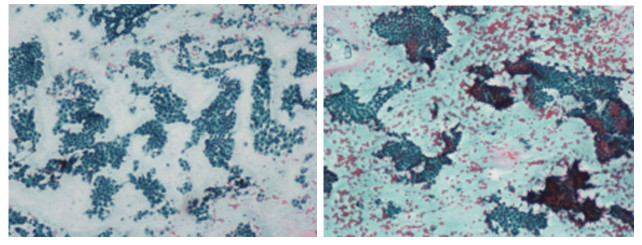
图 1 甲状腺左侧叶及峡部结节穿刺涂片(HE, ×200)
患者男, 39岁, 病理诊断为甲状腺乳头状癌.
Figure 1. Puncture smear of thyroid nodules in left lobe and isthmus (HE, ×200).
表 1 US-FNAC细胞学检查结果比较
Table 1. Comparison of US-FNAC cytology results (n)
组别 总数 阳性 阴性 无法确诊 阳性率(%) A组 51 15 35 1 29.41 B组 119 47 69 3 39.50 C组 53 22 30 1 41.51 D组 55 34 21 0 61.82 总数 278 118 155 5 42.45 US-FNAC: 超声引导下甲状腺细针穿刺. A组:直径 < 5 mm, B组: 5.1 mm≤直径 < 10 mm, C组: 10.1 mm≤直径 < 20 mm, D组: 直径≥20 mm.  下载: 导出CSV
表 2 US-FNAC细胞学结果无法确诊率统计
Table 2. Undiagnosed rate statistics of US-FNAC cytology results (n)
组别 确诊数 无法确诊数 总数 无法确诊率(%) A组 50 1 51 1.96 B组 116 3 119 2.52 C组 52 1 53 1.89 D组 55 0 55 0.00 总数 273 5 278 1.80
下载: 导出CSV
表 3 US-FNAC细胞学结果与病理诊断结果的符合程度
Table 3. The coincidence degree between US-FNAC cytological results and pathological diagnosis results
组别 总数(n) 符合(n) 不符合(n) 准确率(%) 阳性预测值(%) 阴性预测值(%) 敏感度(%) 特异性(%) A组 51 48 3 94.12 94.44 93.33 87.50 97.14 B组 119 114 5 95.80 95.74 95.83 93.75 97.18 C组 53 51 2 94.34 90.91 96.77 95.24 93.75 D组 55 52 3 94.55 97.06 90.48 94.29 95.00 总数 278 265 13 95.32 94.92 95.63 94.12 96.23
下载: 导出CSV
表 4 超声结节分区定位与病理对照
Table 4. Ultrasonic nodule localization and pathological comparison
结节分区 Ⅱ区 Ⅲ区 Ⅳ区 Ⅵ区 总数 US-FNAC(n) 21 52 29 93 195 病理结果(n) 22 53 25 95 195 符合率(%) 95.45 98.11 86.21 97.89 -
下载: 导出CSV
-
[1] 赵焕, 郭会芹, 张智慧, 等. BRAFV600E基因及蛋白检测细针穿刺标本对辅助细胞学诊断甲状腺乳头状癌的价值[J]. 中华耳鼻咽喉头颈外科杂志, 2019, 54(1): 18-22. doi: 10.3760/cma.j.issn.1673-0860.2019.01.005 [2] 李若暄, 周祖邦, 谢金会, 等. 超声引导下细针穿刺细胞学检查联合BRAF V600E突变检测及声像图特征在评估经典型甲状腺乳头状癌颈部淋巴结转移中的应用[J]. 中华超声影像学杂志, 2019, 28(12): 1056-60. doi: 10.3760/cma.j.issn.1004-4477.2019.12.009 [3] 张颖, 罗渝昆, 张艳, 等. 超声引导下细针穿刺细胞学检查联合BRAFV600E检测与甲状腺乳头状癌侵袭性病理特征的相关性[J]. 中国医学科学院学报, 2019, 41(4): 517-23. https://www.cnki.com.cn/Article/CJFDTOTAL-ZYKX201904012.htm [4] 张晓芳, 刘志艳. 2018版甲状腺细针穿刺活检细胞病理学Bethesda报告系统解读[J]. 中华病理学杂志, 2018, 47(9): 729-32. doi: 10.3760/cma.j.issn.0529-5807.2018.09.019 [5] 朱晓静, 施中元, 魏燕华, 等. 甲状腺乳头状癌的超声及细针穿刺细胞学检查特征分析[J]. 天津医药, 2017, 45(5): 533-5, 563. https://www.cnki.com.cn/Article/CJFDTOTAL-TJYZ201705022.htm [6] Motamedfar A, Momen Gharibvand M, Sametzade M, et al. The use of neck sonography in identifying cervical lymph node: a single center study from 2019 to 2020[J]. Jundishapur J Chronic Dis Care, 2021, 10(2): 52-7. [7] Bi W, Huang JY, Nie CL, et al. CircRNA circRNA_102171 promotes papillary thyroid cancer progression through modulating CTNNBIP1-dependent activation of β-catenin pathway[J]. J Exp Clin Cancer Res, 2018, 37: 275. doi: 10.1186/s13046-018-0936-7 [8] 陈燕, 钟洁愉, 林晓娜, 等. FNAC对弥漫硬化型甲状腺乳头状癌的诊断价值[J]. 中国超声医学杂志, 2020, 36(4): 362-4. doi: 10.3969/j.issn.1002-0101.2020.04.024 [9] Cho JW, Lee YM, Lee YH, et al. Dynamic risk stratification system in post-lobectomy low-risk and intermediate-risk papillary thyroid carcinoma patients[J]. Clin Endocrinol, 2018, 89(1): 100-9. doi: 10.1111/cen.13721 [10] Li RX, Hao JP, Zhu Z, et al. Correlation between US- FNAC with BRAF V600E mutation analysis and central neck lymph node metastasis in cN0 papillary thyroid cancer[J]. Biomed Res Int, 2021, 2021: 9937742. DOI: 10.1155/2021/9937742. [11] Jiang HJ, Wu CW, Chiang FY, et al. Reliable sonographic features for nodal thyroglobulin to diagnose recurrent lymph node metastasis from papillary thyroid carcinoma[J]. Clin Otolaryngol, 2018, 43(4): 1065-72. doi: 10.1111/coa.13103 [12] 方达, 马雯婷, 徐露, 等. 超声引导下细针穿刺在不同大小甲状腺结节中的鉴别诊断[J]. 南京医科大学学报: 自然科学版, 2018, 38(12): 1806-9. https://www.cnki.com.cn/Article/CJFDTOTAL-NJYK201812040.htm [13] 汪佳慧, 高力, 寿金朵, 等. 术前细针穿刺洗脱液甲状腺球蛋白在诊断甲状腺乳头状癌淋巴结转移中的应用及影响因素[J]. 中国肿瘤临床, 2018, 45(22): 1142-6. doi: 10.3969/j.issn.1000-8179.2018.22.969 [14] Kim HJ, Lee HJ, Jung JH, et al. Ultrasound assessment of synchronous thyroid nodules in patients with papillary thyroid cancer: a nodule-by-nodule analysis between ultrasound and pathology[J]. Anticancer Res, 2020, 40(3): 1779-86. doi: 10.21873/anticanres.14132 [15] 赵美丽, 杨炜, 李金凤, 等. 超声引导下细针穿刺细胞学检测不同大小可疑甲状腺癌结节的阳性率及准确率[J]. 南方医科大学学报, 2020, 40(5): 693-7. https://www.cnki.com.cn/Article/CJFDTOTAL-DYJD202005013.htm [16] Kim HK, Kim SY, Lee YS, et al. Suspicious thyroid nodules ≥4 cm require diagnostic lobectomy regardless of their benign fine needle aspiration results[J]. Asian J Surg, 2021. DOI: 10.1016/j.asjsur. 2021.08.005. [17] 杨汶士, 张艳, 龙厚隆, 等. 细针穿刺甲状腺球蛋白检测对甲状腺乳头状癌颈淋巴结转移诊断价值探讨[J]. 中国实用外科杂志, 2017, 37 (9): 1032-4, 1038. https://www.cnki.com.cn/Article/CJFDTOTAL-ZGWK201709026.htm [18] 杨汶士, 张艳, 张凤凤, 等. 细针穿刺洗脱液测定甲状腺球蛋白和细针穿刺细胞学诊断甲状腺乳头状癌颈部淋巴结转移的Meta分析[J]. 肿瘤研究与临床, 2022, 34(2): 137-41. doi: 10.3760/cma.j.cn115355-20210711-00304 [19] Liu M, Chen P, Hu HY, et al. Kinase gene fusions: roles and therapeutic value in progressive and refractory papillary thyroid cancer[J]. J Cancer Res Clin Oncol, 2021, 147(2): 323-37. doi: 10.1007/s00432-020-03491-5 [20] Ahn D, Kim JK. Cervical bronchogenic cysts mimicking papillary thyroid carcinoma on ultrasound[J]. Korean J OtorhinolaryngolHead Neck Surg, 2019, 62(12): 735-9. doi: 10.3342/kjorl-hns.2019.00080 -

 点击查看大图
计量
- 文章访问数: 153
- HTML全文浏览量: 117
- PDF下载量: 4
- 被引次数: 0