


Cerebral blood flow in patients with primary dysmenorrhea by arterial spin labeling technique
-
摘要:
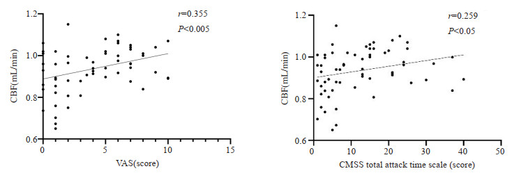
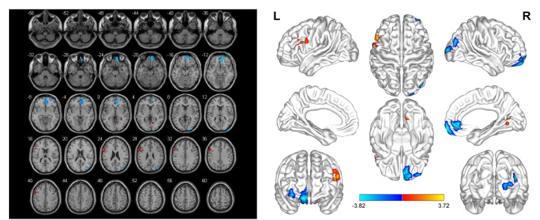
目的 采用基于体素的形态学方法及动脉自旋标记技术,探讨原发性痛经(PDM)患者的脑血流量变化,并分析其与临床量表评分的相关性。 方法 前瞻性纳入陕西中医药大学被确诊为PDM的患者31例作为PDM组,另选取同期32例健康者作为健康对照组,在月经期第1~3天进行高分辨率T1结构像及全脑动脉自旋标记扫描,并采用疼痛视觉模拟量表(VAS)、COX痛经症状量表(CMSS)、焦虑自评量表(SAS)、抑郁自评量表(SDS)进行临床症状评估。采用基于体素的形态学方法,基于Matlab平台的SPM8软件的双样本t检验分析两组间脑血流量差异有统计学意义的脑区,再采用Pearson相关性分析,观察脑血流量差异脑区的脑血流量值与VAS、CMSS、SAS、SDS量表评分之间的相关性。 结果 PDM组与健康对照组在年龄、VAS、CMSS、SAS、SDS量表评分的差异均有统计学意义(P < 0.05)。与健康对照组相比,PDM组脑血流量增高的脑区主要包括左侧三角部额下回,脑血流量减低的脑区包括右侧眶部额上回、右侧眶部额中回、右侧扣带回前部(P < 0.005,团块水平FDR校正,k≥100)。相关性分析结果显示左侧三角部额下回脑血流量值与VAS、CMSS总发作时间量表评分存在正相关(P < 0.05)。 结论 基于动脉自旋标记技术及基于体素的形态学方法能够评估PDM患者的脑血流灌注水平,这些脑血流量改变的脑区主要位于痛觉传导通路,为PDM患者的诊治提供有价值的信息。 Abstract:Objective To explore the changes of cerebral blood flow in patients with primary dysmenorrhea (PDM) and analyze the correlation between them and clinical scale scores by using voxel-based morphological methods and arterial spin labeling technology. Methods Thirty-one patients diagnosed as PDM in Shaanxi University of Traditional Chinese Medicine were prospectively enrolled as PDM group, and 32 healthy people were selected as healthy control group. On the 1st to 3rd day of menstrual period, high-resolution T1 structural images and whole brain arterial spin labeling scans were performed, and the clinical symptoms were evaluated by visual analogue scale for pain (VAS), COX dysmenorrhea symptom scale (CMSS), selfrating anxiety scale (SAS) and self-rating depression scale (SDS). Using voxel-based morphometry method, the double-sample t test of SPM8 software based on Matlab platform was used to analyze the brain regions with significant differences in cerebral blood flow between the two groups, and then Pearson correlation analysis was used to observe the correlation between the cerebral blood flow value of the brain regions with different cerebral blood flow and the scores of VAS, CMSS, SAS and SDS scales. Results There were significant differences in age, VAS, CMSS, SAS and SDS between PDM group and healthy control group (P < 0.05). Compared with the healthy control group, the brain regions with increased cerebral blood flow in PDM group mainly included the left triangular inferior frontal gyrus, while the brain regions with decreased cerebral blood flow included the right orbital superior frontal gyrus, anterior part of right cingulate gyrus and the right orbital middle frontal gyrus (P < 0.005, corrected by FDR of mass level, k≥100). Correlation analysis showed that the cerebral blood flow value of the left triangle inferior frontal gyrus was positively correlated with the scores of VAS and total attack time of CMSS(P < 0.05). Conclusion Based on arterial spin labeling technology and voxel-based morphometry method, the cerebral blood flow perfusion level of patients with primary dysmenorrhea can be evaluated. These brain regions with changes in cerebral blood flow are mainly located in pain conduction pathways, which provides valuable information for the diagnosis and treatment of patients with primary dysmenorrhea. -
Key words:
- primary dysmenorrhea /
- arterial spin labeling /
- cerebral blood flow
-

图 1 痛经组与健康对照组脑血流量差异的脑区分布(红色区域为脑血流量增加的脑区,蓝色为脑血流量减低的脑区)
Figure 1. Distribution of brain regions with different cerebral blood flow between dysmenorrhea group and healthy control group (red region was the brain region with increased cerebral blood flow, and blue region was the brain region with decreased cerebral blood flow).

图 2 PDM组与健康对照组脑血流量差异脑区CBF值与VAS、CMSS总发作时间量表评分之间的相关性
Figure 2. Correlation between CBF value and VAS, CMSS total attack time scale score of PDM group and healthy control group.
表 1 PDM组与健康对照组临床资料比较
Table 1. Comparison of clinical data between PDM group and healthy control group (n=63)
Clinical data PDM group Health group t/Z P Age [year, M(P25, P75)] 23(21, 25) 25(24, 26) -2.42 0.02 Height (cm, Mean±SD) 162.77±5.58 160.41±4.36 1.88 0.07 Body mass (kg, Mean±SD) 51.95±6.26 53.16±6.31 0.78 0.45 Course of disease [year, M(P25, P75)] 5(4, 8) 0(0, 0) -7.32 < 0.01 VAS [M(P25, P75)] 7(5, 8) 1(0, 2) -6.87 < 0.01 CMSS total attack time [M(P25, P75)] 20(14, 25) 4(2, 6) -6.12 < 0.01 CMSS average severity [M(P25, P75)] 15(11, 23) 4(2, 6) -6.29 < 0.01 SAS [M(P25, P75)] 35(28, 40) 24(23, 29) -4.60 < 0.01 SDS [M(P25, P75)] 34(29, 45) 28(24, 32) -3.51 < 0.01 PDM: Primary dysmenorrhea; VAS: Visual analogue scale; CMSS: COX dysmenorrhea symptom scale; SAS: Self-rating anxiety scale; SDS: Self-rating depression scale.  下载: 导出CSV
表 2 两组受试者脑血流量差异脑区分布
Table 2. Difference of cerebral blood flow between two groups and distribution of brain regions
Brain region Hemisphere Lump/voxel MNI coordinate T X Y Z Inferior frontal gyrus of triangle L 334/132 -54 16 36 3.72 Orbital frontal gyrus R 1298/187 8 40 -4 -3.82 Orbital middle frontal gyrus R 1298/429 8 40 -4 -3.82 Anterior cingulate gyrus R 1298/109 8 40 -4 -3.82 P < 0.005, FDR correction of lump level, voxel k≥100.
下载: 导出CSV
-
[1] 王乐, 刘琪. 腹针治疗原发性痛经的临床研究进展[J]. 世界最新医学信息文摘, 2020, 20(87): 96-7, 106. [2] Aouad P, Bui M, Sarraf S, et al. Primary dysmenorrhoea in adolescents and young women: a twin family study of maternal transmission, genetic influence and associations[J]. Aust N Z J Obstet Gynaecol, 2022, 62: 725-31. doi: 10.1111/ajo.13560 [3] Abu Helwa HA, Mitaeb AA, Al-Hamshri S, et al. Prevalence of dysmenorrhea and predictors of its pain intensity among Palestinian female university students[J]. BMC Womens Health, 2018, 18(1): 18. doi: 10.1186/s12905-018-0516-1 [4] Ameade EPK, Amalba A, Mohammed BS. Prevalence of dysmenorrhea among University students in Northern Ghana; its impact and management strategies[J]. BMC Womens Health, 2018, 18(1): 39. doi: 10.1186/s12905-018-0532-1 [5] Yesuf TA, Eshete NA, Sisay EA. Dysmenorrhea among university health science students, northern Ethiopia: impact and associated factors[J]. Int J Reprod Med, 2018, 2018: 1-5. [6] Pokhrel M, Thapa M. Dysmenorrhea among Nursing Staff in a Tertiary Care Center: A Descriptive Cross-sectional Study[J]. J Nepal Med Assoc, 2021, 59(240): 760-2. [7] Bernardi M, Lazzeri L, Perelli F, et al. Dysmenorrhea and related disorders[J]. F1000Res, 2017, 6: 1645. doi: 10.12688/f1000research.11682.1 [8] Fang L, Gu CY, Liu XY, et al. Metabolomics study on primary dysmenorrhea patients during the luteal regression stage based on ultra performance liquid chromatography coupled with quadrupole-time-of-flight mass spectrometry[J]. Mol Med Rep, 2017, 15(3): 1043-50. doi: 10.3892/mmr.2017.6116 [9] Peng SL, Yang HC, Lee YC, et al. Analgesia effect of Verum and sham acupuncture treatments in primary dysmenorrhea: a MRI pilot study[J]. J Pers Med, 2021, 11(12): 1244. doi: 10.3390/jpm11121244 [10] Gracely Richard H, Frank P, Wolf Julie M, et al. Functional magnetic resonance imaging evidence of augmented pain processing in fibromyalgia[J]. Arthritis Rheum, 2002, 46(5): 1333-43. doi: 10.1002/art.10225 [11] Kato Y, Araki N, Matsuda H, et al. Arterial spin-labeled MRI study of migraine attacks treated with rizatriptan[J]. J Headache Pain, 2010, 11(3): 255-8. doi: 10.1007/s10194-010-0215-2 [12] Lefebvre G, Pinsonneault O, Antao V, et al. Primary dysmenorrhea consensus guideline[J]. J Obstet Gynaecol Can, 2005, 27(12): 1117-46. doi: 10.1016/S1701-2163(16)30395-4 [13] 张玉珍. 中医妇科学[M]. 北京: 中国中医药出版社, 2017. [14] 李俊霞. 药物过度使用性头痛患者认知功能与脑磁共振成像研究[D]. 济南: 山东大学, 2018. [15] Zhang YN, Huang YR, Liu JL, et al. Aberrant resting-state cerebral blood flow and its connectivity in primary dysmenorrhea on arterial spin labeling MRI[J]. Magn Reson Imag, 2020, 73: 84-90. doi: 10.1016/j.mri.2020.07.012 [16] Zhang YN, Huo JW, Huang YR, et al. Altered amplitude of low-frequency fluctuation and regional cerebral blood flow in females with primary dysmenorrhea: a resting-state fMRI and arterial spin labeling study[J]. J Pain Res, 2019, 12: 1243-50. doi: 10.2147/JPR.S177502 [17] 杨娅. 基于ASL-MRI技术的针刺治疗对原发性痛经脑血流量的影响研究[D]. 成都: 成都中医药大学, 2019. [18] Liu YL, Xu H, Sun GH, et al. Frequency dependent electrical stimulation of PFC and ACC for acute pain treatment in rats[J]. Front Pain Res, 2021, 2: 728045. doi: 10.3389/fpain.2021.728045 [19] 赵家友. 基于低频振幅算法的脊柱旋转手法治疗下腰痛的fMRI研究[D]. 广州: 广州中医药大学, 2016. [20] Tu CH, Niddam DM, Chao HT, et al. Abnormal cerebral metabolism during menstrual pain in primary dysmenorrhea[J]. NeuroImage, 2009, 47(1): 28-35. doi: 10.1016/j.neuroimage.2009.03.080 [21] Lee YC, Alexander F, Ekaterina P, et al. Brain correlates of continuous pain in rheumatoid arthritis as measured by pulsed arterial spin labeling[J]. Arthritis Care Res, 2019, 71(2): 308-18. doi: 10.1002/acr.23601 [22] Fu T, Liu LD, Huang XB, et al. Cerebral blood flow alterations in migraine patients with and without aura: an arterial spin labeling study[J]. J Headache Pain, 2022, 23(1): 131. doi: 10.1186/s10194-022-01501-0 [23] Apkarian AV, Bushnell MC, Treede RD, et al. Human brain mechanisms of pain perception and regulation in health and disease [J]. Eur J Pain, 2005, 9(4): 463-84. doi: 10.1016/j.ejpain.2004.11.001 [24] Apkarian AV, Baliki MN, Geha PY. Towards a theory of chronic pain[J]. Prog Neurobiol, 2009, 87(2): 81-97. doi: 10.1016/j.pneurobio.2008.09.018 [25] Wiech K, Ploner M, Tracey I. Neurocognitive aspects of pain perception[J]. Trends Cogn Sci, 2008, 12(8): 306-13. doi: 10.1016/j.tics.2008.05.005 -

 点击查看大图
计量
- 文章访问数: 49
- HTML全文浏览量: 31
- PDF下载量: 6
- 被引次数: 0