

 Find Duplicates
 Check Document
 Submission(new)
 Experts Office
 Editorial Office
2024 Vol. 47, No. 1
column
Display Method:
2024, 47(1): 1-6.
doi: 10.12122/j.issn.1674-4500.2024.01.01
Abstract:
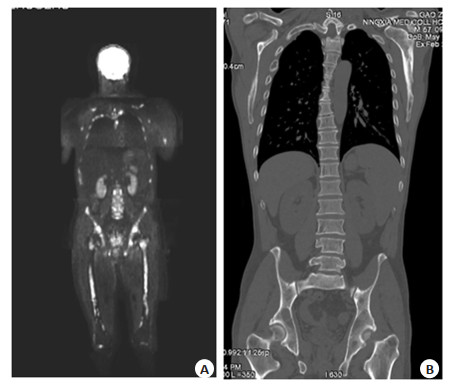
Objective To investigate the application value of PET/CT imaging technology based on 18F-PSMA-1007 in the non-invasive and accurate diagnosis of prostate cancer (PCa). Methods A total of 117 patients with suspected PCa admitted to Meizhou People 's Hospital from November 2020 to April 2022 were examined by 18F-PSMA-1007 PET/CT before biopsy. The maximum standardized uptake value of the lesion and liver was measured by delineating the region of interest, and the tumor background ratio (TBR) was calculated with the liver as the background. Combined with the pathological results after biopsy (64 cases of PCa and 53 cases of benign), the TBR differences between benign and malignant diseases were compared, and the ROC curve was drawn to evaluate their diagnostic efficacy, so as to obtain the best cutoff value. Results The TBR level of PCa patients was higher than that of benign patients, and there was significant difference between the two groups (P < 0.001). The ROC curve was plotted by TBR to diagnose PCa, and the area under the ROC curve was 0.881 (P < 0.001), and the sensitivity and specificity were 78.1% and 94.3%, respectively, when the cutoff value was taken as 0.955. Moreover, among the 14 PCa patients with TBR below the cutoff value, 7 had developed lymph nodes and/or bone metastases, which could be indirectly diagnosed as PCa. Conclusion 18F-PSMA-1007 PET/CT TBR has a high application value in distinguishing benign and malignant prostate lesions. Using TBR=0.955 as the cut-off value can achieve good diagnostic efficiency. Even if the TBR is lower than the cutoff value, the detection of metastases can be used as a supplementary diagnosis of PCa and further improve the diagnostic accuracy. 18F-PSMA-1007 PET/CT examination can noninvasively diagnose and stage the vast majority of PCa.
2024, 47(1): 7-13.
doi: 10.12122/j.issn.1674-4500.2024.01.02
Abstract:
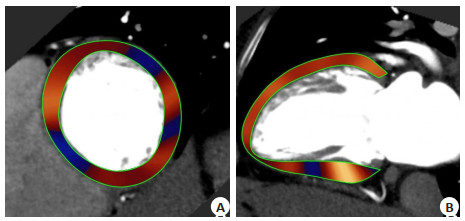
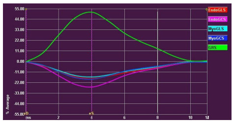
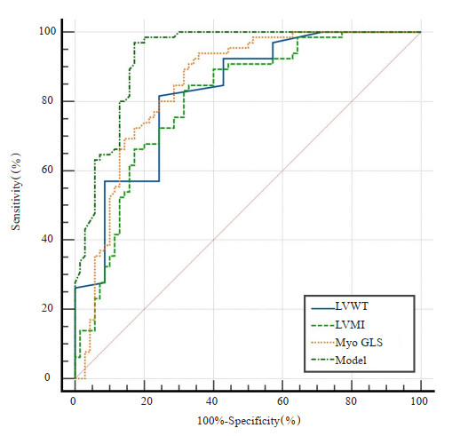
Objective To explore the feasibility of myocardial strain (MS) parameters obtained through CT technology for assessing early left ventricular function in hypertrophic cardiomyopathy (HCM) and hypertensive heart disease (HHD), as well as the capability of this parameter to differentiate between these two diseases, providing clinical reference. Methods This study was a retrospective analysis involving 205 adult participants with negative results from cardiac coronary imaging examinations conducted at the Xijing Hospital of Air Force Medical University from December 2021 to January 2023. Based on inclusion and exclusion criteria, the participants were categorized into three groups: HCM (n=70), HHD (n=65), and healthy control group (n=70). Subsequently, post-processing software was utilized to quantify left ventricular morphological characteristics, traditional cardiac function parameters, and MS parameters among the three groups. The differences in these parameters were compared and their discriminative abilities between the two diseases were assessed. Results Compared to the healthy control group, both the HCM and HHD groups exhibited increased maximal left ventricular wall thickness and left ventricular mass index (9.25±1.68 vs 15.32±1.67 vs 18.01±2.24; 56.64±19.57 vs 86.90±12.31 vs 106.27±19.56, respectively, P < 0.001). Meanwhile, the absolute values of MS were reduced in both groups (myocardial global circumferential strain: -25.80±3.74 vs -23.00±4.49 vs -21.03±4.97; endocardial global circumferential strain: -40.95±8.13 vs -35.86±7.90 vs -31.85±9.16; myocardial global radial strain: 81.26±37.76 vs 66.99±18.37 vs 55.31±23.19, P < 0.001), with the longitudinal strain showing the most significant decrease (myocardial global longitudinal strain: -23.03±3.84 vs -19.86±2.22 vs -15.47±4.28; endocardial global longitudinal strain: -30.35±5.35 vs -25.01±3.62 vs 21.92±8.16, P < 0.001). Multivariate Logistic regression analysis revealed that the combination model of maximal left ventricular wall thickness, left ventricular mass index, and myocardial global longitudinal strain had the largest area under the ROC curve of 0.930 (sensitivity: 97%, specificity: 83%). Conclusion The MS parameters obtained from CT imaging can accurately assess early left ventricular functional impairment in patients with HCM and HHD, with longitudinal strain showing the most significant impairment. Among the parameters obtained, the combination model of maximal left ventricular wall thickness, left ventricular mass index, and myocardial global longitudinal strain demonstrated the best effectiveness in distinguishing between these two diseases. These discoveries hold promise for providing more accurate diagnostic and therapeutic guidance in clinical practice.
2024, 47(1): 14-18.
doi: 10.12122/j.issn.1674-4500.2024.01.03
Abstract:
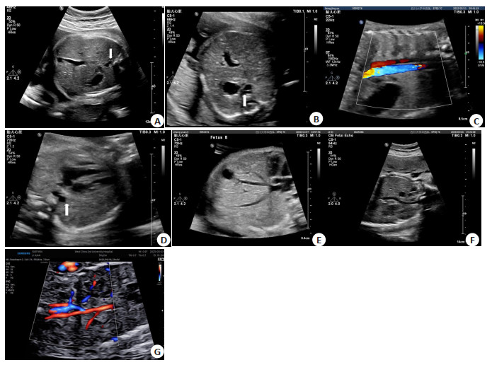
Objective To explore the value and clinical significance of prenatal ultrasound examination by summarizing the ultrasound image features of fetal inferior vena cava (IVC) development abnormalities. Methods A retrospective analysis was conducted on the ultrasound image characteristics and combined intracardiac and extracardiac abnormalities of 88 cases diagnosed with fetal IVC developmental abnormalities in our hospital from January 2018 to February 2023. Multiple tabular chi square tests were performed on the composition ratio of IVC developmental abnormalities with intracardiac and extracardiac abnormalities of different types. The Bonferroni method was used for multiple comparisons between groups. All the results with postpartum MRA, echocardiography, or autopsy results were compared. Results There were 88 fetuses with abnormal IVC development, including 18 ectopic connection of inferior vena cava (ECIVC) patients, 42 left inferior vena cava (LIVC) patients and 28 double inferior vena cava (DIVC) patients. 18 ECIVC fetuses showed abnormalities in both the oblique coronal and four chamber views of the chest and abdomen, with 17 cases showing abnormalities in the transverse section of the abdomen and 1 case showing atypical abnormalities in the transverse section of the abdomen; Two cases showed abnormal longitudinal section of the right atrium; 42 LIVC fetuses showed an "S" sign on the coronal section of the inferior vena cava on ultrasound; 28 cases of DIVC fetuses showed an "h" sign on the coronal section of the inferior vena cava on ultrasound. 88 fetuses with IVC developmental abnormalities were associated with abnormalities in the cardiovascular system, skeletal system, urinary system, digestive system, respiratory system, and sensory system, respectively. There was a statistically significant overall difference in the proportion of isolated and combined intracardiac and extracardiac abnormalities in different types of IVC development (P < 0.001); The ratio of isolated ECIVC to isolated DIVC and isolated LIVC, as well as the ratio of ECIVC with intracardiac abnormalities to LIVC and DIVC with intracardiac abnormalities, and it showed statistically significant differences (P < 0.05). 88 fetuses with abnormal IVC development, of which 5 were confirmed by induced labor; 83 cases were followed up until 6 months after birth, and all underwent MRI and echocardiography examinations, which were consistent with prenatal diagnosis. Conclusion Fetal IVC developmental abnormalities have specificity in ultrasound images. Ultrasound can accurately identify whether there are concomitant abnormalities in the endocardial and endocardial structures. Analyzing the composition ratio of different types of IVC developmental abnormalities combined with abnormalities in the endocardial and endocardial structures can provide theoretical basis for clinical evaluation, prognosis, and management. Improving understanding and early diagnosis have important guiding value for clinical practice.
2024, 47(1): 19-24.
doi: 10.12122/j.issn.1674-4500.2024.01.04
Abstract:
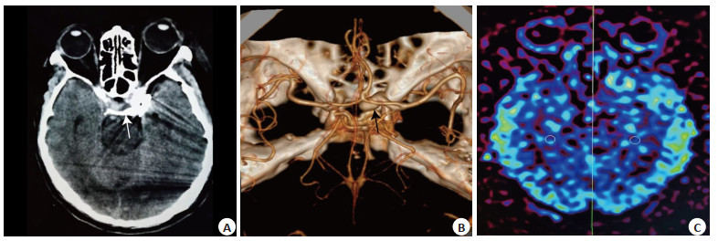
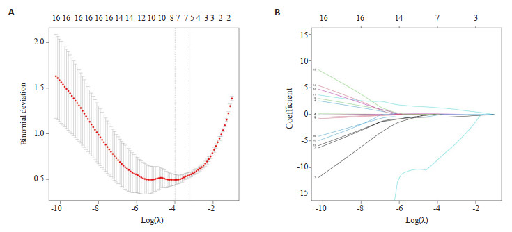
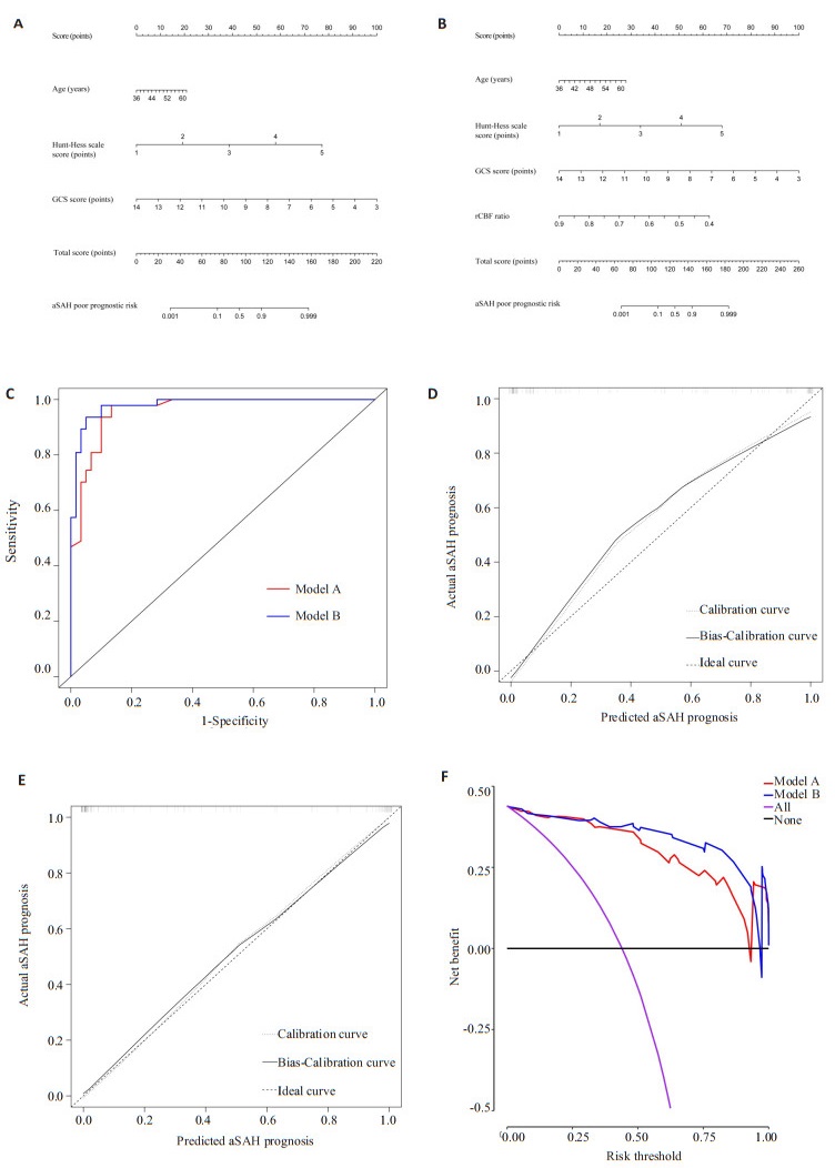
Objective To develop a short-term prognostic model for aneurysmal subarachnoid hemorrhage (aSAH) using arterial spin labeling and to assess the model's efficacy. Methods A total of 107 aSAH patients from our hospital underwent arterial spin labeling examination and were prospectively selected as study subjects. Patients were classified into good and poor prognosis groups based on their modified Rankin scale scores at 90 days post-discharge. Comparisons between the two groups were conducted for age, Hunt-Hess scale score, modified Fisher scale score, Glasgow coma scale (GCS) score, and blood cell parameters. Risk factors for the aSAH prognosis were then analyzed using LASSO-Logistic regression. A prognostic model was developed, and its effectiveness was evaluated using ROC curves, calibration curves, and decision curve analysis. Results Compared to the good prognosis group, the poor prognosis group exhibited higher levels of age, Hunt-Hess scale score, modified Fisher scale score, neutrophils, lymphocytes, neutrophils/lymphocytes and systemic immune inflammation index (P < 0.05). Conversely, the GCS score, platelets/lymphocytes, ipsilateral regional cerebral blood flow (rCBF) values, and rCBF ratio were lower than those in the good prognosis group (P < 0.05). LASSO-Logistic regression analysis revealed that age > 44 years old and a Hunt-Hess scale score of 3-5 were independent risk factors for the prognosis of aSAH (P < 0.05), while a GCS score > 9 and an rCBF ratio > 0.66 were independent protective factors for the prognosis of aSAH (P < 0.05). The model A, consisting of age, Hunt-Hess scale score, and GCS score, had an area under the ROC curve of 0.961, which was not statistically significantly different from the area under the ROC curve of model B, consisting of age, Hunt-Hess scale score, GCS score, and rCBF ratio (0.981) (P > 0.05). While the calibration curve of model A moderately overlapped with the ideal curve, model B exhibited a high overlap with the ideal curve. Moreover, the mean absolute error value was higher in model A (0.042) than in model B (0.014). The net benefit of model B outweighed that of model A across most of the risk threshold range. Conclusion The age, Hunt-Hess scale score, and GCS score of aSAH patients are significant in predicting poor prognosis. Incorporating the rCBF ratio into the model was found to enhance its value in assessing poor prognosis in aSAH patients.
2024, 47(1): 25-30.
doi: 10.12122/j.issn.1674-4500.2024.01.05
Abstract:
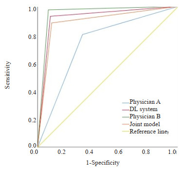
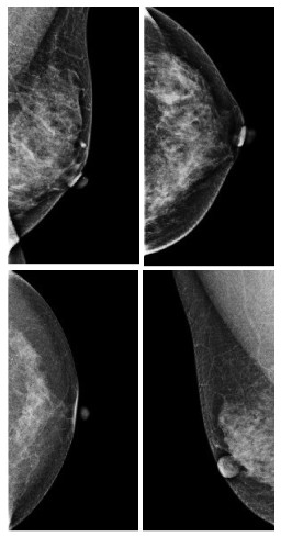
Objective To investigate the clinical value of the deep learning-based mammography-assisted diagnosis (DL) system for breast calcification detection and benign and malignant classification. Methods A retrospective analysis was performed on the craniocaudal and internal and external oblique imaging data of 400 patients who underwent bilateral mammography in Xuzhou Central Hospital from January 2020 to December 2022. The unanimous judgment of two associate chief physicians with more than 15 years of experience in mammography diagnosis was used as the standard group, the images were blinded and independently reviewed by 1 junior resident, 1 senior attending physician, and the DL system, respectively. After a 4-week washout period, the images were blinded and independently reviewed by the combined model (junior resident+DL system) again. Combined with two- way table chi- square test, the effects of different ACR types, morphology and distribution of calcification, and BI-RADS classification on the detection of calcification were evaluated. The area under the curve (AUC) was used to evaluate the difference in the detection of suspicious calcification among junior residents, senior attending physician, DL system and combined model (junior resident +DL system). Results A total of 975 suspicious calcifications of BI-RADS3 grade and above were detected in 1600 images (400 patients). The sensitivities of junior resident A, senior attending physician B, DL system and combined model were 81.95%, 96.62%, 93.03% and 96.41%, respectively. The sensitivity of senior attending physician B, DL system and combined model to calcification detection was not affected by breast ACR type, morphology and distribution of calcification, and BI- RADS classification, while the sensitivity of junior resident A was affected by it. The combined model (junior resident + DL system) had high AUC value, sensitivity and specificity in predicting the benign and malignant nature of calcifications, with 0.891, 90.0% and 88.2%, respectively, which differed from that of the junior resident (P < 0.01). With the help of the DL system, the diagnostic performance of the junior resident was significantly improved, and the AUC value increased from 0.740 to 0.891. Conclusion The DL system is highly sensitive to the detection of suspicious calcifications of BI-RADS 3 grade and above, and has a high classification performance of benign and malignant calcifications, which is comparable to that of senior attending physician. With the help of the DL system, the junior resident can reduce the missed diagnosis of calcification and misdiagnosis, and improve the accuracy of breast cancer screening and diagnosis.
2024, 47(1): 31-35.
doi: 10.12122/j.issn.1674-4500.2024.01.06
Abstract:
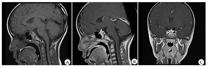
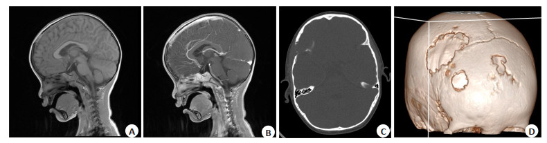
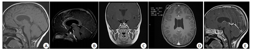
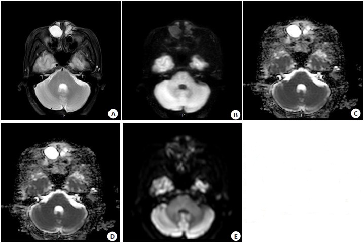
Objective To investigate the MRI and clinical characteristics of Langerhans cell histiocytosis (LCH) of pituitary gland in children. Methods We conducted a retrospective analysis the MRI data of the 23 children with pituitary LCH confirmed by pathological from November 2014 to August 2023, including 12 males and 11 females with an average age of 1 year to 12 years 6 months (6.72±3.33 years old). The lesion location, pituitary height, presence of neurohypophysis, pituitary stalk shape, size, pituitary reinforcement, and pituitary/pineal concomitant lesions were observed. The MRI findings of pituitary LCH were summarized. Results The main reason for the first visit was diabetes insipidus, and other symptoms were head mass in 2 cases, head tenderness in 1 case and neck mass in 1 case. Only pituitary was involved in 16 cases, and the rest were multisystem involved. Craniofacial bones were often involved outside the pituitary, and lungs and ribs were rarely involved. The adenohypophyseal was significantly depressed and thin in 1 case and full/bulging in 4 cases. No neurohypophysial hypersignal was found in all cases. The adenohypophyseal was significantly depressed and thin in 1 case and full/bulging in 4 cases. No neurohypophysial hypersignal was found in all cases. There were 13 cases of pituitary stalk thickening, 2 cases of fine, 1 case of funnel line stenosis, the upper segment nodular thickening, 7 cases without obvious thickening/fine. The pituitary was enhanced uniformly after enhancement. There were 3 cases with rathke cyst/remnant cavity and 2 cases with pineal cyst. Conclusion The MRI manifestations of pituitary LCH in children have certain characteristics, which is helpful for diagnosis.
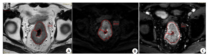
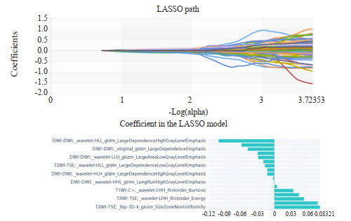
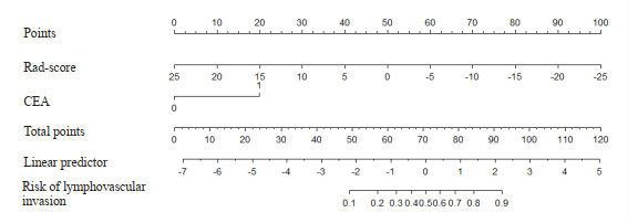
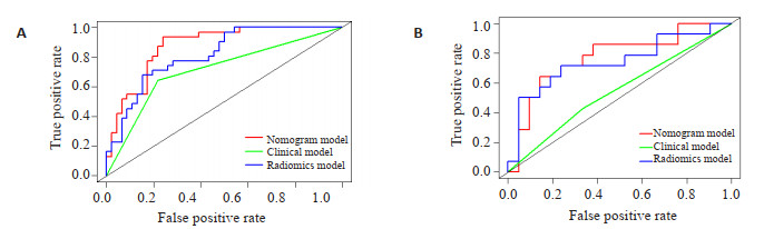
2024, 47(1): 36-41.
doi: 10.12122/j.issn.1674-4500.2024.01.07
Abstract:
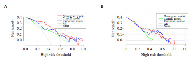
Objective To explore the value of a nomogram model based on multi-parametric MRI radiomics combined with clinical risk factors in preoperative prediction of lymphovascular invasion of rectal cancer. Methods A total of 112 patients who underwent preoperative multi-parametric MRI examination and were confirmed as rectal adenocarcinomas by postoperative pathology in the First Affiliated Hospital of Bengbu Medical College were retrospectively analyzed. The clinical and pelvic imaging data of the patients were collected, and they were randomly divided into training set and validation set at a ratio of 7:3. Clinical independent risk factors related to lymphovascular invasion in rectal cancer were selected through single-multiple Logistic regression analysis. Regions of interest were manually delineated on T2WI, diffusion weighted imaging and T1WI enhanced sequences, and radiomics characteristics were extracted. The optimal radiomics characteristics were selected through characteristics dimension reduction, and a radiomics model was constructed. A nomogram model was built by combining clinical predictive factors with radiomics score labels. The predictive efficiency of the model was evaluated adopting area under the ROC curve, calibration curve and decision curve analysis. Results The nomogram model showed excellent predictive efficiency, with an area under the curve of 0.876(95% CI: 0.799-0.952)and 0.769(95% CI: 0.600-0.938)for the training set and validation set respectively, which was significantly higher than the radiomics model (0.818, 0.741) and clinical model (0.714, 0.548). Conclusion The nomogram model exhibited excellent predictive efficiency in predicting lymphovascular invasion of rectal cancer, which can provide important guidance for clinical decision-making preoperatively.
2024, 47(1): 42-46.
doi: 10.12122/j.issn.1674-4500.2024.01.08
Abstract:
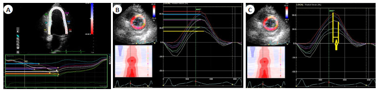
Objective To discuss changes in cardiac function from early stage after aneurysmal subarachnoid hemorrhage (aSAH) to shortly later stage using echocardiography. Methods We prospectively studied 83 patients with aSAH in Beijing Hospital of Traditional Chinese Medicine from May 2018 to May 2023. These patients were divided into the highly positive cTnI group (cTnI≥0.3 ng/mL, n=25) and the mildly positive cTnI group (cTnI < 0.3 ng/mL, n=58) according to cTnI level. Longitudinal strain and radial strain were acquired by strain echocardiography. Global longitudinal strain (GLS), the standard deviation of time-to-peak longitudinal strain from LV segments (LT-Dispersion), the standard deviation of time‑to‑peak radial strain from LV segments (RT‑Dispersion) and the time‑to‑peak radial strain difference between LV anterior septum and posterior wall in left ventricular short axis view at papillary muscle level (APT) were measured and calculated. Results At early stage, LT-Dispersion, RT-Dispersion, APT and GLS in highly positive cTnI group were associated with more serious neurocardiac damage than others (P < 0.05). Improvements (later stage vs early stage) of GLS, LT‑Dispersion, RT‑Dispersion and APT were more significantly better in the mildly positive cTnI group than in the highly positive cTnI group (P < 0.05). Conclusion GLS and left ventricular regional discoordination measured by LT-Dispersion, RT-Dispersion and APT were significantly associated with neurocardiac injury severity after aSAH as reflected by elevated cTnI. Novel echocardiographic analysis to detect neurocardiac injury severity in patients with aSAH has promise for clinical utility.
2024, 47(1): 47-51.
doi: 10.12122/j.issn.1674-4500.2024.01.09
Abstract:
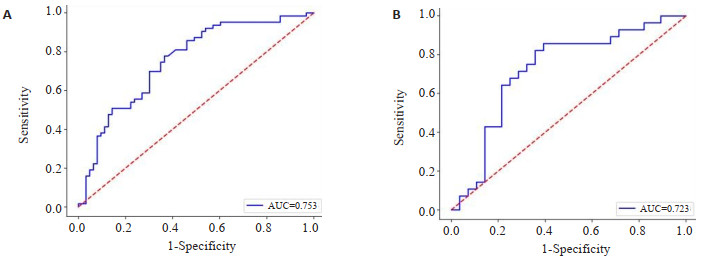
Objective To explore the diagnostic value of whole body magnetic resonance diffusion weighted imaging (WB-DWI) combined with multi-slice spiral CT in multiple myeloma. Methods A total of 80 patients with multiple myeloma admitted to our hospital from June 2019 to January 2023 were selected as study subjects and divided into WB-DWI group (n=25), multi-slice spiral CT group (n=25) and combined group (n=30). The detection rates of WB-DWI and multi-slice spiral CT alone and combined in the involved sites of patients were analyzed, and the detection of multiple myeloma by the two methods alone and combined was analyzed. The clinical value of WB-DWI and multi-slice spiral CT combined in the diagnosis of multiple myeloma was analyzed by ROC curve. Results The results were mainly correlated with Durie-Salmon stage and international myeloma stage (P < 0.05). Compared with the single detection of WB-DWI and multi-slice spiral CT, the detection rate of the involved sites in the combined detection was higher (P < 0.05). The combined detection of WB-DWI and multi-slice spiral CT was higher than that of single detection in the diagnosis of multiple myeloma (P < 0.05). Compared with the single diagnosis of WB-DWI and multi-slice spiral CT, the sensitivity, specificity and accuracy of the combined diagnosis of multiple myeloma were higher (P < 0.05). Conclusion Compared with WB-DWI and multi-slice spiral CT single tests, the combined test can effectively improve the pathological detection of patients and improve the diagnostic value of multiple myeloma.

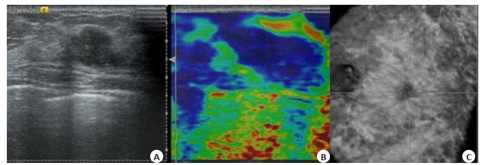
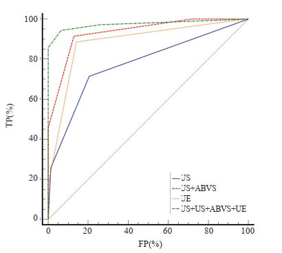
2024, 47(1): 52-56.
doi: 10.12122/j.issn.1674-4500.2024.01.10
Abstract:
Objective To explore the application value of automatic breast volume scanning (ABVS), ultrasound elastography score (UES) and their combination in correcting the BI-RADS 4 breast lesions. Methods A total of 113 patients with 109 breast lesions diagnosed with BI-RADS 4 by conventional ultrasound were collected.After the BI-RADS grading was corrected by ABVS and UES, the ROC curve was compared with the pathological results, and the differences in the diagnosis of BI-RADS 4 breast lesions by conventional ultrasound, ABVS, UES, ABVS combined with UES were compared. Results Among 113 masses in 109 patients, 78 were benign and 35 were malignant.The sensitivity, specificity, accuracy and area under the ROC curve of ABVS combined with UES were 94.29%, 93.59%, 93.80% and 0.975 respectively. Conclusion US+ABVS+UES can significantly improve the diagnostic efficiency and accuracy of US in the diagnosis of BI-RADS 4 breast lesions.
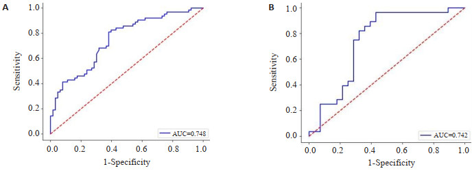
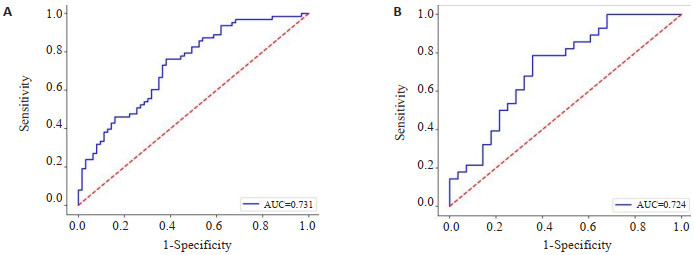
2024, 47(1): 57-63.
doi: 10.12122/j.issn.1674-4500.2024.01.11
Abstract:
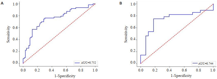
Objective To explore the potential value of radiomics model based on different MRI sequences of breast combined with clinicopathological factors in predicting sentinel lymph node metastasis of breast cancer. Methods We retrospectively analyzed 182 cases of breast cancer with sentinel lymph node metastasis diagnosed by pathology, including 91 in the sentinel lymph node positive group and 91 in the sentinel lymph node negative group, and divided them into a training group (64 positive and 64 negative) and a validation group (27 positive and 27 negative) according to the ratio of 7:3. The clinical, imaging and pathological data of breast cancer patients were analyzed by univariate and multivariate logistic regression, and the independent risk factors related to sentinel lymph node metastasis of breast cancer were screened out. Based on T2WI, diffusion-weighted imaging and dynamic contrast enhancement, the best imaging features were extracted, and several singlesequence and multi- sequence radiomics label scores were constructed respectively, and the combined radiomics prediction model was constructed combined with the above independent risk factors of clinical, pathological and imaging features. The effectiveness of each model in predicting breast cancer sentinel lymph node metastasis was evaluated by plotting the ROC curves and calculating the area under the curve (AUC). Results Peritumoural edema (P < 0.001), tumour long diameter (P < 0.001), tumour short diameter (P < 0.001), pathological grade (P < 0.001) and vascular infiltration (P < 0.001), burr sign (P=0.006), diffusion-weighted imaging rim high signal sign (P=0.028) and ADC value (P < 0.001) were the independent clinicopathological factors of anterior sentinel lymph node metastasis in breast cancer. Among the radiomics label scores, the multi- sequence radiomics label score of T2WI+ diffusion-weighted imaging+dynamic contrast enhancement had the best predictive efficiency, its AUC in the validation group was 0.744, and the predictive efficiency of the combined radiomics prediction model established by combining clinical, pathological and imaging feature independent risk factors had been further improved, and its AUC in the validation group was 0.834. Conclusion The breast MRI-based imaging radiomic model can effectively predict sentinel lymph node metastasis in breast cancer prior to surgery
2024, 47(1): 64-70.
doi: 10.12122/j.issn.1674-4500.2024.01.12
Abstract:
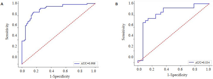
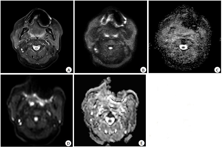
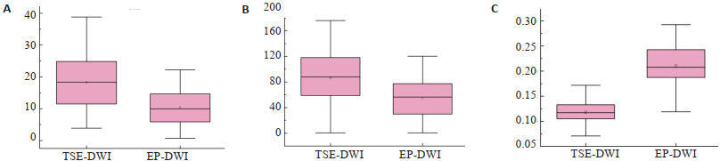
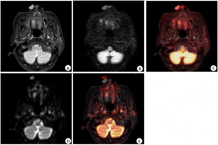
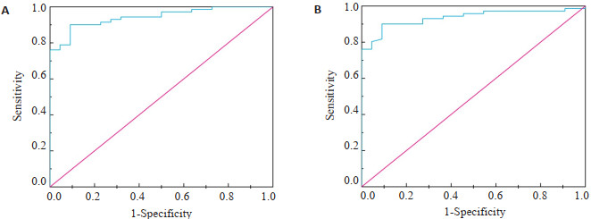
Objective To prospectively compare the difference in image quality between turbo spin-echo diffusion- weighted imaging (TSE- DWI) and planar echo diffusion- weighted imaging (EP-DWI) for nasal sinus lesions. To investigate the diagnostic value of the apparent diffusion coefficient (ADC) values of TSE-DWI and EP- DWI in identifying benign and malignant nasal sinus lesions. Methods Ninety-three patients with nasal sinus lesions seen in our hospital from October 2022 to August 2023 were prospectively selected for the study. A Philips 1.5T MRI scanner was used for scanning, and all patients underwent TSE-DWI and EP-DWI imaging after routine nasal sinus MRI plain scanning. Image quality was compared through subjective and objective evaluation of TSE-DWI and EP-DWI images by two physicians. The agreement between the two physicians, the agreement between the ADC values of the two DWI sequences, and the Pearson correlation coefficient were evaluated. The differences in ADC values of benign and malignant nasal sinus lesions were compared and the diagnostic efficacy of ADC values for identifying benign and malignant nasal sinus lesions was calculated using ROC curves. Results The mean values of all subjective scores, signal-to-noise ratios and contrast-to-noise ratios of TSE-DWI images were higher than those of EP-DWI (P < 0.05), and the distortion rate of TSE-DWI images was lower than that of EP-DWI (P < 0.05). Regarding subjective scoring of images, the agreement between the two physicians was good (Kappa>0.4). For ADC value measurement, the agreement between two physicians was excellent (ICC>0.74). The agreement between the ADC values of the two DWI sequences was excellent (ICC>0.74) and showed a very strong positive correlation (Pearson's correlation coefficient>0.8). The difference in ADC values for benign and malignant lesions was statistically significant (P < 0.05). The ADC values of the two DWI sequences had the same sensitivity, specificity, positive predictive value, negative predictive value and accuracy in identifying benign and malignant lesions of the nasal sinuses, which were 90.1%, 90.9%, 97.0%, 74.1%, 90.3%, respectively. Conclusion The magnetic resonance TSE-DWI sequence can effectively improve the image quality of nasal sinus sites and is suitable for a wide range of clinical applications. The ADC value has high diagnostic efficacy in identifying benign and malignant nasal sinus lesions.
2024, 47(1): 71-77.
doi: 10.12122/j.issn.1674-4500.2024.01.13
Abstract:
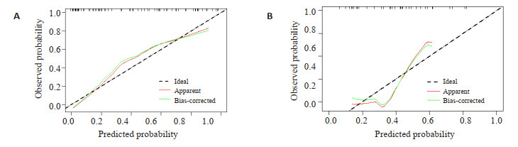
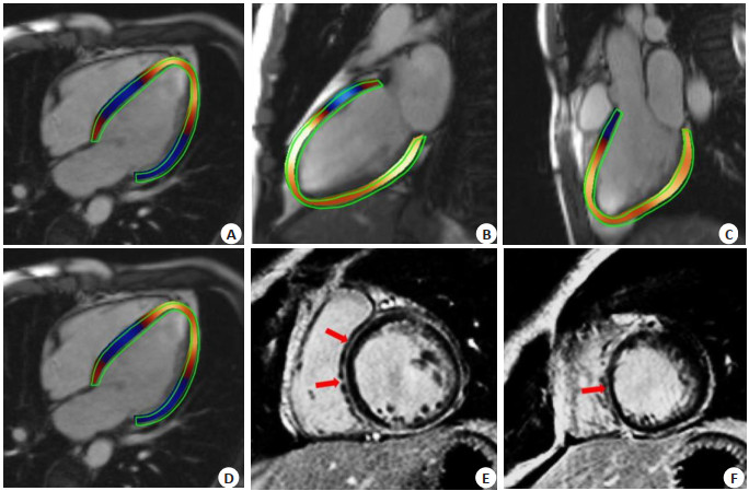
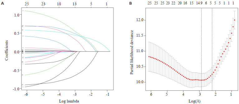
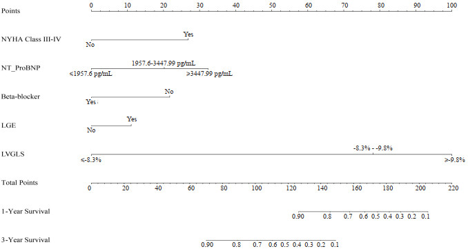
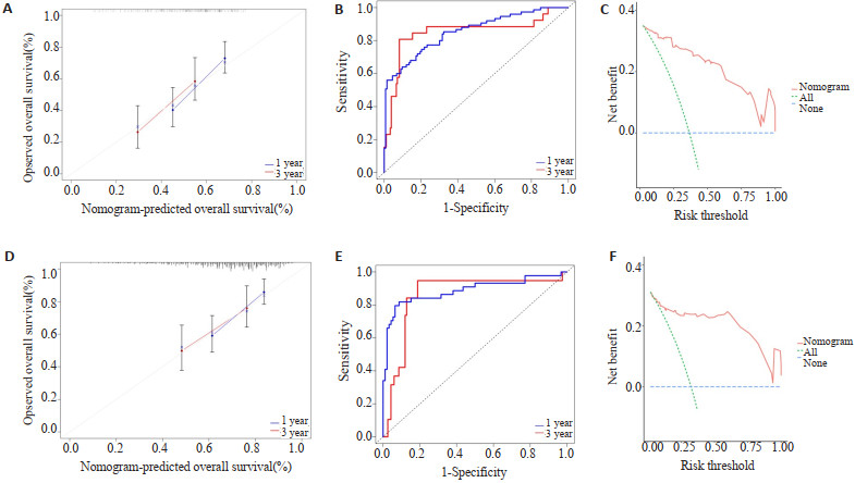
Objective To investigate the predictive value of cardiac magnetic resonance (CMR) parametric nomogram modeling for major adverse cardiovascular events (MACE) in elderly patients with dilated cardiomyopathy (DCM). Methods A total of 173 elderly patients with DCM who underwent CMR at Xiaogan Hospital of Wuhan University of Science and Technology from July 2017 to July 2020 were retrospectively analyzed, and they were randomly divided into a training set (n=104) and a test set (n=69) in a ratio of 6:4. Potential predictors were screened by Lasso regression and multifactorial Cox regression, and the MACE nomogram prediction model for DCM patients was constructed with these factors. The nomogram model was evaluated and validated by calibration curve, ROC curve, decision curve analysis, and Kaplan-Meier survival analysis. Results The median follow-up was 29.7 (16.4, 45.4) months. At the end of follow-up, MACE occurred in 59 (34.1%) patients. A total of 9 potential predictors were screened by LASSO regression and cross-validation. The results of multifactorial Cox regression analysis showed that NYHA class Ⅲ-Ⅳ, N-terminal-brain natriuretic peptide precursor, beta-blocker, CMR late gadolinium enhancement, and overall longitudinal strain of the left ventricle were risk factors for the development of MACE in patients with DCM, and a nomogram prediction model was constructed with these indicators. In the training and test sets, the calibration plots showed that the nomogram predicted 1-year and 3-year survival in good agreement with the actual survival. The area under the ROC curve for 1-year and 3-year survival prediction in the training set was 0.850 (95% CI: 0.748-0.953) and 0.853 (95% CI: 0.797-0.909), respectively, and that for 1-year and 3-year survival prediction in the test set was 0.858 (95% CI: 0.758-0.959) and 0.887 (95% CI: 0.816-0.958), respectively. The results of the decision curve analysis showed that the nomogram model had a higher net clinical benefit rate. The results of Kaplan-Meier survival analysis showed that patients in the high-risk group of the predictive model had a reduced probability of survival compared with the low-risk group (P < 0.05). Conclusion In this study, we constructed a nomogram prediction model for the occurrence of MACE in elderly patients with DCM by clinical and CMR characteristic parameters, which has good calibration, differentiation and clinical application value.
2024, 47(1): 78-82.
doi: 10.12122/j.issn.1674-4500.2024.01.14
Abstract:
Objective To compare the diagnostic value of single and dual phase of enhanced CT in thyroid nodules, and explore the best scanning scheme. Methods A total of 171 nodules in 153 patients were included from January 2020 to June 2022 according to inclusion and exclusion criteria. Image sequence with a delay of 50 seconds was taken for diagnosis in the group of single phase of enhanced CT, then image sequences with a delay of 25 and 50 seconds were taken for diagnosis in the group of dual phase of enhanced CT. Diagnostic accuracy of benign and malignant thyroid nodules, characteristics of thyroid nodules, positive lymph node and enhanced CT features of thyroid nodules in two scanning schemes were the main evaluation indicators. Results There was no statistical significance in diagnostic accuracy of benign and malignant thyroid nodules between single and dual phase of enhanced CT. There was no statistical significance in edge, morphology, capsule, density, calcification and lymph node of thyroid nodules between two scanning scheme. There was no statistical significance in different densities of benign and malignant thyroid nodules between two scanning scheme. There was no statistical significance in enhancement mode of benign and malignant thyroid nodules in dual phase of enhanced CT. Conclusion There is no significant difference in the diagnostic value of single and dual phase of enhanced CT in thyroid nodules, and single phase of enhanced CT would be more suitable for clinical promotion and application by its lower radiation dose.
2024, 47(1): 83-87.
doi: 10.12122/j.issn.1674-4500.2024.01.15
Abstract:
Objective To analyze the multi- slice CT (MSCT) features of patients with smear- negative active pulmonary tuberculosis and construct a diagnostic model. Methods A total of 1016 patients with pulmonary diseases who were admitted to our hospital were enrolled from January 2020 to January 2023, and they were divided into smear- negative pulmonary tuberculosis group (n=478) and non-tuberculous pulmonary disease group (n=538, including 200 cases of lung cancer and 338 cases of pneumonia) according to clinical diagnosis. All patients received MSCT examination, and their MSCT imaging features were analyzed. Logistic regression analysis was conducted to identify the signs related to the diagnosis of smearnegative active pulmonary tuberculosis, and a diagnostic model for smear- negative active pulmonary tuberculosis was constructed. The diagnostic efficiency of the model was evaluated with AUC. Results There were statistically significant differences in MSCT signs such as tree in bud, centrilobular nodule, cavity, calcification and lobulation between the two groups (P < 0.05). Logistic regression analysis found that tree in bud sign, centrilobular nodule and cavity were independent risk factors for smear-negative active pulmonary tuberculosis (P < 0.05). The equation of the combined diagnosis model constructed based on logistic regression analysis was as follow: Log(P)=-1.256+1.455×tree in bud sign+0.982×centrilobular nodule+1.023×cavity. The AUC, sensitivity and specificity of this model were 0.825, 93.94% and 70.97%, respectively. Conclusion The diagnostic model constructed based on MSCT imaging features is of high value in diagnosis of smear- negative active pulmonary tuberculosis and can provide a reliable basis for clinical diagnosis and treatment of the disease.

2024, 47(1): 88-92.
doi: 10.12122/j.issn.1674-4500.2024.01.16
Abstract:
Primary hyperparathyroidism (PHPT) is a common endocrine disease, often accompanied by calcium and phosphorus metabolism disorders and multi-system lesions. Surgical resection is the main method for the treatment of PHPT. With the popularization and application of minimally invasive surgery, preoperative molecular imaging has become an urgent need for accurate localization. Radionuclide imaging of dual-phase 99mTc-MIBI SPECT/CT, 11C-choline and 18F-Fluorocholine PET/CT have always been the focus of preoperative localization research for PHPT. The basic imaging principles of 99mTc-MIBI, 11C-choline and 18F-Fluorocholine are presented. The advantages and disadvantages of these radionuclide imaging methods in the clinical applications of PHPT preoperative localization in ectopic/recurrent lesions, polyglandular lesions, inconsistent between PTH elevation with radionuclide imaging findings, and previous neck surgery history were reviewed.
Primary hyperparathyroidism (PHPT) is a common endocrine disease, often accompanied by calcium and phosphorus metabolism disorders and multi-system lesions. Surgical resection is the main method for the treatment of PHPT. With the popularization and application of minimally invasive surgery, preoperative molecular imaging has become an urgent need for accurate localization. Radionuclide imaging of dual-phase 99mTc-MIBI SPECT/CT, 11C-choline and 18F-Fluorocholine PET/CT have always been the focus of preoperative localization research for PHPT. The basic imaging principles of 99mTc-MIBI, 11C-choline and 18F-Fluorocholine are presented. The advantages and disadvantages of these radionuclide imaging methods in the clinical applications of PHPT preoperative localization in ectopic/recurrent lesions, polyglandular lesions, inconsistent between PTH elevation with radionuclide imaging findings, and previous neck surgery history were reviewed.
2024, 47(1): 93-97.
doi: 10.12122/j.issn.1674-4500.2024.01.17
Abstract:
Hepatocellular carcinoma is one of the common malignant tumours. It has a high degree of malignancy and poor prognosis and is characterized by high morbidity and mortality. Radiomics provides a quantitative analysis method. It transforms histopathology and tumour biology information in medical images into high-dimensional quantitative feature information and combines with artificial intelligence algorithms for data mining and statistical analysis to assist in the early clinical diagnosis and treatment of tumours. This paper reviews the research progress of radiomics in differential diagnosis, pathological grading, microvascular invasion, and immunohistochemical marker prediction of hepatocellular carcinoma. Meanwhile, it discusses the deficiencies in data volume and model reliability. It points out that it can be developed towards multicentre and multitasking to provide a reference for the auxiliary diagnosis of hepatocellular carcinoma.
Hepatocellular carcinoma is one of the common malignant tumours. It has a high degree of malignancy and poor prognosis and is characterized by high morbidity and mortality. Radiomics provides a quantitative analysis method. It transforms histopathology and tumour biology information in medical images into high-dimensional quantitative feature information and combines with artificial intelligence algorithms for data mining and statistical analysis to assist in the early clinical diagnosis and treatment of tumours. This paper reviews the research progress of radiomics in differential diagnosis, pathological grading, microvascular invasion, and immunohistochemical marker prediction of hepatocellular carcinoma. Meanwhile, it discusses the deficiencies in data volume and model reliability. It points out that it can be developed towards multicentre and multitasking to provide a reference for the auxiliary diagnosis of hepatocellular carcinoma.
2024, 47(1): 98-101.
doi: 10.12122/j.issn.1674-4500.2024.01.18
Abstract:
Subjective cognitive decline is considered to be the first clinical manifestation of the Alzheimer's disease continuum, preceding mild cognitive impairment. Its cognitive changes are characterized by subtle cognitive decline and compensatory cognitive effort, and have been shown to be a high-risk stage of Alzheimer's disease. Studying people with subjective cognitive decline is important to understanding the pathological mechanisms of early Alzheimer's disease and identifying biomarkers associated with subjective cognitive decline, and early diagnosis and intervention can effectively improve patient outcomes. With the advent of advanced neuroimaging techniques such as positron emission tomography and MRI, a growing body of evidence is revealing alterations in brain structure and function associated with symptoms of subjective cognitive decline. This study mainly reviewed the current research status of diagnosis and prediction of subjective cognitive decline from the perspectives of structural magnetic resonance imaging, diffusion tensor imaging, functional magnetic resonance imaging and machine learning, in order to reveal its neurophysiological mechanism and provide imaging basis for early diagnosis.
Subjective cognitive decline is considered to be the first clinical manifestation of the Alzheimer's disease continuum, preceding mild cognitive impairment. Its cognitive changes are characterized by subtle cognitive decline and compensatory cognitive effort, and have been shown to be a high-risk stage of Alzheimer's disease. Studying people with subjective cognitive decline is important to understanding the pathological mechanisms of early Alzheimer's disease and identifying biomarkers associated with subjective cognitive decline, and early diagnosis and intervention can effectively improve patient outcomes. With the advent of advanced neuroimaging techniques such as positron emission tomography and MRI, a growing body of evidence is revealing alterations in brain structure and function associated with symptoms of subjective cognitive decline. This study mainly reviewed the current research status of diagnosis and prediction of subjective cognitive decline from the perspectives of structural magnetic resonance imaging, diffusion tensor imaging, functional magnetic resonance imaging and machine learning, in order to reveal its neurophysiological mechanism and provide imaging basis for early diagnosis.
2024, 47(1): 102-106.
doi: 10.12122/j.issn.1674-4500.2024.01.19
Abstract:
In the field of high-intensity focused ultrasound ablation of tumors, the ideal multi-mode and multi-functional nano-therapeutic agent is not only able to target the region of interest, but also achieve specific imaging diagnosis. The combined application can exert synergistic effects and achieve better therapeutic effects. This paper presents a review of the progress of research on inorganic nanoparticles, organic nanoparticles, nanoparticles containing targeted genes, and chemotherapeutic drug-centered nanoparticles in high intensity focused ultrasound ablated tumors.
In the field of high-intensity focused ultrasound ablation of tumors, the ideal multi-mode and multi-functional nano-therapeutic agent is not only able to target the region of interest, but also achieve specific imaging diagnosis. The combined application can exert synergistic effects and achieve better therapeutic effects. This paper presents a review of the progress of research on inorganic nanoparticles, organic nanoparticles, nanoparticles containing targeted genes, and chemotherapeutic drug-centered nanoparticles in high intensity focused ultrasound ablated tumors.