


CT can improve the diagnostic accuracy in the staging of silicosis
-
摘要:
目的 探讨CT对矽肺分期的诊断价值。 方法 选取我院2022年1月~2023年1月的80例矽肺患者,所有患者均行CT检查,对比CT对矽肺分期的诊断结果及其临床诊断结果的一致性,计算CT对矽肺分期的诊断效能。 结果 CT征象分析显示,80例矽肺患者中,20例患者在双肺上、中、下区均存在结节,36例患者在4个肺区存在结节,24例患者在2个肺区存在结节;39例患者无团块,13例患者团块为1~3 cm,28例患者团块 > 3 cm;54例患者存在肺结节密度增加;80例患者存在不同程度淋巴结肿大,57例患者发生于两肺门区淋巴结,23例患者发生于纵隔;60例患者存在胸膜增厚,累及叶间胸膜和脏壁胸膜。临床诊断结果显示,80例矽肺患者中,Ⅰ期24例,Ⅱ期36例,Ⅲ期20例;CT检查结果显示,Ⅰ期26例,Ⅱ期35例,Ⅲ期19例,与临床诊断结果的Kappa值分别为0.884、0.823、0.898,提示两者具有良好的一致性。CT对矽肺分期诊断的总准确度91.25%,CT对Ⅰ期、Ⅱ期、Ⅲ期分期诊断的准确率分别为95.00%、91.25%、96.25%,敏感度分别为95.83%、88.89%、90.00%,特异性分别为94.64%、93.18%、98.33%,阳性预测值分别为88.46%、91.43%、94.74%,阴性预测值分别为98.15%、91.11%、96.72%。 结论 CT在矽肺患者诊断中起着积极作用,能提高矽肺分期的诊断准确性,具有良好的诊断效能。 Abstract:Objective To explore the diagnostic value of CT in the staging of silicosis. Methods Eighty silicosis patients from our hospital from January 2022 to January 2023 were selected, and all patients underwent CT examination. The diagnostic results of CT for silicosis staging and the consistency of clinical diagnostic results were compared, and the diagnostic efficacy of CT for silicosis staging was calculated. Results The CT sign analysis showed that among 80 silicosis patients, 20 patients had nodules in the upper, middle, and lower regions of both lungs, 36 patients had nodules in 4 lung regions, and 24 patients had nodules in 2 lung regions; 39 patients had no masses, 13 patients had masses ranging from 1 to 3cm, and 28 patients had masses greater than 3cm; 54 patients had increased pulmonary nodule density; 80 patients had different degrees of Lymphadenopathy, 57 patients had lymph nodes in hilar region, 23 patients had in mediastinum; 60 patients had pleural thickening, involving interlobular pleura and visceral pleura. The clinical diagnosis results showed that there were 24 cases in stage Ⅰ, 36 cases in stage Ⅱ, and 20 cases in stage Ⅲ among 80 silicosis patients; The CT examination results showed that there were 26 cases in Phase Ⅰ, 35 cases in Phase Ⅱ, and 19 cases in Phase Ⅲ. The Kappa value was 0.884, 0.823, 0.898, respectively, indicating good consistency between the two. The overall accuracy of CT in staging diagnosis of silicosis was 91.25%. The accuracy of CT in staging diagnosis of stage Ⅰ, stage Ⅱ and stage Ⅲ was 95.00%, 91.25%, 96.25%, respectively. The sensitivity was 95.83%, 88.89%, 90.00%, respectively. The specificity was 94.64%, 93.18%, 98.33%, respectively. The positive predictive value was 88.46%, 91.43%, 94.74%, respectively, and the negative predictive values was 98.15%, 91.11%, 96.72%, respectively. Conclusion CT plays a positive role in the diagnosis of silicosis patients, can improve the diagnostic accuracy of silicosis staging, and has good diagnostic efficacy. -
Key words:
- silicosis /
- CT /
- clinical staging /
- diagnostic value
-

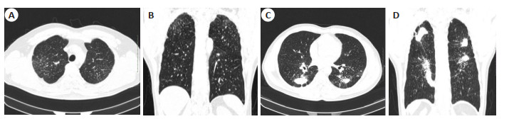
图 1 矽肺患者CT图像
Figure 1. CT images of silicosis. A, B: View of axial and coronal showed that patients had nodules in the upper, middle, and lower regions of both lungs; C, D: Another patients, view of axial and coronal showed that patients had masses and diffuse small nodules.
表 1 CT对矽肺分期的诊断结果及其与临床诊断结果的一致性分析
Table 1. The consistency analysis of the diagnostic results of CT for silicosis staging and the clinical diagnostic results (n)
CT results Clinical diagnosis results Total Kappa value Stage Ⅰ Stage Ⅱ Stage Ⅲ Stage Ⅰ 23 3 0 26 0.884 Stage Ⅱ 1 32 2 35 0.823 Stage Ⅲ 0 1 18 19 0.898 Total 24 36 20 80  下载: 导出CSV
表 2 CT对矽肺分期的诊断效能
Table 2. The diagnostic efficacy of CT for silicosis staging (%)
CT examination results Accuracy Sensitivity Specificity Positive predictive value Negative predictive value Stage Ⅰ 95.00(76/80) 95.83(23/24) 94.64(53/56) 88.46(23/26) 98.15(53/54) Stage Ⅱ 91.25(73/80) 88.89(32/36) 93.18(41/44) 91.43(32/35) 91.11(41/45) Stage Ⅲ 96.25(77/80) 90.00(18/20) 98.33(59/60) 94.74(18/19) 96.72(59/61)
下载: 导出CSV
-
[1] Carneiro APS, da Silva LL, Silva FDCL, et al. Volume-based tomography for the diagnosis of incipient silicosis in former gold miners[J]. Occup Environ Med, 2022, 79(6): 427-32. doi: 10.1136/oemed-2021-107922 [2] Ferrante P. Costs of asbestosis and silicosis hospitalization in Italy (2001–2018)[J]. Int Arch Occup Environ Health, 2021, 94(4): 763-71. doi: 10.1007/s00420-020-01637-z [3] 庞勇, 潘力平, 唐煌, 等. 四川省江油市矽肺流行病学和影像学特征及其与矽肺分期的关联性[J]. 职业与健康, 2020, 36(2): 161-4. https://www.cnki.com.cn/Article/CJFDTOTAL-ZYJK202002006.htm [4] Dixit R, Jalutharia J, Gupta A, et al. Measurement of diffusion lung capacity (DLCO) in silicosis patients: correlation with radiographic abnormalities on high-resolution CT scan chest[J]. Lung Ind, 2022, 39(4): 352. doi: 10.4103/lungindia.lungindia_280_21 [5] 王成霞, 柳澄, 仇路, 等. 铝尘肺与矽肺患者胸部CT影像特征的对照研究[J]. 中华劳动卫生职业病杂志, 2021, 39(7): 534-7. [6] Jones CM, Pasricha SS, Heinze SB, et al. Silicosis in artificial stone workers: spectrum of radiological high-resolution CT chest findings [J]. J Med Imag Rad Onc, 2020, 64(2): 241-9. doi: 10.1111/1754-9485.13015 [7] 孙良璋, 张晋, 李建忠, 等. 呼吸门控定量CT在评估矽肺功能中的价值[J]. 中国医学装备, 2022, 19(12): 60-4. https://www.cnki.com.cn/Article/CJFDTOTAL-YXZB202212013.htm [8] 胡必锋, 朱胜康, 翟荣存, 等. CT小阴影密集度判定方法及参考片在矽肺诊断中的应用[J]. 中华放射学杂志, 2021, 55(11): 1172-7. [9] 陆聪. DR、CT、HRCT对矽肺的影像学诊断价值分析[J]. 中国卫生标准管理, 2018, 9(22): 115-6. https://www.cnki.com.cn/Article/CJFDTOTAL-WSBZ201822051.htm [10] 中华人民共和国国家卫生和计划生育委员会. 职业性尘肺病的诊断: GBZ 70-2015[S]. 北京: 中国标准出版社, 2016. [11] Xue CJ, Wu N, Fan YL, et al. Distinct metabolic features in the plasma of patients with silicosis and dust-exposed workers in China: a case-control study[J]. BMC Pulm Med, 2021, 21(1): 91. doi: 10.1186/s12890-021-01462-1 [12] Knight D, Ehrlich R, Cois A, et al. Predictors of silicosis and variation in prevalence across mines among employed gold miners in South Africa[J]. BMC Public Health, 2020, 20(1): 829. doi: 10.1186/s12889-020-08876-2 [13] 梁倩, 龙莹, 李蕊丹, 等. 四川省某院矽肺住院患者流行病学特征及结核潜伏感染率分析[J]. 现代预防医学, 2021, 48(15): 2730-3, 2754. https://www.cnki.com.cn/Article/CJFDTOTAL-XDYF202115010.htm [14] Wang XH, Yu JZ, Zhu Q, et al. Potential of deep learning in assessing pneumoconiosis depicted on digital chest radiography[J]. Occup Environ Med, 2020, 77: 597-602. doi: 10.1136/oemed-2019-106386 [15] 朱怡, 刘荣荣, 刘静, 等. 能谱CT成像在肺结核与矽肺结节鉴别诊断中的价值[J]. 中国防痨杂志, 2020, 42(3): 240-4. https://www.cnki.com.cn/Article/CJFDTOTAL-ZFLZ202003012.htm [16] Çankaya BY, Polat G, Tezcan A, et al. Evaluation of lung densitometric and volumetric changes in silicosis patients using three-dimensional software for multidetector CT and the relationship with profusion scores[J]. Clin Radiol, 2021, 76(5): e19-24. [17] Masanori A. Imaging diagnosis of classical and new pneumoconiosis: predominant reticular HRCT pattern[J]. Insights Imaging, 2021, 12(1): 33. doi: 10.1186/s13244-021-00966-y [18] Leso V, Fontana L, Romano R, et al. Artificial stone associated silicosis: a systematic review[J]. Int J Environ Res Public Health, 2019, 16(4): 568. doi: 10.3390/ijerph16040568 [19] 李娜, 朱林平, 李智贤. 矽肺兔模型肺超声评分与胸部高分辨率CT相关性的初步研究[J]. 广西医科大学学报, 2020, 37(2): 209-12. https://www.cnki.com.cn/Article/CJFDTOTAL-GXYD202002012.htm [20] Yang F, Tang ZR, Chen J, et al. Pneumoconiosis computer aided diagnosis system based on X-rays and deep learning[J]. BMC Med Imag, 2021, 21(1): 189. doi: 10.1186/s12880-021-00723-z [21] 李斌. MSCT冠状位多平面重组在早期矽肺诊断价值的探讨[J]. 工业卫生与职业病, 2022, 48(2): 149-50, 156. https://www.cnki.com.cn/Article/CJFDTOTAL-GYWZ202202017.htm [22] 刘静, 李敏, 刘荣荣, 等. 基于CT图像放射组学的矽肺和肺结核结节鉴别诊断预测模型的建立[J]. 中华劳动卫生职业病杂志, 2019, 37 (9): 707-10. [23] 曾敏, 胡茂能, 含笑, 等. 胸部DR高仟伏成像与CT成像在尘肺病诊断中的价值比较[J]. 安徽医学, 2020, 41(10): 1147-50. https://www.cnki.com.cn/Article/CJFDTOTAL-AHYX202010010.htm [24] 张雅娟, 曾凤霞, 吴天琼, 等. 多层螺旋CT多平面重建技术与数字化X线摄影对照分析在叁期尘肺诊治中的应用[J]. 中华劳动卫生职业病杂志, 2021, 39(9): 681-4. [25] 曹子文, 杨丽文, 林锦明, 等. 多层螺旋CT肺密度测定与陶工尘肺患者肺功能损伤的相关性分析[J]. 中国现代医生, 2020, 58(8): 113-6. https://www.cnki.com.cn/Article/CJFDTOTAL-ZDYS202008044.htm -

 点击查看大图
计量
- 文章访问数: 51
- HTML全文浏览量: 12
- PDF下载量: 5
- 被引次数: 0