


Value of the model constructed by abbreviated MRI combined with ultrasound S-Detect for the differentiation of benign and malignant breast masses
-
摘要:
目的 探究简化MRI联合超声S-Detect模型对乳腺肿块良恶性鉴别的价值。 方法 选取华北理工大学附属唐山市妇幼保健院2021年3月~2023年1月行乳腺超声和MRI检查的154例患者(159个病灶)作为研究对象。以病理活检结果为金标准。简化MRI Ⅰ为乳腺影像报告和数据系统(BI-RADS)分类模型;简化MRI Ⅱ将BI-RADS分类4A及以下定义为良性,将BI-RADS分类4B及以上定义为恶性。采用Kappa检验分析不同方法鉴别乳腺肿块良恶性结果与病理结果的一致性;采用Logistic回归构建简化MRI Ⅰ和超声S-Detect+简化MRI Ⅰ鉴别乳腺肿块良恶性的模型;采用ROC曲线和决策曲线分析评价不同方法鉴别乳腺肿块良恶性的价值。 结果 病理结果显示,乳腺肿块中良性43例,恶性116例。超声S-Detect+简化MRI Ⅰ鉴别乳腺肿块良恶性的准确率高于超声S-Detect(P < 0.05),与简化MRI Ⅰ、简化MRI Ⅱ和超声S-Detect+简化MRI Ⅱ的准确率差异无统计学意义(P > 0.05)。超声S-Detect+简化MRI Ⅰ的Kappa值高于超声S-Detect、简化MRI Ⅰ、简化MRI Ⅱ和超声S-Detect+简化MRI Ⅱ。超声S-Detect+简化MRI Ⅰ鉴别乳腺肿块良恶性的ROC曲线下面积高于超声S-Detect、简化MRI Ⅰ、简化MRI Ⅱ和超声S-Detect+简化MRI Ⅱ(P < 0.05)。决策曲线分析结果显示,在全风险阈值范围内,超声S-Detect+简化MRI Ⅰ鉴别乳腺肿块良恶性的净收益高于超声S-Detect和简化MRI Ⅱ;在绝大部分风险阈值范围内,超声S-Detect+简化MRI Ⅰ鉴别乳腺肿块良恶性的净收益高于简化MRI Ⅰ和超声S-Detect+简化MRI Ⅱ。 结论 简化MRI联合超声S-Detect模型有助于乳腺肿块良恶性鉴别,其价值高于单纯简化MRI和超声S-Detect。 -
关键词:
- 乳腺肿块 /
- 简化MRI /
- 超声S-Detect /
- 模型 /
- 鉴别
Abstract:Objective To investigate the value of abbreviated MRI combined with ultrasound S-Detect model for benign and malignant differentiation of breast masses. Methods A total of 154 patients (159 lesions) who underwent breast ultrasound and MRI from March 2021 to January 2023 at Tangshan Maternal and Child Health Hospital affiliated to North China University of Science and Technology were selected as study subjects. Pathologic biopsy results were used as the gold standard. The abbreviated MRI Ⅰ represented the Breast Imaging Reporting and Data System (BI-RADS) classification model. For abbreviated MRI Ⅱ, BI-RADS classifications of 4A and below were deemed benign, while classifications of 4B and above were deemed malignant. The consistency between the results of different methods for identifying benign and malignant breast masses and pathology results was analyzed with the Kappa test. Logistic regression was used to construct models for identifying benign and malignant breast masses by abbreviated MRI Ⅰ and ultrasound S-Detect + abbreviated MRI Ⅰ. The value of different methods to identify benign and malignant breast masses was evaluated using the ROC curve and decision curve analysis. Results Pathologic findings showed 43 benign and 116 malignant breast masses. The accuracy of ultrasound S-Detect+abbreviated MRI Ⅰ in identifying benign and malignant breast masses was higher than that of ultrasound S-Detect alone (P < 0.05). It was also comparable to the accuracy of abbreviated MRI Ⅰ, abbreviated MRI Ⅱ, and ultrasound S-Detect + abbreviated MRI Ⅱ (P > 0.05). The Kappa value of ultrasound S-Detect+abbreviated MRI Ⅰ was higher than that of ultrasound S-Detect, abbreviated MRI Ⅰ, abbreviated MRI Ⅱ, and ultrasound S-Detect + abbreviated MRI Ⅱ. The area under the ROC curve for identifying benign and malignant breast masses with ultrasound S-Detect + abbreviated MRI Ⅰ was higher than that with ultrasound S-Detect, abbreviated MRI Ⅰ, abbreviated MRI Ⅱ and ultrasound S-Detect + abbreviated MRI Ⅱ (P < 0.05). Decision curve analysis results showed that within the full risk threshold, the net benefit of identifying breast masses with ultrasound S-Detect+abbreviated MRI Ⅰ was higher than that with ultrasound S-Detect and abbreviated MRI Ⅱ. In the vast majority of the risk threshold range, the net benefit of ultrasound S-Detect + abbreviated MRI Ⅰ in identifying benign and malignant breast masses was higher than that of abbreviated MRI Ⅰ and ultrasound S-Detect + abbreviated MRI Ⅱ. Conclusion The model constructed by abbreviated MRI combined with ultrasound S-Detect can help identify benign and malignant breast masses with a higher value than abbreviated MRI and ultrasound S-Detect alone. -
Key words:
- breast mass /
- abbreviated MRI /
- ultrasound S-Detect /
- model /
- differentiation
-

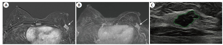
图 1 典型图像
Figure 1. Typical images. A: First post contrast subtracted; B: Maximum intensity projection; C: Ultrasound S-Detect image.

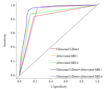
图 2 不同方法鉴别乳腺肿块良恶性的ROC曲线
Figure 2. ROC curves for different methods of identifying benign and malignant breast masses.

图 3 不同方法鉴别乳腺肿块良恶性的DCA结果
Figure 3. DCA results of different methods to identify benign and malignant breast masses.
表 1 MRI扫描参数
Table 1. MRI scan parameters
Sequence Repetition time (ms) Echo time (ms) Field of view (mm2) Slice thickness (mm) Number of slices (slices) Number of excitation (times) Transverse axis T2 5080.0 75.00 380×380 5.0 30 1 Axial T1 7.6 4.56 360×360 1.6 104 1 Sagittal T2 4290.0 80.00 180×180 5.0 18 1 DWI 5600.0 66.00 380×380 5.0 30 1 Enhanced axial T1 4.0 1.89 360×360 1.6 112 1  下载: 导出CSV
表 2 不同方法鉴别乳腺肿块良恶性的结果
Table 2. Results of different methods to identify benign and malignant breast masses (n)
Pathological results Ultrasound S-Detect Abbreviated MRIⅠ Abbreviated MRI Ⅱ Ultrasound S-Detect + Abbreviated MRI Ⅰ Ultrasound S-Detect + Abbreviated MRI Ⅱ Benign Malignancy Benign Malignancy Benign Malignancy Benign Malignancy Benign Malignancy Benign (n=43) 35 8 25 18 37 6 36 7 33 10 Malignancy (n=116) 19 97 2 114 15 101 5 111 4 112 Accuracy rate (%) 83.02 87.42 86.79 92.45 91.19 Kappa value 0.602 0.639 0.686 0.806 0.767
下载: 导出CSV
表 3 不同方法鉴别乳腺肿块良恶性的区分度比较
Table 3. Comparison of the discrimination of different methods for identifying the benign and malignant nature of breast masses
Methods Area under curve 95% CI Optimal cutoff P Sensitivity (%) Specificity (%) Ultrasound S-Detect 0.825 0.757-0.893 - < 0.001 83.62 81.40 Abbreviated MRI Ⅰ 0.910 0.857-0.964 2 < 0.001 87.07 86.05 Abbreviated MRI Ⅱ 0.866 0.805-0.926 - < 0.001 87.07 86.05 Ultrasound S-Detect + Abbreviated MRI Ⅰ 0.929 0.873-0.984 0.682 < 0.001 92.24 88.37 Ultrasound S-Detect + Abbreviated MRI Ⅱ 0.866 0.800-0.933 - < 0.001 96.55 76.74
下载: 导出CSV
-
[1] Siegel RL, Miller KD, Fuchs HE, et al. Cancer statistics, 2022[J]. CAA Cancer J Clinicians, 2022, 72(1): 7-33. doi: 10.3322/caac.21708 [2] Sung H, Ferlay J, Siegel RL, et al. Global cancer statistics 2020: GLOBOCAN estimates of incidence and mortality worldwide for 36 cancers in 185 countries[J]. CA Cancer J Clin, 2021, 71(3): 209-49. doi: 10.3322/caac.21660 [3] Wilkinson L, Gathani T. Understanding breast cancer as a global health concern[J]. Br J Radiol, 2022, 95(1130): 20211033. doi: 10.1259/bjr.20211033 [4] Zhang SW, Sun KX, Zheng RS, et al. Cancer incidence and mortality in China, 2015[J]. J Natl Cancer Cent, 2021, 1(1): 2-11. doi: 10.1016/j.jncc.2020.12.001 [5] Wang XR, Wang C, Guan JH, et al. Progress of Breast Cancer basic research in China[J]. Int J Biol Sci, 2021, 17(8): 2069-79. doi: 10.7150/ijbs.60631 [6] 张雪, 董晓平, 管雅喆, 等. 女性乳腺癌流行病学趋势及危险因素研究进展[J]. 肿瘤防治研究, 2021, 48(1): 87-92. https://www.cnki.com.cn/Article/CJFDTOTAL-ZLFY202101017.htm [7] Ren WH, Chen MY, Qiao YL, et al. Global guidelines for breast cancer screening: a systematic review[J]. Breast, 2022, 64: 85-99. doi: 10.1016/j.breast.2022.04.003 [8] Mann RM, Hooley R, Barr RG, et al. Novel approaches to screening for breast cancer[J]. Radiology, 2020, 297(2): 266-85. doi: 10.1148/radiol.2020200172 [9] Rahman WT, Helvie MA. Breast cancer screening in average and high-risk women[J]. Best Pract Res Clin Obstet Gynaecol, 2022, 83: 3-14. doi: 10.1016/j.bpobgyn.2021.11.007 [10] 曹力, 张会萍, 周毓青, 等. 不同年资超声科医师应用S-Detect技术诊断乳腺结节的价值研究[J]. 肿瘤影像学, 2022, 31(4): 439-43. https://www.cnki.com.cn/Article/CJFDTOTAL-YXYX202204013.htm [11] Jones LI, Marshall A, Elangovan P, et al. Evaluating the effectiveness of abbreviated breast MRI (abMRI) interpretation training for mammogram readers: a multi-centre study assessing diagnostic performance, using an enriched dataset[J]. Breast Cancer Res, 2022, 24(1): 55. doi: 10.1186/s13058-022-01549-5 [12] 李娜, 彭梅, 詹韵韵, 等. 血管指数VIMV联合BI-RADS在乳腺肿块良恶性鉴别中的价值[J]. 中国超声医学杂志, 2022, 38(10): 1093-6. https://www.cnki.com.cn/Article/CJFDTOTAL-ZGCY202210004.htm [13] 张蒙, 崔永春, 王春平, 等. 1990-2019年中国女性乳腺癌疾病负担及其危险因素变化趋势分析[J]. 中华肿瘤防治杂志, 2022, 29 (7): 456-62. https://www.cnki.com.cn/Article/CJFDTOTAL-QLZL202207002.htm [14] Cao W, Chen HD, Yu YW, et al. Changing profiles of cancer burden worldwide and in China: a secondary analysis of the global cancer statistics 2020[J]. Chin Med J, 2021, 134(7): 783-91. doi: 10.1097/CM9.0000000000001474 [15] 张晨滢, 姜洪标, 盛美红, 等. 乳腺MRI检查中双上肢体位对患者舒适度影响的探讨[J]. 医学影像学杂志, 2022, 32(5): 786-90. https://www.cnki.com.cn/Article/CJFDTOTAL-XYXZ202205015.htm [16] Bougias H, Stogiannos N. Breast MRI: where are we currently standing?[J]. J Med Imag Radiat Sci, 2022, 53(2): 203-11. doi: 10.1016/j.jmir.2022.03.072 [17] Harms SE. Abbreviated breast magnetic resonance imaging (MRI): first postcontrast subtracted images and maximum-intensity projection-a novel approach to breast cancer screening with MRI [J]. Breast Dis, 2015, 26(1): 38-40. [18] Comstock CE, Gatsonis C, Newstead GM, et al. Comparison of abbreviated breast MRI vs digital breast tomosynthesis for breast cancer detection among women with dense breasts undergoing screening[J]. JAMA, 2020, 323(8): 746-56. doi: 10.1001/jama.2020.0572 [19] Kwon MR, Choi JS, Won H, et al. Breast cancer screening with abbreviated breast MRI: 3-year outcome analysis[J]. Radiology, 2021, 299(1): 73-83. doi: 10.1148/radiol.2021202927 [20] Rueda MA, Bedoya Murillo ND, Cardona Ortegón JD, et al. Abbreviated screening MRI in breast cancer: promising but not widely available[J]. Radiology, 2022, 305(1): E58. doi: 10.1148/radiol.221727 [21] 闫虹, 李响, 程慧芳, 等. S-Detect技术应用于超声诊断乳腺包块的影响因素及与超声医师联合诊断的分析[J]. 中国临床医学影像杂志, 2020, 31(1): 24-9. https://www.cnki.com.cn/Article/CJFDTOTAL-LYYX202001010.htm [22] Xing B, Chen X, Wang Y, et al. Evaluating breast ultrasound S-detect image analysis for small focal breast lesions[J]. Front Oncol, 2022, 12: 1030624. doi: 10.3389/fonc.2022.1030624 [23] Zhang D, Jiang F, Yin R, et al. A review of the role of the S-detect computer-aided diagnostic ultrasound system in the evaluation of benign and malignant breast and thyroid masses[J]. Med Sci Monit, 2021, 27: e931957. [24] 梁晓芸. 乳腺磁共振简化方案在乳腺病变检查中的价值[D]. 唐山: 华北理工大学, 2022. [25] 潘加珍, 刘心培, 查海玲, 等. S-Detect在乳腺肿块诊断中出现假阴性和假阳性的影响因素[J]. 肿瘤影像学, 2023, 32(1): 64-72. https://www.cnki.com.cn/Article/CJFDTOTAL-YXYX202301011.htm -


 点击查看大图
计量
- 文章访问数: 36
- HTML全文浏览量: 14
- PDF下载量: 1
- 被引次数: 0