


Comparison of free breathing ultrashort echo time and star volume interpolation sequence in pulmonary nodules
-
摘要:
目的 比较星状容积内插屏气检查(star VIBE)序列和螺旋超短回波时间序列(spiral UTE)两种磁共振自由呼吸序列在肺部检查中的图像质量,肺结节检出及形态学征象显示能力的差别。 方法 收集2019年11月~2022年10月胸部CT发现肺结节且进行胸部MRI检查的患者,CT和MRI检查间隔时间为48 h。MRI检查采用star VIBE序列和spiral UTE序列进行扫描。2位放射科医师采用5分法独立对MRI序列进行图像质量评价,并比较其肺结节检出率及形态学征象的显示能力。 结果 图像质量评价方面,spiral UTE图像的血管清晰度评分高于star VIBE序列(P < 0.05),而star VIBE图像的运动伪影评分低于spiral UTE图像(P < 0.05)。在结节的检测能力上,spiral UTE序列的磨玻璃结节检出率高于star VIBE序列(P < 0.05),且在中下叶的结节总检出率高于star VIBE序列(P < 0.05)。Spiral UTE序列的分叶、毛刺、棘突、空泡和空洞检出率(90.90%、85.71%、88.88%、100%、100%)高于star VIBE序列(86.36%、71.42%、77.77%、50%、83.33%),但差异无统计学意义(P>0.05)。 结论 Star VIBE序列和spiral UTE序列各有优势。Star VIBE序列不易受呼吸和心脏运动影响。Spiral UTE序列能更好地检出肺中叶和下叶结节,尤其是磨玻璃结节,且spiral UTE序列能更好地清晰肺血管。对于呼吸幅度较大不规律患者,建议使用star VIBE序列扫描;对于磨玻璃结节和中下叶结节患者,推荐使用spiral UTE序列。 -
关键词:
- 磁共振成像 /
- 超短回波时间 /
- 肺结节 /
- star VIBE序列
Abstract:Objective To compare the image quality and the ability of showing nodules and morphology of lung nodules in MRI free breathing sequence: star volumetric interpolated breath-hold examination (star VIBE) sequence and spiral ultrashort echo time sequence (spiral UTE). Methods Patients with pulmonary nodules detected by chest CT and examined by chest MRI from November 2019 to September 2022 were collected. The interval between CT and MRI was 48 h. MRI examination was performed with star VIBE sequence and spiral UTE sequence. Two radiologists used the 5-point method to independently evaluate the image quality of MRI sequences, and compared the detection rate of pulmonary nodules and the ability to display morphological signs. Results In terms of image quality evaluation, the vascular sharpness score of spiral UTE image was higher than that of star VIBE sequence (P < 0.05), while the motion artifact score of star VIBE image was lower than that of spiral UTE image. In terms of nodule detection ability, the ground glass nodule detection rate of spiral UTE sequence was higher than that of star VIBE sequence (P < 0.05), and the total detection rate of nodules in the middle and lower lobe was higher than that of star VIBE sequence (P < 0.05). The detection rates of lobulation, burr, spinous process, vacuole and cavity in Spiral UTE sequence (90.90%, 85.71%, 88.88%, 100%, 100%) were higher than those in star VIBE sequence (86.36%, 71.42%, 77.77%, 50%, 83.33%), but the difference was not statistically significant (P>0.05). Conclusion Star VIBE sequence and spiral UTE sequence have their own advantages. Star VIBE sequence is not easily affected by respiration and cardiac movement. Spiral UTE sequence can better detect middle and lower lobe nodules, especially ground glass nodules, and spiral UTE sequence can better clarify pulmonary vessels. Therefore, star VIBE sequence scan is recommended for patients with large and irregular breathing, and spiral UTE sequence is recommended for patients with ground glass nodules and middle and lower lobe nodules. -

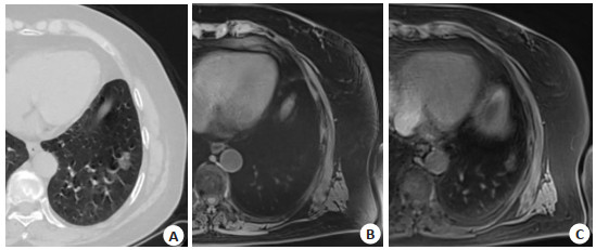
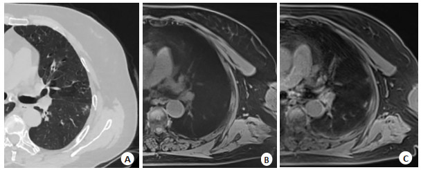
图 1 结节CT和MRI影像图
Figure 1. The CT and MRI images of the nodules. A 68-year-old male showed part-solid nodules in the left lower lobe on CT (A), unclear on star VIBE (B) images but clear on spiral UTE (C) images.

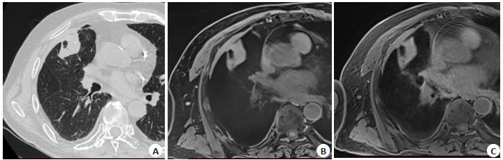
图 2 结节CT和MRI影像图
Figure 2. The CT and MRI images of the nodules. A 73-year-old patient's CT showed ground glass nodules in the lower left lung (A), not shown on star VIBE (B) image, and ground glass nodules showed on spiral UTE (C) image.

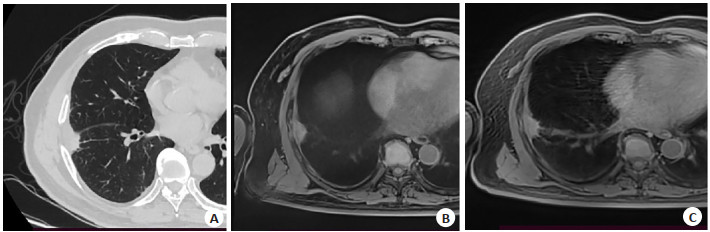
图 3 结节形态学征象的CT和MRI影像图
Figure 3. CT and MRI images of the morphological signs of the nodules. In a 69-year-old male, CT image (A) clearly showed the cavity in the nodule, and the cavity was also clearly displayed in star VIBE (B) and spiral UTE (C).

图 4 结节形态学征象的CT和MRI影像图
Figure 4. CT and MRI images of the morphological signs of the nodules. A 77-year-old male patient, CT (A) and spiral UTE (C) images clearly showed the burr sign, while the star VIBE (B) sequence did not show clearly.
表 1 图像质量5分主观评分法
Table 1. The five-point subjective scoring method of image quality
Parameter 1 2 3 4 5 Clarity of vascular Uacceptable Poor Acceptable Good Excellent Clarity of lesions Unacceptable Poor Acceptable Good Excellent Contrast Unacceptable Poor Acceptable Good Excellent Movement artifact No artifacts Mild artifacts Moderate artifacts Acceptable Unacceptable  下载: 导出CSV
表 2 不同MRI序列的图像质量评估
Table 2. Image quality assessment of the different MRI sequences (Mean±SD)
Parameter Image quality Z P Star VIBE Spiral UTE Clarity of vascular 3.29±0.73 3.74±0.44 2.562 0.010 Clarity of lesions 3.45±0.56 3.54±0.62 0.655 0.513 Contrast 3.54±0.50 3.64±0.48 0.728 0.467 Movement artifact 3.12±0.88 3.64±0.60 2.664 0.008 star VIBE: Star volumetric interpolated breath-hold examination; UTE: Ultrashort echo time.
下载: 导出CSV
表 3 不同MRI序列结节的检出能力比较
Table 3. Comparing the ability to detect nodules in different MRI sequences [n(%)]
Detection result Star VIBE(+)/ CT(+) Spiral UTE(+)/ CT(+) χ2 P Maximum diameter of the nodule axis (mm) Diameter≤6 9/19(47.36) 15/19(78.94) 3.125 0.077 6 < diameter≤10 16/24(66.66) 19/24(79.16) 0.100 0.752 10 < diameter≤30 38/45(84.44) 41/45(91.11) 1.778 0.182 Nodule type Solid nodules 46/47(97.87) 45/47(95.74) 0.000 >0.999 Part-solid nodules 10/14(71.42) 11/14(78.57) 0.000 >0.999 Non-solid 7/27(25.92) 19/27(70.37) 6.667 0.010 Number of nodules per location Upper lung 36/41(87.80) 34/41(82.92) 0.083 0.773 Middle and lower lung 27/47(57.44) 41/47(87.23) 7.682 0.006
下载: 导出CSV
表 4 比较不同MRI序列形态学征象检出能力
Table 4. To compare the detection ability of morphological signs of different MRI sequences [n(%)]
Morphological signs Star VIBE(+)/ CT(+) Spiral UTE(+)/ CT(+) χ2 P Lobulation 19/22(86.36) 20/22(90.90) 0.000 >0.999 Burr 10/14(71.42) 12/14(85.71) 0.167 0.683 Pleural indentation 17/17(100) 17/17(100) - - Spinous process 7/9(77.77) 8/9(88.88) 0.000 >0.999 Vacuole 2/4(50) 4/4(100) 0.500 0.480 Cavity 5/6(83.33) 6/6(100) 0.000 >0.999 Enlarged lymph node 26/26(100) 26/26(100) - -
下载: 导出CSV
-
[1] Grzegorz B, Lee Nam G, Ye T, et al. Submillimeter lung MRI at 0.55 T using balanced steady-state free precession with half-radial dual-echo readout (bSTAR)[J]. Magn Reson Med, 2023, 90(5): 1949-57. doi: 10.1002/mrm.29757 [2] Sanchez F, Tyrrell P, Cheung P, et al. Detection of solid and subsolid pulmonary nodules with lung MRI: performance of UTE, T1 gradient-echo, and single-shot T2 fast spin echo[J]. Cancer Imaging, 2023, 23(1): 17. doi: 10.1186/s40644-023-00531-4 [3] 邓和平, 杨柳, 龚辉, 等. 自由呼吸状态下star-VIBE技术在肺癌动态对比增强MRI检查中的价值[J]. 实用放射学杂志, 2023, 39(3): 479-83. doi: 10.3969/j.issn.1002-1671.2023.03.033 [4] Yu N, Duan HF, Yang CB, et al. Free-breathing radial 3D fat-suppressed T1-weighted gradient echo (r-VIBE) sequence for assessment of pulmonary lesions: a prospective comparison of CT and MRI[J]. Cancer Imaging, 2021, 21(1): 68. doi: 10.1186/s40644-021-00441-3 [5] 胡俊蛟, 刘梅桃, 赵伟, 等. T1WI star-VIBE增强结合TWIST-VIBE动态增强MRI对肺结节诊断的应用价值[J]. 中南大学学报: 医学版, 2023, 48(4): 581-93. https://www.cnki.com.cn/Article/CJFDTOTAL-HNYD202304012.htm [6] Huang YS, Niisato E, Su MY M, et al. Applying compressed sensing volumetric interpolated breath-hold examination and spiral ultrashort echo time sequences for lung nodule detection in MRI [J]. Diagnostics, 2021, 12(1): 93. doi: 10.3390/diagnostics12010093 [7] Wang FN, Lin X, Lin C, et al. Ability of three-dimensional 3-Tesla ultrashort echo time magnetic resonance imaging to display the morphological characteristics of pulmonary nodules: a sensitivity analysis[J]. Quant Imaging Med Surg, 2023, 13(3): 1792-801. doi: 10.21037/qims-22-118 [8] Benlala I, Berger P, Girodet PO, et al. Automated volumetric quantification of emphysema severity by using ultrashort echo time MRI: validation in participants with chronic obstructive pulmonary disease[J]. Radiology, 2019, 292(1): 216-25. doi: 10.1148/radiol.2019190052 [9] Ohno Y, Takenaka D, Yoshikawa T, et al. Efficacy of ultrashort echo time pulmonary MRI for lung nodule detection and lungRADS classification[J]. Radiology, 2022, 302(3): 697-706. doi: 10.1148/radiol.211254 [10] 范丽, 夏艺, 刘士远. 肺部磁共振成像机遇与挑战: 中国十年来发展成果及展望[J]. 磁共振成像, 2022, 13(10): 61-5. https://www.cnki.com.cn/Article/CJFDTOTAL-CGZC202210008.htm [11] Dournes G, Yazbek J, Benhassen W, et al. 3D ultrashort echo time MRI of the lung using stack-of-spirals and spherical k-Space coverages: evaluation in healthy volunteers and parenchymal diseases[J]. J Magn Reson Imaging, 2018, 48(6): 1489-97. doi: 10.1002/jmri.26212 [12] Cha MJ, Park HJ, Paek MY, et al. Free-breathing ultrashort echo time lung magnetic resonance imaging using stack-of-spirals acquisition: a feasibility study in oncology patients[J]. Magn Reson Imag, 2018, 51: 137-43. doi: 10.1016/j.mri.2018.05.002 [13] Darçot E, Delacoste J, Dunet V, et al. Lung MRI assessment with high-frequency noninvasive ventilation at 3 T[J]. Magn Reson Imag, 2020, 74: 64-73. doi: 10.1016/j.mri.2020.09.006 [14] Cha M, Ahn H, Choi H, et al. Accelerated stack- of- spirals freebreathing three-dimensional ultrashort echo time lung magnetic resonance imaging: a feasibility study in patients with breast cancer [J]. Front Oncol, 2021, 11: 746059. doi: 10.3389/fonc.2021.746059 [15] Torres L, Kammerman J, Hahn AD, et al. "structure-function imaging of lung disease using ultrashort echo time MRI"[J]. Acad Radiol, 2019, 26(3): 431-41. doi: 10.1016/j.acra.2018.12.007 [16] Bruckmann NM, Kirchner J, Morawitz J, et al. Free-breathing 3D Stack of Stars GRE (StarVIBE) sequence for detecting pulmonary nodules in 18F-FDG PET/MRI[J]. EJNMMI Phys, 2022, 9(1): 11. doi: 10.1186/s40658-022-00439-1 [17] Feng H, Shi G, Liu HZ, et al. The application and value of 3T magnetic resonance imaging in the display of pulmonary nodules[J]. Front Oncol, 2022, 12: 844514. doi: 10.3389/fonc.2022.844514 [18] Ohno Y, Koyama H, Yoshikawa T, et al. Standard-, reduced-, and No-dose thin-section radiologic examinations: comparison of capability for nodule detection and nodule type assessment in patients suspected of having pulmonary nodules[J]. Radiology, 2017, 284 (2): 562-73. doi: 10.1148/radiol.2017161037 [19] Meier-Schroers M, Homsi R, Gieseke J, et al. Lung cancer screening with MRI: evaluation of MRI for lung cancer screening by comparison of LDCT-and MRI-derived Lung-RADS categories in the first two screening rounds[J]. Eur Radiol, 2019, 29(2): 898-905. doi: 10.1007/s00330-018-5607-8 [20] Bae K, Jeon KN, Hwang MJ, et al. Comparison of lung imaging using three-dimensional ultrashort echo time and zero echo time sequences: preliminary study[J]. Eur Radiol, 2019, 29(5): 2253-62. doi: 10.1007/s00330-018-5889-x [21] Ohno Y, Kauczor HU, Hatabu H, et al. MRI for solitary pulmonary nodule and mass assessment: current state of the art[J]. J Magn Reson Imag, 2018, 47(6): 1437-58. doi: 10.1002/jmri.26009 [22] Wielpütz MO, Triphan SMF, Ohno Y, et al. Outracing lung signal decay-potential of ultrashort echo time MRI[J]. RoFo, 2019, 191 (5): 415-23. doi: 10.1055/a-0715-2246 [23] Renz DM, Herrmann KH, Kraemer M, et al. Ultrashort echo time MRI of the lung in children and adolescents: comparison with nonenhanced computed tomography and standard post-contrast T1w MRI sequences[J]. Eur Radiol, 2022, 32(3): 1833-42. doi: 10.1007/s00330-021-08236-7 [24] 王福南, 朱柳红, 周建军. 探讨3.0T磁共振UTE序列对肺结节的显示能力: 与CT图像对比[J]. 放射学实践, 2021, 36(3): 357-60. https://www.cnki.com.cn/Article/CJFDTOTAL-FSXS202103019.htm [25] 任占丽, 张敏, 雷雨欣, 等. T1加权STAR VIBE序列与CT成像在评估肺实质疾病中的对比研究[J]. 磁共振成像, 2019, 10(6): 440-4. https://www.cnki.com.cn/Article/CJFDTOTAL-CGZC201906012.htm [26] Wielpütz MO, Lee HY, Koyama H, et al. Morphologic characterization of pulmonary nodules with ultrashort TE MRI at 3T[J]. Am J Roentgenol, 2018, 210(6): 1216-25. doi: 10.2214/AJR.17.18961 -

 点击查看大图
计量
- 文章访问数: 61
- HTML全文浏览量: 18
- PDF下载量: 3
- 被引次数: 0