


Early diagnostic efficiency of coronary heart disease based on multi-slice spiral CT measurement of pericardial fat volume combined with lipoprotein α and apolipoprotein B detection
-
摘要:
目的 探讨多层螺旋CT(MSCT)测量心周脂肪体积(PAT)联合脂蛋白α(Lp-α)、载脂蛋白B(Apo-B)检测在早期冠心病患者中的诊断评估价值。 方法 选取本院2020年3月~2022年4月收治的早期冠心病患者160例纳入观察组,再选取同期健康体检者110例为对照组,检测Lp-α、Apo-B水平及利用MSCT测量PAT,分析3项指标联合检测对冠心病的诊断评估价值。 结果 观察组PAT及Lp-α、水平均高于对照组(P < 0.05);PAT及Lp-α、Apo-B水平与冠心病的发生呈正相关性(P < 0.05);PAT及Lp-α、Apo-B水平与冠状动脉SYNTAX评分呈正相关性(P < 0.05);ROC曲线显示:PAT及Lp-α、Apo-B联合诊断曲线下面积为0.920,高于各单项指标曲线下面积(P < 0.05)。 结论 PAT及Lp-α、Apo-B水平在冠心病早期患者中升高,与冠心病的发生和病情程度密切相关,并对冠心病具有较高的诊断价值。 Abstract:Objective To explore the diagnostic value of multi-slice spiral CT (MSCT) measurement of pericardial adipose tissue (PAT) combined with lipoprotein α (Lp-α) and apolipoprotein B (Apo-B) detection in patients with early coronary heart disease. Methods A total of 160 patients with early coronary heart disease who were treated in our hospital from March 2020 to April 2022 were selected as the observation group. The 110 healthy subjects were selected during the same period. We detected the levels of Lp-α and Apo-B, and used MSCT to measure PAT to analyze the value of the combined detection of these three indicators in the diagnosis and evaluation of coronary heart disease. Results The levels of PAT, Lp-α in the observation group were significantly higher than those in the control group (P < 0.05). Spearman correlation showed that the levels of PAT, Lp-α and Apo-B were positively correlated with the occurrence of coronary heart disease (P < 0.05). PAT, Lp-α, Apo-B levels were positively correlated with coronary SYNTAX score (P < 0.05). ROC curve showed that PAT, Lp-α, Apo-B levels combined diagnostic the area under the curve was 0.920, which was significantly higher than the area under the curve of each individual index (P < 0.05). Conclusion The PAT, Lp-α and Apo-B are elevated in patients with early coronary heart disease, which are closely related to the occurrence and severity of coronary heart disease. It has a high diagnostic value for coronary heart disease. -

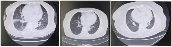
图 1 冠心病患者CT影像学特征
Figure 1. CT imaging features of patients with coronary heart disease.

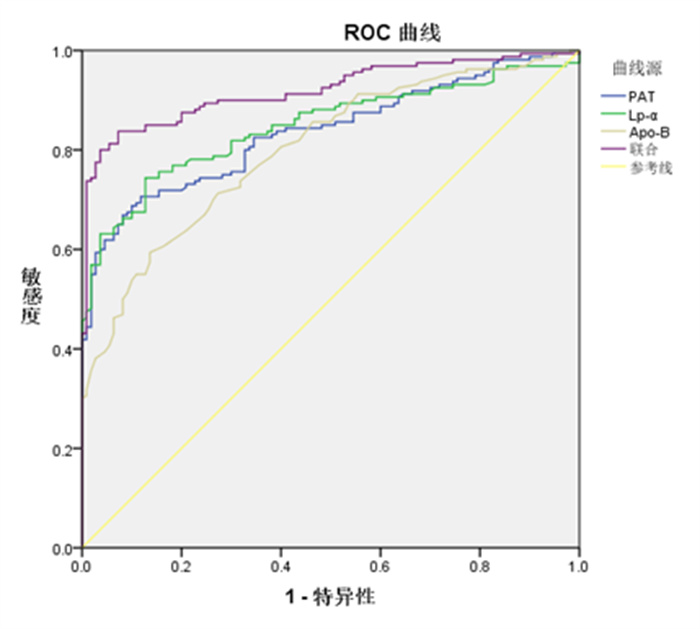
图 2 PAT及Lp-α、Apo-B水平对早期冠心病的诊断价值
Figure 2. The diagnostic value of PAT and Lp-α and Apo-B levels for early coronary heart disease.
表 1 两组患者PAT及Lp-α、Apo-B水平比较
Table 1. Comparison of PAT, LP-α and Apo-B of two groups (Mean±SD)
组别 PAT(cm3) Lp-α(ng/mL) Apo-B(g/L) 观察组(n=160) 183.87±27.84 22.60±6.09 0.91±0.24 对照组(n=110) 149.37±17.10 15.56±3.26 0.65±0.19 t 11.58 11.086 9.465 P < 0.001 < 0.001 < 0.001 PAT: 心周脂肪组织; Lp-α: 脂蛋白α; Apo-B: 载脂蛋白B  下载: 导出CSV
表 2 PAT及Lp-α、Apo-B水平与冠心病的相关性
Table 2. Correlation between PAT and Lp-α, Apo-B levels with coronary heart disease
指标 r P PAT 0.572 < 0.001 Lp-α 0.592 < 0.001 Apo-B 0.513 < 0.001
下载: 导出CSV
表 3 PAT及Lp-α、Apo-B水平与冠状动脉SYNTAX评分的关系
Table 3. Correlation between PAT and Lp- α and Apo- B levels and coronary SYNTAX scores
指标 r P PAT 0.544 < 0.001 Lp-α 0.454 < 0.001 Apo-B 0.505 < 0.001
下载: 导出CSV
表 4 PAT及Lp-α、Apo-B水平对早期冠心病的诊断价值
Table 4. The diagnostic value of PAT and Lp-α and Apo-B levels for early coronary heart disease.
指标 曲线下面积 最佳截断值 敏感度 特异性 95% CI 约登指数 PAT 0.836* 169.81 cm3 0.706 0.882 0.789~0.883 0.588 Lp-α 0.848* 18.670 ng/mL 0.744 0.873 0.802~0.894 0.617 Apo-B 0.801* 0.84 g/L 0.594 0.864 0.750~0.852 0.458 联合诊断 0.92 - 0.838 0.927 0.887~0.952 0.765 *P < 0.05 vs联合诊断.
下载: 导出CSV
-
[1] Li HY, Sun K, Zhao RP, et al. Inflammatory biomarkers of coronary heart disease[J]. Front Biosci (Schol Ed), 2018, 10(1): 185-96. doi: 10.2741/s508 [2] Temple NJ. Fat, sugar, whole grains and heart disease: 50 years of confusion[J]. Nutrients, 2018, 10(1): 39. doi: 10.3390/nu10010039 [3] Erdmann E. Koronare herzerkrankung: fortschritte in diagnostik und therapie[J]. Dtsch Med Wochenschr, 2017, 142(21): 1565. doi: 10.1055/s-0042-112806 [4] 王莹屏, 徐新华, 刘晶哲. 64层螺旋CT冠状动脉成像与冠状动脉造影诊断冠状动脉狭窄的对照研究[J]. 中西医结合心脑血管病杂志, 2019, 17(11): 1753-5. doi: 10.12102/j.issn.1672-1349.2019.11.044 [5] 潘兴邦, 李凡, 陈晨, 等. 脂蛋白α、载脂蛋白B/A-1与颈动脉Crouse积分联合检测对冠状动脉病变程度的预测价值[J]. 中国分子心脏病学杂志, 2021, 21(2): 3856-60. doi: 10.16563/j.cnki.1671-6272.2021.04.015 [6] 肖日国, 许晓杰. 双源CT测量心周脂肪体积联合血尿酸、同型半胱氨酸监测对冠心病早期诊断的价值分析[J]. 中国CT和MRI杂志, 2020, 18(1): 54-6. doi: 10.3969/j.issn.1672-5131.2020.01.018 [7] 热那提·肉孜, 吴娜琼, 孙荻, 等. 脂蛋白α水平与冠心病发生及严重程度的相关性探究[J]. 中国心血管病研究, 2020, 18(3): 206-11. doi: 10.3969/j.issn.1672-5301.2020.03.004 [8] 王鹏飞, 乔海霞, 王瑞鹃, 等. 冠心病患者血脂、血流变及血清超敏C反应蛋白表达水平的监测价值分析[J]. 标记免疫分析与临床, 2018, 25(12): 1835-8. https://www.cnki.com.cn/Article/CJFDTOTAL-BJMY201812024.htm [9] 高润霖. 中华医学百科全书: 心血管病学[M]. 北京: 中国协和医科大学出版社, 2017: 883-4. [10] Yu M, Yang F, Cheng X. Anti- inflammatory therapy of coronary heart disease: where is the way?[J]. Zhonghua Xin Xue Guan Bing Za Zhi, 2021, 49(2): 111-4. [11] Liu YH, Ye T, Chen L, et al. Systemic immune-inflammation index predicts the severity of coronary stenosis in patients with coronary heart disease[J]. CoronArtery Dis, 2021, 32(8): 715-20. doi: 10.1097/MCA.0000000000001037 [12] Ong KL, McClelland RL, Allison MA, et al. Lipoprotein (a) and coronary artery calcification: prospective study assessing interactions with other risk factors [J]. Metabolism, 2021, 116: 154706. doi: 10.1016/j.metabol.2021.154706 [13] Shaya GE, Leucker TM, Jones SR, et al. Coronary heart disease risk: low-density lipoprotein and beyond[J]. Trends Cardiovasc Med, 2022, 32(4): 181-94. [14] Zhao XM, Wang DY, Qin LJ. Lipid profile and prognosis in patients with coronary heart disease: a meta- analysis of prospective cohort studies [J]. BMC Cardiovasc Disord, 2021, 21(1): 69. [15] Ugovšek S, Šebeštjen M. Lipoprotein(a) the crossroads of atherosclerosis, atherothrombosis and inflammation[J]. Biomolecules, 2021, 12(1): 26. [16] Tian M, Li R, Shan ZL, et al. Comparison of Apolipoprotein B/A1 ratio, Framingham risk score and TC/HDL-c for predicting clinical outcomes in patients undergoing percutaneous coronary intervention[J]. Lipids Health Dis, 2019, 18(1): 202. [17] Marston NA, Giugliano RP, Melloni GEM, et al. Association of apolipoprotein B-containing lipoproteins and risk of myocardial infarction in individuals with and without atherosclerosis: distinguishing between particle concentration, type, and content[J]. JAMACardiol, 2022, 7(3): 250-6. [18] Muscella A, Stefàno E, Marsigliante S. The effects of exercise training on lipid metabolism and coronary heart disease[J]. Am J Physiol Heart Circ Physiol, 2020, 319(1): H76-H88. [19] Chang TY, Chen JD. Low- density lipoprotein cholesterol/apolipoprotein B ratio is superior to apolipoprotein B alone in the diagnosis of coronary artery calcification[J]. Coron Artery Dis, 2021, 32(6): 561-6. [20] 翁静飞, 何宇情, 杨彬, 等. 256层螺旋CT测量心脏周围脂肪体积与冠心病的相关性研究[J]. 海南医学, 2022, 33(7): 906-9. https://www.cnki.com.cn/Article/CJFDTOTAL-HAIN202207023.htm [21] Gać P, Macek P, Poręba M, et al. Thickness of epicardial and pericoronary adipose tissue measured using 128- slice MSCT as predictors for risk of significant coronary artery diseases[J]. Ir J Med Sci, 2021, 190(2): 555-66. [22] Goeller M, Achenbach S, Duncker H, et al. Imaging of the pericoronary adipose tissue (PCAT) using cardiac computed tomography: modern clinical implications[J]. J Thorac Imaging, 2021, 36(3): 149-61. [23] Yang TT, Fish AF, Kong WM, et al. Correlates of pericardial adipose tissue volume using multidetector CT scanning in cardiac patients in China[J]. Int J Cardiol, 2017, 244: 285-9. [24] 张成辉, 张瑜, 胡珊. 颈动脉彩超结合脂蛋白相关磷脂酶A2水平对冠心病高危人群的预测价值[J]. 分子影像学杂志, 2021, 44(4): 686-90. doi: 10.12122/j.issn.1674-4500.2021.04.22 -

 点击查看大图
计量
- 文章访问数: 122
- HTML全文浏览量: 55
- PDF下载量: 2
- 被引次数: 0