

 Find Duplicates
 Check Document
 Submission(new)
 Experts Office
 Editorial Office
2022 Vol. 45, No. 5
Display Method:
2022, 45(5): 631-636.
doi: 10.12122/j.issn.1674-4500.2022.05.01
Abstract:
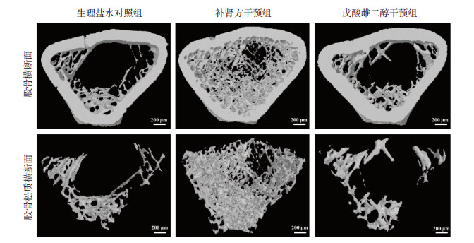
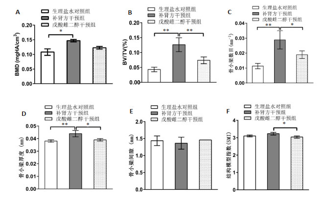
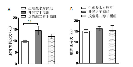
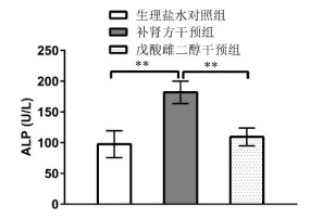
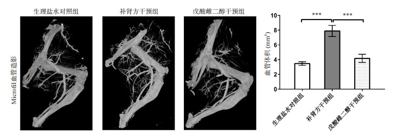
Objective To investigate the effects of Bushen decoction on femur's bone-microarchitecture, vascular volume around the bone, biomechanics and alkaline phosphatase (ALP) activity of ovariectomized rat with osteoporosis based on micro-CT. Methods Thirty healthy Sprague-Dawley female rats aged three months were randomly divided into control group, Bushen decoction intervention group and estradiol valerate intervention group, with 10 rats/group. Both ovaries of rats were removed. Rats were intragastrically administered for 4 weeks after the model creation 3 months. Femur's bone-microarchitecture and vascular volume around the bone were observed by Micro-CT scan. Bone fracture stress and structural strength of each femoral bone were measured. Serum ALP activity were detected. Results Micro-CT showed an increase in morphometry such as bone mineral density (P=0.0114), bone volume fraction (P=0.0006), trabecular number (P=0.0016), trabecular thickness (P= 0.0050), vascular volume (P < 0.0001) in Bushen decoction intervention group as compared with control group. Bushen decoction significantly enhanced bone fracture stress (P=0.0044) as compared with normal saline. Serum ALP activity in Bushen decoction group were improved (P=0.0181) as compared with control group. Conclusion Bushen decoction can improve bone-microarchitecture of femurs, increase vascular volume, strengthen bone fracture stress and higher the activity of ALP. Therefore, Bushen decoction should have a good effect on treating osteoporosis in future clinical studies.
2022, 45(5): 637-642.
doi: 10.12122/j.issn.1674-4500.2022.05.02
Abstract:
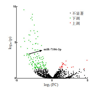
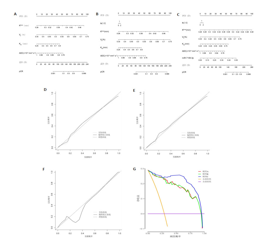
Objective To construct a nomogram regression model based on MRI and microRNA-7106-3p (miR-7106-3p), and investigate its value in evaluating the efficacy of neoadjuvant chemoradiotherapy (NCRT) for advanced rectal cancer. Methods A total of 127 patients with rectal cancer were prospectively enrolled from March 2019 to February 2022 at Hebei General Hospital For Veterans who underwent total rectal mesenteric resection after NCRT. MRI was performed within 1 week before NCRT. Peripheral blood was extracted from patients at the same time. The serum miR-7106-3p level was detected by real-time fluorescence quantitative polymerase chain reaction. Risk factors for the efficacy of NCRT in advanced rectal cancer were analysed by Logistic regression. The nomogram regression model for risk factors was constructed. Consistency index, calibration curve and decision curve analysis were used to evaluate the value of the model. Results There were 37 (29.13%) patients achieved pathological complete remission (pCR) after NCRT (pCR group), and 90 patients (70.87%) did not achieved pCR (non-pCR group). The levels of volume transport constant (Ktrans), extravascular extracellular space volume ratio (Ve), reflux rate constant (Kep) and miR-7106-3p were higher in the pCR group than in the non-pCR group, and apparent dispersion coefficient (ADC) was lower than in the non-pCR group (P < 0.05). The logistic regression analysis showed that higher N stage and ADC > 0.92×10⁻³ mm2/s were independent risk factors for pCR after NCRT in progressive rectal cancer (P < 0.05), and Ktrans > 0.33 min, Ve > 0.55%, Kep > 0.54 min and miR-7106-3p > 0.31 were independent protective factors for pCR after NCRT in progressive rectal cancer (P < 0.05). The consistency index of model C (consisting of N-stage, Ktrans, Ve, Kep, ADC and miR-7106-3p) was 0.977, which was higher than that of model A (consisting of Ktrans, Ve, Kep and ADC, 0.957) and model B (consisting of N-stage, Ktrans, Ve, Kep and ADC, 0.956). The mean absolute error of model B was 0.015, which was lower than that of model A (0.017) and model C (0.024). When the threshold probabilities ranged from 0.10-1.0 for most of the range, the net benefit of model C was higher than that of model A and model B. Conclusion The model can better evaluate the efficacy of NCRT in progressive rectal cancer based on MRI and miR-7106-3p.

2022, 45(5): 643-647.
doi: 10.12122/j.issn.1674-4500.2022.05.03
Abstract:
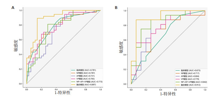
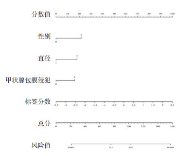
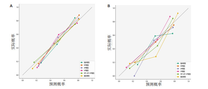
Objective To explore the value of six models established according to clinical data, CT imaging radiomics, and combining both for predicting preoperatively central lymph node metastasis (CLNM) in patients with papillary thyroid carcinoma (PTC). Methods A total of 103 PTC patients were enrolled and divided into non-CLNM group (n=50) and CLNM group (n=53) according to pathology. The clinical data and CT signatures were compared between groups. The patients in each group were randomly divided into training set and test set according at the ratio of 7:3. CT radiomics features of PTC were selected from training set, and clinical model, non-contrast phase (NP) model, arterial phase (AP) model, venous phase (VP) model, NP + AP + VP model and the combining model were constructed, respectively. AUC, sensitivity and specificity were calculated to evaluate the effectiveness of these six models. Results In clinical data, there was significant difference in gender between the two groups (P=0.002). The lesion diameter (P=0.001) and thyroid capsule invasion (P=0.024) of two groups were significant. Among the NP model, AP model, VP model and NP + AP + VP model these four radiomics models, NP + AP + VP model had the best efficacy for predicting CLNM in PTC patients. Compared with the clinical model and NP+AP+VP model, the combining model performed best and had the highest AUC, sensitivity and specificity in both training set and test set. Conclusion All of six models, the combining model high efficacy for predicting preoperatively CLNM, which is expected to provide an effective auxiliary method for preoperative the CLNM in PTC patients.
2022, 45(5): 648-655.
doi: 10.12122/j.issn.1674-4500.2022.05.04
Abstract:
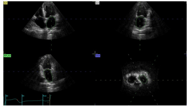
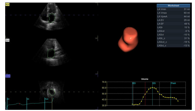
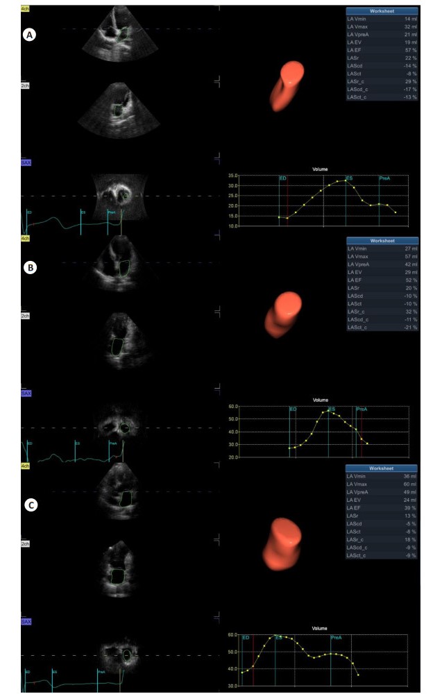
Objective To evaluate left atrial function in essential hypertensive (EH) patients with left ventricular hypertrophy (LVH) based on four-dimensional echocardiographic quantitative (4DLAQ). Methods Eighty EH patients were selected and divided into NLVH group and LVH group according to the new LVH standard. At the same time, 36 healthy subjects were selected as the control group. The participants in the three groups were measured by two-dimensional echocardiography, and the left atrial volume parameters were measured and analyzed by 4DLAQ technology, including left atrial minimum volume (LAVmin), left atrial maximum volume (LAVmax), left atrial pre systolic volume (LAVpreA) and left atrial global ejection fraction (LAEF). The left atrial strain parameters include left atrial reserve long axial strain, left atrial conduit long axial strain, left atrial systolic long axial strain, left atrial reserve circular strain, left atrial conduit circular strain and left atrial systolic circular strain. The differences between the two-dimensional and four-dimensional data of the three groups of participants were compared. Results There were significant differences in mean systolic blood pressure and mean diastolic blood pressure among the three groups (P < 0.05). There were significant differences in left atrial diameter, Biplane LVEF, interventricular septal thickness, left ventricular end-diastolic diameter, left ventricular posterior wall thickness and E/e' among the three groups (P < 0.05). There was significant difference in LAVmin, LAVpreA, LAEF, left atrial passive ejection fraction and left atrial active ejection fraction among the three groups (P < 0.05), but there was no significant difference in LAVmax (P > 0.05). There were significant differences in left atrial strain parameters among the three groups (P < 0.05). The consistency between and within observers was good (ICC > 0.90). Conclusion The increase of left atrial diameter in EH patients is earlier than the impairment of left ventricular systolic function. With the progress of the disease, the left atrial volume increases, the storage function and pipeline function are damaged, and the blood pumping function increases when the blood pumping function is in NLVH. With the aggravation of LVH, the blood pumping function of the left atrium decreases. 4DLAQ can find subtle changes in the left atrial function of EH patients and supplement the technical weakness of the current left atrial research. Its strain parameters are more sensitive to evaluate the left atrial function. Among them, the circular strain is more advantageous than the longitudinal strain in indicating the left atrial conduit function.
2022, 45(5): 656-660.
doi: 10.12122/j.issn.1674-4500.2022.05.05
Abstract:
Objective To explore the clinical application of 3.0T MRI in carotid atherosclerotic plaque composition examination of ischemic stroke. Methods Sixty-two patients with ischemic stroke caused by carotid atherosclerosis in our hospital and Shanghai Jiaotong University affiliated sixth People's Hospital from June 2020 to April 2021 were selected. 3.0T MRI was used to observe and analyze the plaque composition and signal characteristics of different types of plaques in all patients with carotid artery stenosis. The difference between stable plaque and unstable plaque composition was compared. Results A total of 119 carotid atherosclerotic plaques were found in 62 patients. Patch classification: 11 cases were type Ⅰ-Ⅱ (9.24%), 31 cases were type Ⅲ (26.05%), 24 cases were type Ⅳ-Ⅴ (20.17%), 37 cases were type Ⅵ (31.09%), 16 cases were type Ⅶ (13.45%). All plaques showed annular or eccentric wall thickening on MRI, and some of them were accompanied by local crescent or semilunar signal penetrating into the lumen. The manifestations of plaques with different components were different on MRI. There were 62 stable plaques and 57 unstable plaques in all detected carotid plaques. There was no significant difference in the proportion of calcification between stable plaque and unstable plaque (P > 0.05). The proportion of lipid core, hemorrhage, thrombosis and fibrous cap rupture in unstable plaque was significantly higher than that in stable plaque (P < 0.05). Conclusion 3.0T MRI can accurately evaluate the internal composition and stability of carotid atherosclerotic plaques in patients with ischemic stroke. It provide important reference for clinical prediction and disease evaluation of ischemic stroke.
2022, 45(5): 661-666.
doi: 10.12122/j.issn.1674-4500.2022.05.06
Abstract:
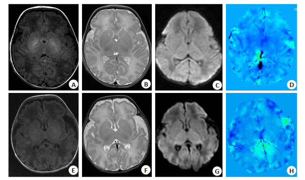
Objective To explore the value of conventional MRI signal changes and quantitative susceptibility mapping (QSM) in quantitative assessment of potential brain injury in neonates with hyperbilirubinemia. Methods Fifty neonates (group Ⅰ) who were clinically diagnosed with neonatal hyperbilirubinemia in our hospital were retrospectively analyzed from January 2019 to December 2020. Forty-eight neonates without hyperbilirubinemia and brain-related diseases were the control group (group Ⅱ). All children underwent MR plain scan and diffusion weighted imaging scan before treatment, and 38 children underwent QSM examination. The differences between the MR signal intensities of children in different groups and their correlation with serum total bilirubin levels were compared. The differences between the magnetic susceptibility of different nuclei in children with elevated bilirubin and the control group were compared, and their correlation with serum total bilirubin levels were analyzed. Results Children with hyperbilirubinemia were more inclined to have high signal intensity of the globus pallidus on T1WI (36/12 vs 13/37, P < 0.001). The difference of SI1GP/SI2GP was statistically significant (P=0.034), while the difference of other indicators were not statistically significant (P > 0.05). There was a very weak correlation between the SI1GP/SI2GP with serum total bilirubin level (r=0.261, P=0.009).When the age and birth weight were controlled, the SI1 GP/SI2GP, ΔSI1(GP-P)/ΔSI1(GP-FGM) was a very weak correlated(r=0.230, P=0.014; r=-0.184, P=0.014). There were statistically significant differences in the magnetic susceptibility of the globus pallidus, putamen, subthalamic nucleus and brainstem between the elevated bilirubin group and the control group(P < 0.05).The magnetic susceptibility of putamen and subthalamic nucleus were slightly correlated with serum total bilirubin levels (r=-0.419, P=0.011; r=-0.391, P=0.018), while there was a moderate correlation between the magnetic susceptibility of globus pallidus and brainstem and serum bilirubin levels (r=-0.620, P < 0.001; r=-0.630, P < 0.001). Conclusion The evaluating signal intensity of the globus pallidus on T1WI is helpful for thediagnosis of neonates with hyperbilirubinemia. There is no clear correlation between the signal intensity of conventional MR and the increase of serum bilirubin. The magnetic susceptibility measured by QSM in the globus pallidus, putamen, subthalamic nucleus and brainste may earlier indicate the potential brain damage in neonates with hyperbilirubinemia.
2022, 45(5): 667-672.
doi: 10.12122/j.issn.1674-4500.2022.05.07
Abstract:
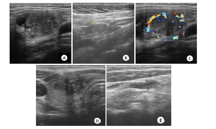
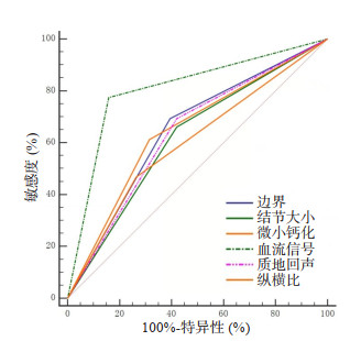
Objective To investigate the diagnostic value of high-frequency ultrasound in cervical lymph node metastasis (CLNM) of thyroid carcinoma. Methods A total of 100 patients with thyroid carcinoma admitted to our hospital were selected as the research subjects. According to the postoperative pathological results, cervical lymph node metastasis was divided into metastasis group (n=62) and non-metastasis group (n=38). The differences of preoperative ultrasound features between the two groups were compared, and the risk factors for CLNM of thyroid cancer were established by Logistic regression analysis. The ROC curve of ultrasound characteristics for CLNM diagnosis of thyroid cancer was plotted. Taking postoperative pathology as the gold standard, the diagnostic efficacy parameters such as sensitivity and specificity of ultrasonic diagnosis of CLNM were calculated. Results In the metastasis group, the nodule size≥1.5 cm, unclear boundary, low echo of texture, microcalcification, aspect ratio≥1 and the incidence of abundant blood flow signals were higher than those in the non-metastasis group (P < 0.05). Logistic regression analysis showed that nodule size≥1.5 cm, unclear boundary, low echo of texture, microcalcification, aspect ratio≥1 and abundant blood flow were independent risk factors for cervical lymph node metastasis of thyroid carcinoma (P < 0.05). ROC curve analysis showed that nodule size≥1.5 cm, unclear boundary, low echo of texture, microcalcification, aspect ratio and abundant blood flow signal had diagnostic value for CLNM of thyroid cancer (AUC=0.620, 0.649, 0.636, 0.649, 0.602 and 0.808, respectively). The positive predictive value of ultrasound in the diagnosis of thyroid cancer CLNM was 84.38%, the negative predictive value was 77.78%, the sensitivity was 87.10%, the specificity was 73.64%, and the diagnostic accuracy was 82.00%. Conclusion High-frequency ultrasound is helpful to predict the risk of CLNM of thyroid cancer and has good diagnostic efficacy.
2022, 45(5): 673-677.
doi: 10.12122/j.issn.1674-4500.2022.05.08
Abstract:
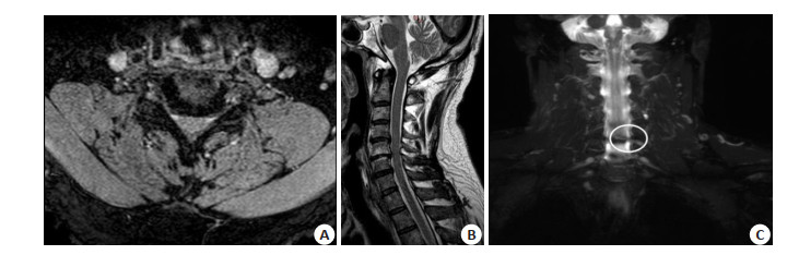
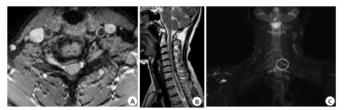
Objective To investigate the three-dimensional spin echo with short time inversion recovery (3D-STIR) sequence for detecting compression of cervical anterior ganglia nerve roots. To evaluate the diagnostic value of this technique in different segments (C6 nerve root and C7 nerve root). Methods From September 2021 to March 2022, we selected 100 patients with cervical spondylotic radiculopathy with neck pain and upper limb numbness and clinically highly suspected cervical spondylotic radiculopathy. The patients underwent conventional magnetic resonance imaging (sagittal T2WI, T1WI, STIR, Axial T2WI) and 3D-STIR sequence scanning, as well as the combination of the two scanning methods to determine whether nerve root compression, and the imaging manifestations of nerve root compression in patients with C5-7 unilateral and single-segment nerve root conduction dysfunction. All images in the 3D-STIR sequence were postprocessed by 3D maximum signal emphasis projection and surface reconstruction. The images were reconstructed by two MRI diagnostic physicians with intermediate and senior professional title, and the images showed the morphology, movement, compression and intervertebral disc and nerve root relationship. To evaluate the clinical application value of 3D-STIR sequence in the diagnosis of cervical spondylotic radiculopathy. Results The C6/C7 nerve roots were divided into two groups according to whether the nerve roots were compressed or not. The diagnostic results of conventional MRI, 3D-STIR sequence and conventional MRI combined with 3D-STIR sequence were counted. The consistency was 0.578, 0.758 and 0.838 according to whether the nerve roots were compressed or not. According to whether C7 nerve root was compressed or not, the statistical method was the same as that of C6 nerve root, and the consistency was 0.559, 0.779 and 0.839, respectively. The consistency of conventional sequence scanning in both groups was greater than 0.40 and less than 0.75, indicating moderate consistency. The consistency of 3D-STIR and combination with conventional sequence was greater than 0.75, indicating high consistency. The final significance test of the two groups showed statistically significant difference (P < 0.05). 3D-STIR sequences have a significant advantage over conventional MR sequences in revealing nerve root compression. Conclusion 3.0 T MR 3D-STIR sequence and conventional MRI combined with 3D-STIR have some advantages in the diagnosis of C6/C7 nerve root compression compared with conventional MRI plain scan.
2022, 45(5): 678-682.
doi: 10.12122/j.issn.1674-4500.2022.05.09
Abstract:
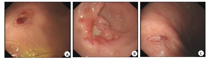
Objective To analyze the efficacy of endoscopic injection of snake venom hemocoagulase combined with intravenous drip of omeprazole in the treatment of ulcerative upper gastrointestinal bleeding. Methods A total of 126 patients with ulcerative upper gastrointestinal bleeding who were treated in our hospital from May 2019 to May 2022 were selected. They were divided into the control group (n=63) and the research group (n=63) according to the order of admission. The control group was given omeprazole intravenous drip treatment, 40 mg/time, 2 times per day. The research group was given intravascular injection of snake venom hemocoagulase around the bleeding site or the broken end on this basis, 1-2 U/d, both groups received continuous treatment for 5 d. Hemostasis time, rebleeding rate, the rate of transfer to surgery and hospitalization time between the two groups were compared, the coagulation function indexes [prothrombin time (PT), partially activated prothrombin time (APTT), D-dimer (D-D), fibrinogen (FIB)] levels, hemodynamic indexes [systolic blood pressure (SBP), diastolic blood pressure (DBP), heart rate (HR)] changes were compared between the two groups before and after treatment, and the clinical efficacy and safety were analyzed. Results The hemostasis time, rebleeding rate, rate of transfer to surgery, and hospital stay in the research group were significantly lower than those in the control group (P < 0.05). After treatment, the PT and APTT of the two groups were lower than those before treatment, and the D-D and FIB were higher than those before treatment (P < 0.05). The levels of PT and APTT in the research group were lower than those in the control group, while the levels of D-D and FIB were higher than those in the control group (P < 0.05). After treatment, the SBP, DBP and HR values of the two groups were all lower than those before treatment (P < 0.05). The SBP, DBP and HR values of the research group were significantly higher than those of the control group (P < 0.05). The total effective rate of the research group was significantly higher than that of the control group (P < 0.05). The total incidence of adverse reactions in the research group was not significantly different from the control group (P > 0.05). Conclusion Endoscopic injection of snake venom hemocoagulase combined with intravenous infusion of omeprazole has a significant hemostatic effect in the treatment of ulcerative upper gastrointestinal bleeding. It can significantly reduce the rate of rebleeding and transfer to surgery, effectively improve the coagulation function indexes of patients, and maintain the stability of blood pressure and heart rate of patients.
2022, 45(5): 683-687.
doi: 10.12122/j.issn.1674-4500.2022.05.10
Abstract:
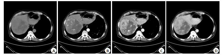
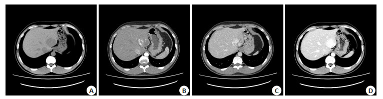
Objective To explore serum thymidine kinase 1 (STK1), soluble B7-H3 (sB7-H3) and extracellular matrix protein-1 (ECM1) combined with multi-slice spiral CT in the differential diagnosis of hepatocellular carcinoma and hepatic hemangioma. Methods Fifty-nine patients with hepatocellular carcinoma and 45 patients with hepatic hemangioma were selected. The imaging characteristics of the two lesions and levels of serum STK1, sB7-H3 and ECM1 were compared and the diagnostic efficiency of multi-slice spiral CT three-phase enhanced scan on the above two diseases was analyzed. Results CT plain scan results showed that there were significant differences in the morphology, type and boundary between hepatocellular carcinoma lesions and hepatic hemangioma lesions (P < 0.05). The levels of serum STK1, sB7-H3 and ECM1 in patients with hepatocellular carcinoma were higher than those in patients with hepatic hemangioma (P < 0.05). There was a statistical difference in the enhanced CT value of uniform high-density lesions in the arterial phase between two kinds of patients (P < 0.05). Multi-slice spiral CT three-phase enhanced scan in the diagnosis of hepatocellular carcinoma and hepatic hemangioma had good consistency with pathological results (Kappa=0.844). Conclusion Multi-slice spiral CT three-phase enhanced scan is effective in distinguishing hepatocellular carcinoma from hepatic hemangioma. Enhanced CT value of uniform high-density lesions in the arterial phase and increased levels of serum STK1, sB7-H3 and ECM1 are helpful for disease diagnosis.
2022, 45(5): 688-692.
doi: 10.12122/j.issn.1674-4500.2022.05.11
Abstract:
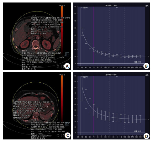
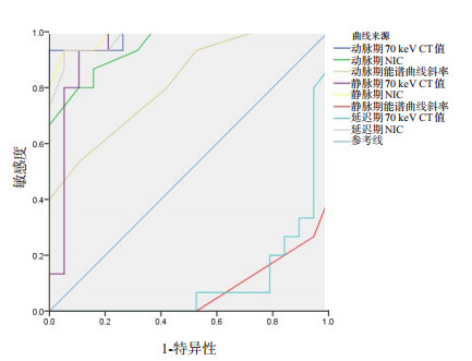
Objective To explore the predictive value of energy spectrum CT multimodal parameters combined with clinical characteristic parameters for gastric stromal tumor (GST) Ki-67. Methods Thirty-four patients with gastric stromal tumor in our hospital who met the inclusion criteria from September 19 to February 22, 2019 were retrospectively analyzed. The patients were divided into groups according to the percentage of Ki-67 in postoperative immunohistochemistry. Ki-67 percentage ≥6% was high expression group (n=15), and Ki-67 < 6% was low expression group (n=19). Dual-source CT scan was performed to measure and collect 70 keV CT value, standardized iodine concentration, energy spectrum curve slope and clinical characteristics, and the correlation between the above data and the expression of Ki-67 in gastric stromal tumor was analyzed. Results The correlation between diameter, age and GST Ki-67 expression was significant (P < 0.05). The lag period NIC value and energy spectrum slope of GST Ki-67 low expression group had some differences within the group (ICC < 0.75), and the other parameters had small differences within the group (ICC > 0.75). Except for the energy spectrum slope value of delay period, there were no significant differences between groups (P > 0.05), other parameters were significantly different between groups (P < 0.05). Ki-67 expression in GST was positively correlated with 70keVCT value in arteriovenous phase, NIC in arterial phase and energy spectrum slope (P < 0.05). Ki-67 expression of GST was negatively correlated with 70 keV CT value in delayed period and slope of energy spectrum curve in venous period (P < 0.05). The expression of Ki-67 in GST was predicted by 70 keV CT value of arteriovenous phase, NIC of phase iii and energy spectrum slope of arterial phase, and the highest AUC value of NIC in venous phase was 0.984. Conclusion The three-phase 70 keV CT value, NIC value, energy spectrum slope and diameter of arteriovenous phase and age have certain value in predicting the expression of GST Ki-67.
2022, 45(5): 693-696.
doi: 10.12122/j.issn.1674-4500.2022.05.12
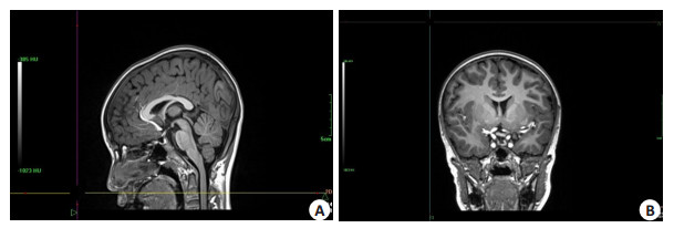
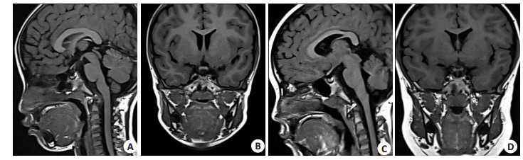
Abstract:
Objective To explore the monitoring value of MRI in the diagnosis and treatment of growth hormone deficiency in children. Methods We retrospectively analyzed the clinical data of 100 patients with growth hormone deficiency in our hospital from January 2018 to April 2019. The serum growth hormone levels of all patients were analyzed. The changes of pituitary MRI images before and after treatment were compared. Results Among the 100 cases of growth hormone deficiency, 45 cases had the peak value of growth hormone less than 5-10 μg/L and 55 cases had the peak value of growth hormone less than 5 μg/L. In the 100 patients with growth hormone deficiency, the main pituitary lesion was anterior pituitary dysplasia (n= 42, 42%), and pituitary stalk interrupt syndrome (n=25, 25%). In addition, 14 cases of empty sella syndrome (14%), long disease of grow in quantity of Hans cells invasive pituitary 12 cases (12%) and 7 cases of craniopharyngioma (7%). Children with growth hormone deficiency of pituitary high sagittal diameter, sagittal diameter after index greater than normal children, and the difference was statistically significant (P < 0.05). After growth hormone replacement therapy, The pituitary gland was smaller in different degrees at all ages, and the change was more significant at 3 - 10 years old and 11 - 16 years old (P < 0.05). Conclusion MRI can accurately detect growth hormone deficiency caused by pituitary disease, and can also accurately monitor the change of pituitary size after growth hormone replacement therapy.
2022, 45(5): 697-700.
doi: 10.12122/j.issn.1674-4500.2022.05.13
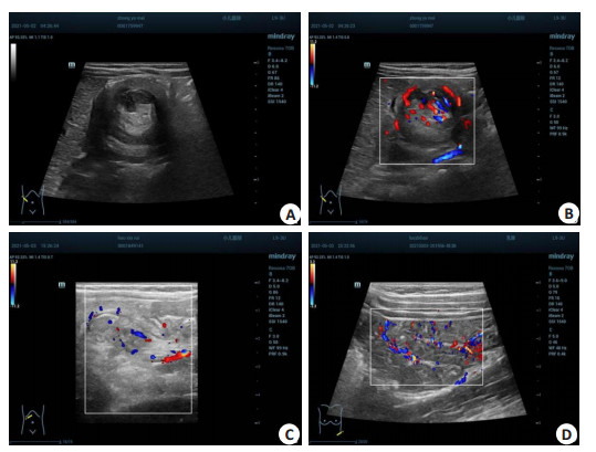
Abstract:
Objective To explore the application value of high and low frequency ultrasound in the diagnosis of intussusception in children. Methods The clinical data of 60 children suspected of intussusception in the hospital were retrospectively analyzed from Jun 2018 to Jun 2021. All patients underwent high frequency and low frequency color Doppler ultrasound examination, and the operation pathology was the gold standard. Accuracy, sensitivity, specificity, imaging characteristics and blood flow signals were analyzed, and the specific treatment of the children was analyzed. Results Clinical diagnosis results showed 52 positive cases and 8 negative cases in the 60 children with intussusception. Compared with clinical diagnosis results, the sensitivity, specificity, and accuracy of high-frequency two-dimensional ultrasound and color Doppler ultrasound in diagnosing intussusception in children were 86.54%, 100.00% and 86.15%, The diagnostic accuracy rates of simple, mixed and ischemic intussusception were 93.75%, 90.00% and 100.00% by high and low frequency ultrasound.The accuracy of the diagnosis of gyronodal lesions, gyronodal lesions, caecal nodal lesions, nodal lesions and small intestinal lesions were 80.00%, 96.55%, 100.00%, 100.00%, 100.00%. Under low frequency probe, low echo mass and distention of proximal intestine were found in ultrasound. Under the high frequency probe, the boundary of the lumps is clear, the transverse section is alternating with strong and weak echo, and the periphery is low echo, showing "concentric circle" sign. Longitudinal section of strong and weak alternate echo, anterior and posterior low echo, showing a "sleeve sign", proximal intestinal dilatation. Of the 45 children diagnosed with intussusception in this study, 43 had clear blood flow images in high frequency ultrasound scan, showing striped and rod-shaped blood flow. The remaining 2 cases showed no abnormal blood flow signals, and the spectrum could not be detected, which indicated the probability of intestinal necrosis. Finally, the 2 cases were diagnosed as intestinal necrosis by surgery or enema. Under the guidance of color Doppler ultrasound, 45 cases were reset with normal saline enema. After treatment, all the children had better reduction of the intussed bowel, and their symptoms were better relieved, without other uncomfortable reactions. Conclusion High and low frequency ultrasound can improve the accuracy, sensitivity and specificity of the diagnosis of intussusception in children, and clarify the type, location, ultrasonic imaging characteristics and blood flow signal of intussusception.
2022, 45(5): 701-704.
doi: 10.12122/j.issn.1674-4500.2022.05.14
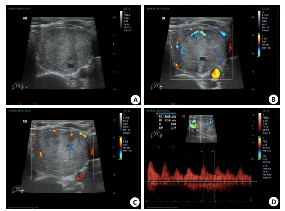
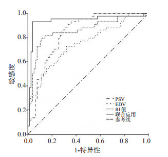
Abstract:
Objective To explore the sensitivity and specificity of ultrasound in the evaluation of benign and malignant thyroid nodules by hemodynamic parameters. Methods A total of 122 patients with thyroid nodules treated from June 2020 to January 2022 were selected. They were divided into benign nodule group (n=78) and malignant nodule group (n=44) according to the pathological results. The patients were examined by color ultrasound, and the resected tissues were examined by routine pathology. Taking the pathological diagnosis results as the gold standard, the ROC curve was drawn to analyze the sensitivity and specificity of each index used alone and in combination to evaluate the benign and malignant thyroid nodules. Results the values of peak systolic velocity (PSV) and resistance index (RI) in the lesion area in the benign nodule group were lower than the malignant nodule group, and the level of end diastolic velocity (EDV) was higher than the malignant nodule group (P < 0.05). The predictive value of PSV, EDV and RI in predicting benign and malignant thyroid nodules was log(P)=-0.654×PSV+ 0.627× EDV-0.608× RI +0.613; The sensitivity, specificity and AUC of combined application of PSV, EDV and RI in predicting benign and malignant thyroid nodules were significantly higher than those of single application (P < 0.05). Conclusion Ultrasonographic examination of hemodynamic parameters has high sensitivity and specificity in evaluating benign and malignant thyroid nodules.
2022, 45(5): 705-708.
doi: 10.12122/j.issn.1674-4500.2022.05.15
Abstract:
Objective To investigate the diagnostic value of dynamic contrast enhancement MRI (DCE-MRI) with diffusion weighted imaging (DWI) sequence for benign and malignant breast nodules. Methods A total of 123 patients with small breast nodules admitted to our hospital were collected. Among 123 patients, there were 46 cases of benign tumor (benign group) and 77 cases of malignant tumor (malignant group). MRI scan characteristics were analyzed. The morphology and MRI parameters of small breast nodules were compared, and the diagnostic value of DWI and DCE-MRI were analyzed. Results In the benign group, the edges were smooth, the shape was regular, and the internal enhancement was mainly uniform, the TIC type Ⅰ was common, and the early enhancement rate was less than 60%. The malignant group was the opposite (P < 0.05). Compared with the malignant group, the benign group had higher ADC value, lower Slope and SlopeR values (P < 0.05). The differences in maximum signal intensity and peak height between the two groups were not significant (P > 0.05). The sensitivity, specificity and accuracy of DWI for differential diagnosis of benign and malignant breast nodules were 62.34%, 65.22% and 63.41%, respectively. The DCE-MRI were 71.43%, 80.43% and 74.80%, respectively, which were significantly lower than 87.01%, 91.30% and 88.62% of DWI+DCE-MRI. Conclusion DCE-MRI combined with DWI can improve the diagnostic sensitivity, specificity and accuracy of benign and malignant breast nodules.

2022, 45(5): 709-717.
doi: 10.12122/j.issn.1674-4500.2022.05.16
Abstract:
Objective To explore the correlation between ultrasonographic signs of invasive ductal carcinoma of breast and the expression of immunohistochemical factors. Methods We retrospectively analyzed 524 patients with primary invasive ductal carcinoma of breast confirmed by surgical pathology in our hospital from January 2019 to December 2021. Preoperative ultrasound examination was performed and standard ultrasound images were retained. Univariate analysis χ2 test and binary logistic regression were used to analyze the correlation between ultrasonographic signs and the expression of ER, PR, HER-2, P53, Ki-67, TOPIIa and CK5/6 immunohistochemical factors. The logistic regression equation was established. Results Logistic regression analysis showed that positive expression of ER was correlated with tumor shape, tumor margin, posterior echo, hyperecho halo and abundant blood supply (P < 0.05). The positive expression of PR was associated with tumor margin, hyperechoic halo, diameter > 2 cm. Positive HER-2 expression and internal microcalcification, aspect ratio diameter greater than 1 (P < 0.05). The high expression of Ki-67 was correlated with lymph node metastasis and posteriorechogenicity (P < 0.05). The positive expression of P53 was correlated with internal calcification, lymph node metastasis, tumor shape and hyperechoic halo (P < 0.05). The positive expression of CK5/6 was correlated with internal microcalcification and hyperechoic halo (P < 0.05). The positive expression of TOPIIa was associated with lymph node metastasis, posterior echo, and tumor diameter > 2 cm (P < 0.05). The corresponding regression equation was obtained and ROC curve was used to test the efficiency of the equation. Conclusion There are some differences in the signs of invasive ductal carcinoma with different immunohistochemical expressions. These ultrasound signs can provide important reference for the diagnosis, clinical treatment and prognosis assessment of breast cancer, and further link imaging and pathology together.
Effects of transjugular intrahepatic portosystemic shunt and endoscopic therapy on the prevention of
2022, 45(5): 718-722.
doi: 10.12122/j.issn.1674-4500.2022.05.17
Abstract:
Objective To analyze the the preventive effects of transjugular intrahepatic portosystemic shunt (TIPS) and endoscopic therapy on esophageal and gastric variceal bleeding (EGVB), and provide a reference for early rational treatment regimen for patients with liver cirrhosis. Methods Eighty-nine patients with liver cirrhosis complicated with EGVB who received primary prevention in Suining Central Hospital from January 2018 to February 2020 were selected. They were divided into two groups according to different surgical methods. Among the patients, the cases undergoing endoscopic variceal ligation were set as EVL group (n=43) and the cases receiving TIPS were included in TIPS group (n=46). The improvement effects of varicose veins, changes in portal vein diameter and liver function before and after treatment as well as occurrence of EGVB, complications and death after treatment were compared between the two groups. Results The effective rate of varicose veins improvement was 90.70% in TIPS group and 86.96% in EVL group (P > 0.05). The liver function Child-Pugh classification and score in the two groups showed no significant changes (P > 0.05). The portal-systemic pressure gradient in TIPS group after surgery was significantly reduced (P < 0.05). At 3, 6, 12, 24 months after treatment, the portal vein diameter in TIPS group was smaller than that in EVL group (P < 0.05). The incidence rates of EGVB in the two groups after treatment were 32.56% and 17.39% respectively (P > 0.05), and the incidence rate of long-term bleeding after 3 months of treatment was significantly lower in TIPS group than that in EVL group (P < 0.05). The incidence rates of postoperative complications and the death rates between 2 groups were not significantly differrent (P > 0.05). Conclusion Both early TIPS and EVL for primary prevention of patients with liver cirrhosis can effectively improve the esophageal and gastric varices and help to reduce the risk of EGVB. TIPS has certain advantages in reducing the portal venous pressure and incidence rate of long-term bleeding.
2022, 45(5): 723-728.
doi: 10.12122/j.issn.1674-4500.2022.05.18
Abstract:
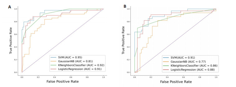
Objective To predict 1p/19q deletion status in low-grade gliomas by construction of the radiomics model based on magnetic resonance T2WI. Methods A total of 154 patients with low- grade gliomas confirmed by pathology in our hospital (including 100 cases of 1p/19q deletion and 54 cases of non-deletion of 1p/19q) were retrospectively analyzed and divided into training set and validation set according to stratified sampling at 7∶3. The tumor region was segmented manually by 3D-Slicer software and the features were extracted by pyradiomics. The clinical data were analyzed by t-test and χ2 test, and the radiomics features were screened by variance method and 10-fold cross- validation LASSO algorithm. Finally, the models of support vector machine, gaussian naïve bayes、K-nearest neighbor and logistic regreesion were established, and the efficacy was evaluated by the area under the ROC curve and the reference indexes (accuracy, sensitivity, specificity, F1 score) in sklearn classification report. Results Among the four models, the AUC value of support vector machine was the highest, the training set and verification set were 0.95 and 0.91 respectively, the best reference index was K-nearest neighbor, its accuracy, sensitivity, specificity and F1 score were 0.87, 0.97, 0.70, 0.91 respectively, followed by support vector machine, each index was equal to the average value of the model. Conclusion Based on the T2WI radiomics model, the state of 1p/19q deletion in low-grade gliomas can be effectively predicted.
2022, 45(5): 729-732.
doi: 10.12122/j.issn.1674-4500.2022.05.19
Abstract:
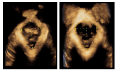
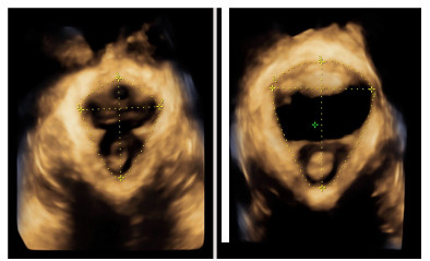
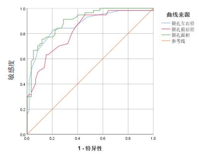
Objective To quantitatively analyze the deformation of levator ani hiatus in middle-aged and elderly parturients by 4D pelvic floor ultrasound, and to explore the feasibility of diagnosing cystocele. Methods Fifty-seven middle-aged and elderly women with cystocele in our hospital from January 2020 to June 2022 were selected as the experimental group, and 59 middle-aged and elderly women without cystocele in the same period as the control group. 4D pelvic floor ultrasonography was performed in both groups. The deformation of levator ani muscle hiatus at rest and Valsalva was observed, and the anterior and posterior diameter, left and right diameter and area of the hiatus were recorded, and the relationship between hiatus and cystocele was analyzed. Results In the case group, the structure was loose, the internal arrangement was disordered, and the levator hiatus was significantly increased(P < 0.05). There was no significant difference in the parameters between the two groups at rest. Under the condition of Valsalva, the AUC of the case group was significantly higher than that of the control group (P < 0.05). The best cutoff values of the anterior and posterior diameter, left and right diameter and area of levator hiatus were 4.45 cm, 5.25 cm and 19.05 cm2, respectively. The AUC of the area was the largest (AUC=0.898), sensitivity was 0.754 and specificity was 0.881. It was better than front and back diameter and transverse diameter. Conclusion The hiatus of levator ani muscle is reconstructed by four-dimensional ultrasound. Its deformation and parameter changes have high diagnostic value in the evaluation of cystocele in the middle-aged and elderly.
2022, 45(5): 733-736.
doi: 10.12122/j.issn.1674-4500.2022.05.20
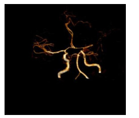
Abstract:
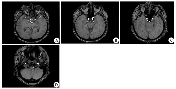
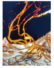
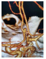
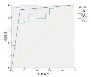
Objective To evaluate the diagnostic value of computed tomography angiography (CTA) combined with interleukin (IL-6) and tumor necrosis factor-α (TNF-α) in intracranial aneurysms. Methods A total of 108 suspected cases of intracranial aneurysm admitted to our hospital from March 2019 to March 2022 were selected. The diagnostic value of CTA combined with IL-6 and TNF-α were analyzed. Results Among 108 suspected patients, 93 were diagnosed as intracranial aneurysm by DSA and 87 were diagnosed by CTA. The Kappa value of CTA was 0.669, accuracy was 90.74%, sensitivity was 0.914 and specificity was 0.867. The serum levels of IL-6 and TNF-α in patients with intracranial aneurysm were significantly higher than those in patients without intracranial aneurysm (P < 0.05). Spearman correlation analysis showed that IL-6 and TNF-α were positively correlated with the occurrence of intracranial aneurysm (r=0.377, 0.453, P < 0.05). ROC curve showed that the area under the curve of CTA combined with IL-6 and TNF-α was 0.974, which was significantly higher than the area under the curve of each single index (P < 0.05). Conclusion CTA combined with IL-6 and TNF-α has high diagnostic value for intracranial aneurysm.
2022, 45(5): 737-743.
doi: 10.12122/j.issn.1674-4500.2022.05.21
Abstract:
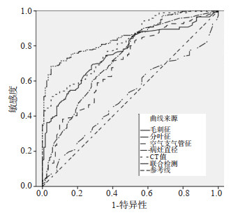
Objective To explore the predictive value of CT images in the degree of pure ground-glass nodule (PGGN) infiltration and the pathological properties of pulmonary nodules in patients with lung adenocarcinoma. Methods A total of 122 patients with pggn hospitalized in our hospital from January 2017 to January 2021 were selected. According to the types of lung adenocarcinoma, 82 patients in the pre invasion group [39 patients in adenocarcinoma in situ (AIS) group and 43 patients in microinvasive adenocarcinoma (MIA) group] and 40 patients in the invasion group. According to the classification of benign and malignant lung adenocarcinoma, there were 80 cases in benign group and 42 cases in malignant group. We compared the differences of lesion diameter, CT value, shape, tumor lung interface, hairpin sign, vacuole sign, three-dimensional shape, lobulation sign, air bronchus sign and pleural indentation between the pre invasion group and the invasion group, AIS group and MIA group, benign group and malignant group. Results The differences of lesion diameter, CT value, shape, burr sign, lobulation sign and air bronchogram sign between the pre invasion group and the invasion group were significant (P < 0.05). The differences of lobation sign, vacuole sign and CT value between AIS and MIA groups were statistically significant (P < 0.05). The differences of tumor lung interface, lesion diameter, CT value, shape, burr sign, lobulation sign and air bronchial sign between benign group and malignant group were significant (P < 0.05). The diagnostic sensitivity of combined detection of lesion diameter, CT value, shape, burr sign, lobulation sign and air bronchogram sign in patients with lung cancer invasion and malignant group was significantly higher than that of single detection. ROC curve analysis showed that the area under the curve of the combined detection of lesion diameter, CT value, shape, burr sign, lobulation sign and air bronchial sign were significantly higher than that of the single detection. Conclusion CT images of patients with lung adenocarcinoma have significant predictive value for the degree of PGGN infiltration and the pathological properties of pulmonary nodules.


2022, 45(5): 744-748.
doi: 10.12122/j.issn.1674-4500.2022.05.22
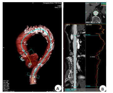
Abstract:
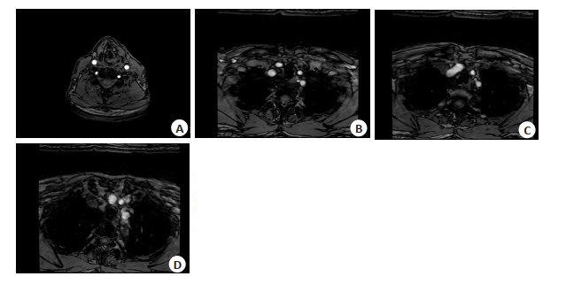
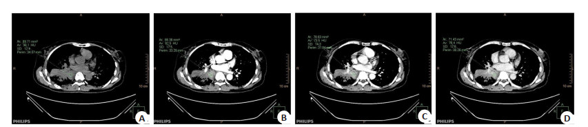
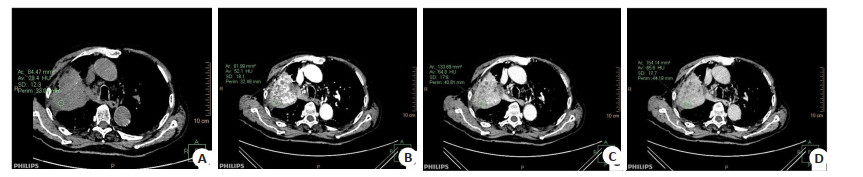
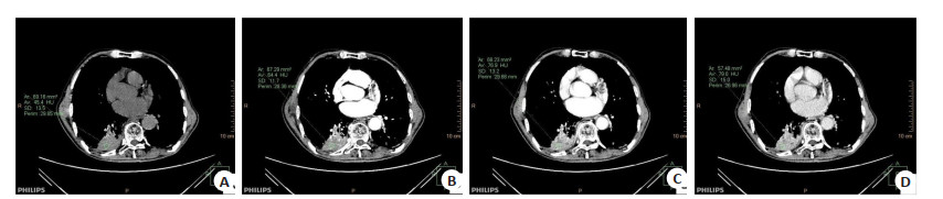
Objective To observe the characteristics and clinical significance of thoracic aorta CT reconstruction in valvular heart disease. Methods A total of 300 patients admitted to radiology department of Beibei District Hospital of Traditional Chinese Medicine were enrolled from October 2018 to December 2021, including 95 cases with valvular heart disease in case group and other 205 ordinary cases in control group. All patients underwent CT scan and multi-phase reconstruction. The general clinical data and CT measurement data (length of thoracic aorta, diameter of aortic isthmus, aortic diameter at diaphragm level) were compared between the two groups. The influencing factors of thoracic aorta length were analyzed by Logistics analysis. Results The length of thoracic aorta, diameter of aortic isthmus and aortic diameter at diaphragm level in cases were longer than those in control group (P < 0.05). The main CT characteristics in patients with heart valve disease included aortic dilatation and valve thickening. The differences in length of thoracic aorta among patients with different age, genders, hypertension, aortic wall plaques and valvular heart diseases were statistically significant (P < 0.05). Age (OR=2.121), aortic wall plaque (OR= 2.234) and valvular heart disease(OR=1.964) were influencing factors of thoracic aorta length. Conclusion The thoracic aorta is increased, aortic isthmus and aortic diameter at diaphragm level are thickened in patients with valvular heart disease. The length of thoracic aorta is affected by age, aortic wall plaques and heart valve disease. Understanding the morphological characteristics has certain guiding significance for aortic surgical treatment.
2022, 45(5): 749-753.
doi: 10.12122/j.issn.1674-4500.2022.05.23
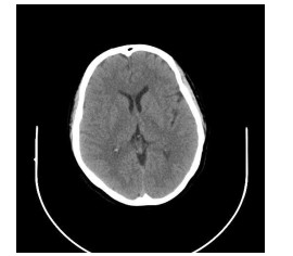
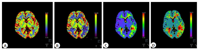
Abstract:
Objective To explore the evaluated value of CT perfusion imaging (CTPI) on cerebral tissue blood perfusion, cerebral collateral blood circulation status and responsible vessel stenosis in patients with acute ischemic stroke. Methods Fifty- one patients with acute ischemic stroke who were treated in Panzhihua Hospital of Integrated Traditional Chinese and Western Medicine were selected from March 2021 to March 2022, and they underwent CT plain scan and CT perfusion imaging after admission. The cerebral blood flow (CBF), cerebral blood volume (CBV), mean transit time (MTT) and time to peak (TTP) in different regions of patients were analyzed. CTPI parameters in different collateral blood circulation status and different stenosis degrees of responsible vessels were compared. Results CTPI of patients with AIS showed abnormal cerebral tissue blood perfusion in 46 cases, with the positive rate of CTPI was 90.20% (46/51). The CBF and CBV of the infarcted lesion area and ischemic penumbra of the affected side were lower than those of the healthy side. The MTT and TTP were longer than those of the healthy side (P < 0.05). Among patients with good collateral circulation, the CBF value of the affected side was lower than that of the healthy side, and the MTT and TTP were longer than those of the healthy side (P < 0.05). But there was no statistical significance in CBV between the affected side and the healthy side (P > 0.05). The CBF and CBV of the affected side in patients with poor collateral circulation were lower than those of the healthy side, and the MTT and TTP were longer than those of the healthy side (P < 0.05). The incidence rate of severe stenosis or occlusion of responsible vessels in patients with abnormal cerebral tissue blood perfusion indicated by CTPI was higher than that in patients with normal blood perfusion indicated by CTPI (P < 0.05). Conclusion CTPI can timely and accurately reflect the blood perfusion in the infarcted lesion area and ischemic penumbra in patients with acute ischemic stroke. It is helpful for clinically grasping the collateral circulation status, objectively assessing the responsible vessel stenosis degree.
2022, 45(5): 754-758.
doi: 10.12122/j.issn.1674-4500.2022.05.24
Abstract:
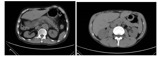
Objective To investigate the changes of copeptin (CPT), C- reactive protein (CRP) and D- dimer (D- D) levelsbefore and after the treatment of acute biliary pancreatitis by endoscopic retrograde cholangiopancreatography (ERCP). Methods Ninety- five patients with acute biliary pancreatitis in our hospital from May 2018 to March 2020 were selected and divided into control group (n=47) and observation group (n=48) according to the random number table method . The control group was treated with traditional open surgery, and theobservation group was treated with ERCP. The clinicalefficacy, clinical symptoms recovery time and complications of the two groups were compared, and changes in CPT, CRP, and D-D levels were observed before and after treatment. Results Compared with the control group (87.23%), the total effective rate of observation group (97.92%) was significantly higher (P < 0.05). The levels of CPT, CRP, and D-D at 1 d and 4 d after the operation were lower than before the operation, and the decrease in the observation group was larger than that of the control group (P < 0.05). Compared with the control group, the observation group had shorter abdominal pain relief time, gastrointestinal function and liver function recovery time (P < 0.05). Compared with the control group, the observation group had shorter hospital stays and lower hospitalization costs, lower complication rate (P < 0.05). Conclusion ERCP surgery has a good effect in the treatment of acute biliarypancreatitis. Postoperative CPT, CRP and D-D levels and clinical symptomsare significantly improved, with fewercomplications and high safety.
2022, 45(5): 759-762.
doi: 10.12122/j.issn.1674-4500.2022.05.25
Abstract:
Objective To analyze the relationship between dual-layer detector spectral CT quantitative parameters and lung cancer and its pathological characteristics. Methods The clinical data of 87 patients with lung cancer (lung cancer group) were collected from March 2021 to January 2022. Thirty-six patients with lung inflammatory nodules who were admitted to the hospital during the same period were included in control group. All patients in the two groups received dual-layer detector spectral CT within 2 weeks before treatment. The differences in conventional CT parameters and spectral CT quantitative parameters were compared between the two groups. The differences in spectral CT quantitative parameters were analyzed among patients in lung cancer group with different pathological characteristics. Results There were no statistical differences in the spinous process sign, CT value and iodine content (IC)、normalized IC (NIC), effective atomic number (Zeff) in the arterial phase between the two groups (P > 0.05). But the CT signs of lobulation sign, spiculation sign, pleural indentation and vessel convergence sign in lung cancer group were more than those in control group (P < 0.05). The IC, NIC and Zeff in the venous phase were lower in lung cancer group than those in control group (P < 0.05). There were no statistical differences in the IC, NIC and Zeff in the arterial phase among patients with different pathological characteristics in lung cancer group (P > 0.05). The IC, NIC and Zeff in the venous phase of adenocarcinoma were higher than those of squamous cell carcinoma and small cell carcinoma (P < 0.05). The three indicators of squamous cell carcinoma were higher than those of small cell carcinoma (P < 0.05). Conclusion The quantitative parameters of dual-layer detector spectral CT are beneficial to distinguishing lung cancer from lung inflammatory nodules. It can assist in judging the pathological characteristics of lung cancer, with a good application prospect.
2022, 45(5): 767-773.
doi: 10.12122/j.issn.1674-4500.2022.05.27
Abstract:
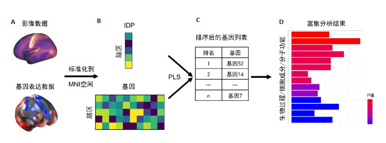
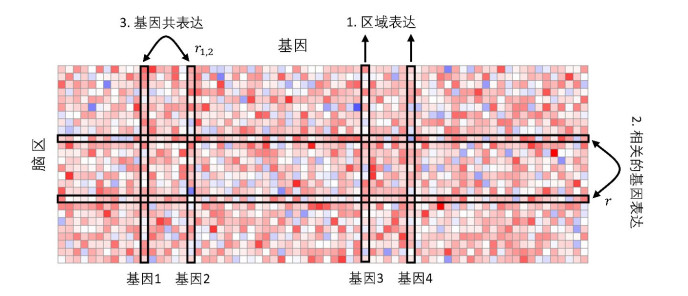
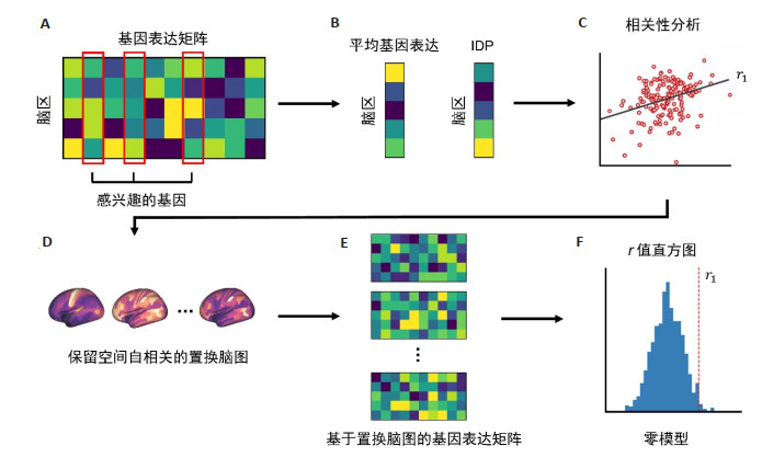
With the advent of brain-wide transcriptomics data, i.e. brain-wide gene expression atlases such as the Allen human brain atlas, imaging transcriptomics has opened new opportunities for understanding the relationship between spatial variations on molecular scale of the brain and macroscopic neuroimaging phenotypes. A growing body of literature is demonstrating relationships between gene expression and different properties of brain structure and function. This article introduces the gene expression dataset widely used in the field of imaging transcriptomics, as well as the basic steps and the commonly used toolbox for transcriptomics data processing, and outlines the basic workflow and three kinds of analysis methods for associating gene expression data with image data. In recent years, imaging transcriptomics has been widely used to understand brain neurodevelopment and various neuropsychiatric disorders. However, the field is still nascent, and several methodological challenges must be overcome to ensure the robustness of the findings. As the filed develops and existing methodologies are refined, future studies can be combined with increasingly more comprehensive and precise transcriptional atlas data, which will offer a powerful and reliable framework for identifying the molecular correlates of disease-related brain changes observed in vivo.
With the advent of brain-wide transcriptomics data, i.e. brain-wide gene expression atlases such as the Allen human brain atlas, imaging transcriptomics has opened new opportunities for understanding the relationship between spatial variations on molecular scale of the brain and macroscopic neuroimaging phenotypes. A growing body of literature is demonstrating relationships between gene expression and different properties of brain structure and function. This article introduces the gene expression dataset widely used in the field of imaging transcriptomics, as well as the basic steps and the commonly used toolbox for transcriptomics data processing, and outlines the basic workflow and three kinds of analysis methods for associating gene expression data with image data. In recent years, imaging transcriptomics has been widely used to understand brain neurodevelopment and various neuropsychiatric disorders. However, the field is still nascent, and several methodological challenges must be overcome to ensure the robustness of the findings. As the filed develops and existing methodologies are refined, future studies can be combined with increasingly more comprehensive and precise transcriptional atlas data, which will offer a powerful and reliable framework for identifying the molecular correlates of disease-related brain changes observed in vivo.
2022, 45(5): 774-778.
doi: 10.12122/j.issn.1674-4500.2022.05.28
Abstract:
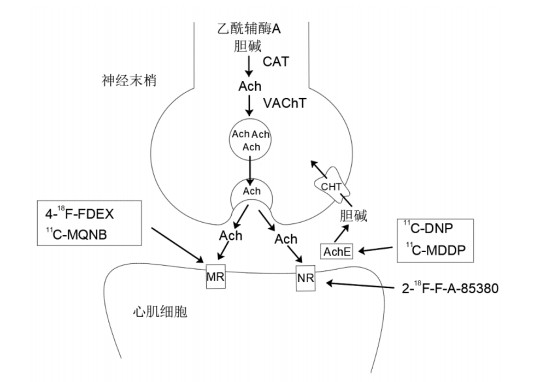
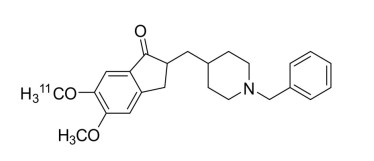
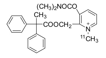
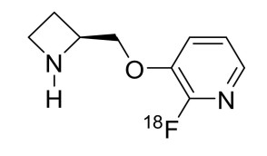
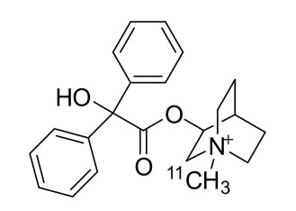
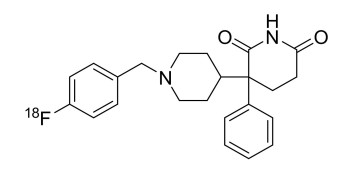
The development of cardiac parasympathetic positron imaging agents plays an immeasurable role in the evaluation of cardiovascular disease, and positron- emission tomography can reflect changes in the parasympathetic nerves of the heart with high sensitivity. 2-[18F]fluoro-3-[2(S)-2-azetidinyl-methoxy]pyridine (2-18F-F-A-85380), (R, S)-N-[11C]-Methyl-quinuclidin-3-yl benzilate (11C-MQNB) and 4-18F- fluorobenzyl-dexetimide (4-18F-FDEX) are nicotinoid and muscarinic parasympathetic imaging agents, 11C-donepezil (11C-DNP) and N-[11C]methyl-3-[[(dimethylamino)carbonyl]oxy]-2-(2', 2'-diphenylpropio-noxymethyl) pyridinium (11C-MDDP) are acetylcholinesterase parasympathetic imaging agents. Among them, 11C-MQNB and 2-18F- F-A-85380 are more intensively studied, while 11C-DNP, 11C-MDDP and 4-18F-FDEX are imaging agents with good potential for clinical application.
The development of cardiac parasympathetic positron imaging agents plays an immeasurable role in the evaluation of cardiovascular disease, and positron- emission tomography can reflect changes in the parasympathetic nerves of the heart with high sensitivity. 2-[18F]fluoro-3-[2(S)-2-azetidinyl-methoxy]pyridine (2-18F-F-A-85380), (R, S)-N-[11C]-Methyl-quinuclidin-3-yl benzilate (11C-MQNB) and 4-18F- fluorobenzyl-dexetimide (4-18F-FDEX) are nicotinoid and muscarinic parasympathetic imaging agents, 11C-donepezil (11C-DNP) and N-[11C]methyl-3-[[(dimethylamino)carbonyl]oxy]-2-(2', 2'-diphenylpropio-noxymethyl) pyridinium (11C-MDDP) are acetylcholinesterase parasympathetic imaging agents. Among them, 11C-MQNB and 2-18F- F-A-85380 are more intensively studied, while 11C-DNP, 11C-MDDP and 4-18F-FDEX are imaging agents with good potential for clinical application.
2022, 45(5): 779-789.
doi: 10.12122/j.issn.1674-4500.2022.05.29
Abstract:
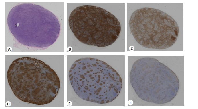
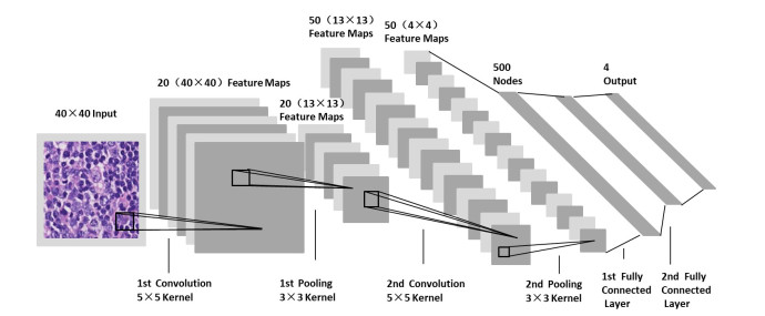
Based on the development of machine vision in recent years, the application of deep learning artificial intelligence methods in histopathology has greatly promoted pathologists to solve clinical diagnostic problems. This method can be called computer pathology. Artificial intelligence can help pathologists sift through most benign data, aid in diagnosis, predict efficacy, identify biomarkers, and even monitor therapeutic efficacy and identify unknown signals for drugs. Based on the in-depth study of deep learning in the field of pathology, it is possible for the computer to process pathological data automatically. Artificial intelligence diagnostic decisions are based on big data, and it is possible to personalize the management of each patient. It has the advantage of more rapid and accurate diagnosis for most common diseases. However, it is worth considering that the development of digital pathology is still limited by some problems, so that it is not widely used in digital pathology diagnostic platform at the present stage. We summarized the recent progress of artificial intelligence in the field of pathological diagnosis in this paper. We discussed the feasibility of this technology, added the difficulties and challenges encountered in digital pathology, and put forward the prospect of its practicality in this field.
Based on the development of machine vision in recent years, the application of deep learning artificial intelligence methods in histopathology has greatly promoted pathologists to solve clinical diagnostic problems. This method can be called computer pathology. Artificial intelligence can help pathologists sift through most benign data, aid in diagnosis, predict efficacy, identify biomarkers, and even monitor therapeutic efficacy and identify unknown signals for drugs. Based on the in-depth study of deep learning in the field of pathology, it is possible for the computer to process pathological data automatically. Artificial intelligence diagnostic decisions are based on big data, and it is possible to personalize the management of each patient. It has the advantage of more rapid and accurate diagnosis for most common diseases. However, it is worth considering that the development of digital pathology is still limited by some problems, so that it is not widely used in digital pathology diagnostic platform at the present stage. We summarized the recent progress of artificial intelligence in the field of pathological diagnosis in this paper. We discussed the feasibility of this technology, added the difficulties and challenges encountered in digital pathology, and put forward the prospect of its practicality in this field.
2022, 45(5): 790-794.
doi: 10.12122/j.issn.1674-4500.2022.05.30
Abstract:
Liver iron overload is the main histological feature of hereditary hemochromatosis and transfusion hemosiderinosis, excess iron can result in liver damage, with the slow development of cirrhosis, liver failure, and hepatocellular carcinoma if left untreated.Therefore, assessment of liver iron content is critical for detection, quantitative grading of iron overload and monitoring of iron chelation therapy.Gold standard liver biopsy is invasive and prone to sampling bias, while the non-invasive nature of MRI and its high sensitivity to iron make it a widely used method. The research progress of signal intensity ratio method, T2/R2 relaxation, magnetic resonance spectroscopy, T2*/R2* relaxation, ultrashort echo time imaging technology and quantitative susceptibility mapping to assess liver iron quantification will be reviewed in this article.
Liver iron overload is the main histological feature of hereditary hemochromatosis and transfusion hemosiderinosis, excess iron can result in liver damage, with the slow development of cirrhosis, liver failure, and hepatocellular carcinoma if left untreated.Therefore, assessment of liver iron content is critical for detection, quantitative grading of iron overload and monitoring of iron chelation therapy.Gold standard liver biopsy is invasive and prone to sampling bias, while the non-invasive nature of MRI and its high sensitivity to iron make it a widely used method. The research progress of signal intensity ratio method, T2/R2 relaxation, magnetic resonance spectroscopy, T2*/R2* relaxation, ultrashort echo time imaging technology and quantitative susceptibility mapping to assess liver iron quantification will be reviewed in this article.
2022, 45(5): 795-799.
doi: 10.12122/j.issn.1674-4500.2022.05.31
Abstract:
Glioma is a common malignant tumor in the central nervous system. It has the characteristics of diffuse infiltration and growth and it is difficult to treat it. The tumor has a high degree of malignancy, easy recurrence and poor prognosis. Multimodal magnetic resonance imaging is often used in the preliminary diagnosis of tumors before surgery. It has an important value in grading and predicting the prognosis of gliomas. This article reviews the research progress of multimodal magnetic resonance imaging including diffusion weighted magnetic resonance imaging, diffusion tensor magnetic resonance imaging, dynamic magnetic sensitive contrast perfusion weighted imaging, dynamic contrast- enhanced magnetic resonance imaging, arterial spin labeling imaging and magnetic resonance spectroscopy imaging in preoperative grading diagnosis and prognosis evaluation of glioma.
Glioma is a common malignant tumor in the central nervous system. It has the characteristics of diffuse infiltration and growth and it is difficult to treat it. The tumor has a high degree of malignancy, easy recurrence and poor prognosis. Multimodal magnetic resonance imaging is often used in the preliminary diagnosis of tumors before surgery. It has an important value in grading and predicting the prognosis of gliomas. This article reviews the research progress of multimodal magnetic resonance imaging including diffusion weighted magnetic resonance imaging, diffusion tensor magnetic resonance imaging, dynamic magnetic sensitive contrast perfusion weighted imaging, dynamic contrast- enhanced magnetic resonance imaging, arterial spin labeling imaging and magnetic resonance spectroscopy imaging in preoperative grading diagnosis and prognosis evaluation of glioma.