DenseNet network deep learning analysis of CT image in differentiating benign and malignant pulmonary nodules
-
摘要:
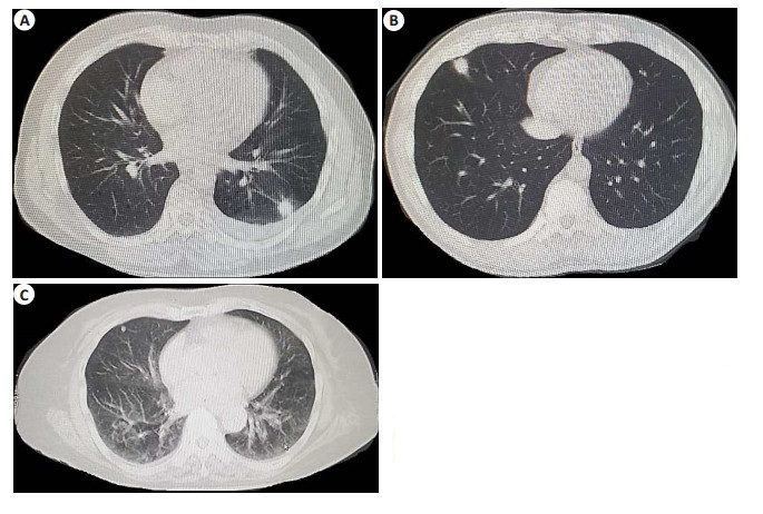
目的 探究DenseNet网络深度学习分析CT图像鉴别肺结节良恶性的价值。 方法 选取2017年2月~2019年5月我院收治的疑似肺结节患者80例,患者均进行CT扫描和DenseNet网络深度学习的人工智能系统诊断其良恶性,以病理结果作为金标准。分析CT图像、DenseNet网络深度学习分析联合CT图像对肺结节良恶性的诊断价值。 结果 CT图像表现肺密度增高影,有云雾状阴影,可清晰显示支气管内血管情况,评估结节良恶性准确率为88.75%,敏感度为76.92%,特异性为94.44%,与病理诊断的Kappa值为0.736(P < 0.001);DenseNet网络深度学习联合CT评估结节良恶性的敏感度为96.15%,特异性为88.89%,DenseNet网络深度学习联合CT评估准确率高于单纯CT评估准确率(91.25% vs 88.75%),且与病理诊断一致性较好(Kappa= 0.810,P < 0.001)。 结论 DenseNet网络深度学习分析CT图像鉴别肺结节良恶性准确性较高,且与病理结果具有较好的一致性。 Abstract:Objective To explore the value of DenseNet network deep learning analysis in differentiating benign and malignant pulmonary nodules. Methods Eighty patients with suspected pulmonary nodules in our hospital from February 2017 to May 2019 were selected. All patients underwent CT scan and artificial intelligence system of DenseNet network deep learning to diagnose benign and malignant, and pathological results were taken as gold standard. We analyzed the diagnostic value of CT images, DenseNet network deep learning analysis combined with CT images in benign and malignant pulmonary nodules. Results CT images showed increased lung density and cloudy shadow, which could clearly show the situation of bronchial vessels. The accuracy rate of benign and malignant nodules was 88.75%, the sensitivity was 76.92%, and the specificity was 94.44%, Kappa value with pathological diagnosis was 0.736 (P < 0.001). The sensitivity and specificity of DenseNet network depth learning combined with CT in evaluating benign and malignant nodules were 96.15% and 88.89%, respectively. The accuracy of densenet network deep learning combined with CT in evaluating benign and malignant nodules was 91.25%, which was higher than that of CT alone (88.75%). It had good consistency with pathological diagnosis (Kappa=0.810, P < 0.001). Conclusion DenseNet network deep learning analysis of CT images in differentiating benign and malignant pulmonary nodules has high accuracy and good consistency with pathological results. -
Key words:
- DenseNet /
- deep learning /
- CT /
- pulmonary nodules /
- benign and malignant
-
表 1 CT与病理诊断肺结节良恶性结果比较
Table 1. Comparison between CT and pathological diagnosis of benign and malignant pulmonary nodules (n)
CT诊断 病理诊断 合计 恶性 良性 恶性 20 3 23 良性 6 51 57 合计 26 54 80 表 2 DenseNet网络深度学习联合CT与病理诊断肺结节良恶性结果比较
Table 2. Comparison of results of deep learning of DenseNet network combined with CT and pathological diagnosis of benign and malignant pulmonary nodules (n)
DenseNet网络深度学习联合CT诊断 病理诊断 合计 恶性 良性 恶性 25 6 31 良性 1 48 49 合计 26 54 80 表 3 CT、DenseNet网络深度学习及联合诊断价值比较
Table 3. Comparison of CT and DenseNet network in-depth learning and joint diagnosis value
诊断方法 敏感度(%) 特异性(%) 阳性预测值(%) 阴性预测值(%) 准确率(%) Kappa CT 76.92 94.44 86.96 89.47 88.75 0.736 DenseNet网络深度学习联合CT 96.15 88.89 80.65 97.96 91.25 0.810 -
[1] 戴志京, 孙蓉, 钱小建. 多层螺旋CT联合磁共振扩散加权成像在肺部结节良恶性病变鉴别诊断中的应用[J]. 中国CT和MRI杂志, 2019, 17(12): 62-4. https://www.cnki.com.cn/Article/CJFDTOTAL-CTMR201912020.htm [2] 王风, 王磊, 李囡, 等. 基于三维卷积神经网络深度学习的肺结节良恶性的鉴别诊断[J]. 中国医学影像学杂志, 2019, 27(10): 779-82, 787. doi: 10.3969/j.issn.1005-5185.2019.10.016 [3] Digumarthy SR, Padole AM, Lo Gullo R, et al. CT texture analysis of histologically proven benign and malignant lung lesions[J]. Medicine, 2018, 97(26): e11172. doi: 10.1097/MD.0000000000011172 [4] 戴垚均, 闫士举, 宋成利. 基于密集网络改进的肺结节良恶性分类模型[J]. 中国医学影像技术, 2018, 34(7): 1104-9. doi: 10.13929/j.1003-3289.201710013 [5] Yang KQ, Liu JS, Tang W, et al. Identification of benign and malignant pulmonary nodules on chest CT using improved 3D U-Net deep learning framework[J]. Eur J Radiol, 2020, 129: 109013. doi: 10.1016/j.ejrad.2020.109013 [6] 吕文晖, 周长圣, 李新宇, 等. 利用深度学习模型判断基线胸部平扫CT肺结节的良恶性[J]. 中华放射学杂志, 2019, 53(11): 957-62. [7] Wu LY, Cao GQ, Zhao L, et al. Spectral CT analysis of solitary pulmonary nodules for differentiating malignancy from benignancy: the value of iodine concentration spatial distribution difference[J]. Biomed Res Int, 2018, 2018: 4830659. [8] 刘露, 杨培亮, 孙巍巍, 等. 深度置信网络对孤立性肺结节良恶性的分类[J]. 哈尔滨理工大学学报, 2018, 23(3): 9-15. https://www.cnki.com.cn/Article/CJFDTOTAL-HLGX201803003.htm [9] Li H, Zhuang SS, Li DA, et al. Benign and malignant classification of mammogram images based on deep learning[J]. Biomed Signal Process Control, 2019, 51: 347-54. doi: 10.1016/j.bspc.2019.02.017 [10] 陶广昱, 叶剑定, 叶晓丹, 等. 基于深度学习的肺结节良恶性判别模型在靶扫描CT数据的效能验证[J]. 中华放射学杂志, 2019, 53(11): 952-6. [11] Fujioka T, Kubota K, Mori M, et al. Distinction between benign and malignant breast masses at breast ultrasound using deep learning method with convolutional neural network[J]. Jpn J Radiol, 2019, 37 (6): 466-72. [12] 郭芳芳, 李欣菱, 王欣悦, 等. 亚实性肺结节CT征象在良恶性鉴别及腺癌恶性侵袭程度评估中的价值[J]. 中国肺癌杂志, 2018, 21(6): 451-7. https://www.cnki.com.cn/Article/CJFDTOTAL-FAIZ201806005.htm [13] 王俊, 曾庆华, 李永红, 等. C T影像学检查对肺部磨玻璃样小结节样良、恶性的鉴别诊断价值及影响因素分析[J]. 中国CT和MRI杂志, 2021, 19(6): 62-4. https://www.cnki.com.cn/Article/CJFDTOTAL-CTMR202106020.htm [14] 张俊, 侯聪, 刘新疆. 基于深度学习的人工智能在肺结节诊断领域的进展[J]. 分子影像学杂志, 2020, 43(3): 365-8. doi: 10.12122/j.issn.1674-4500.2020.03.01 [15] 南岩东, 李玉娟, 刘苗苗, 等. 人工智能在肺结节良恶性鉴别诊断中的价值分析[J]. 中华肺部疾病杂志: 电子版, 2020, 13(6): 760-3. https://www.cnki.com.cn/Article/CJFDTOTAL-ZFBD202006009.htm [16] 刘凯, 张荣国, 涂文婷, 等. 深度学习技术对胸部X线平片亚实性结节的检测效能初探[J]. 中华放射学杂志, 2017, 51(12): 918-21. [17] 马圆, 王风, 韩勇, 等. 基于深度信念网络检测PET/CT图像肺结节良恶性[J]. 中国医学影像技术, 2020, 36(1): 77-80. https://www.cnki.com.cn/Article/CJFDTOTAL-ZYXX202001027.htm [18] Tao YT, Xu MZ, Lu ZY, et al. DenseNet-based depth-width double reinforced deep learning neural network for high-resolution remote sensing image per-pixel classification[J]. Remote Sens, 2018, 10(5): 779. [19] Zhou H, Jin YH, Dai L, et al. Differential diagnosis of benign and malignant thyroid nodules using deep learning radiomics of thyroid ultrasound images[J]. Eur J Radiol, 2020, 127: 108992. [20] Kim DH, Lee MK, Lee SH, et al. Macro unit-based convolutional neural network for very light-weight deep learning[J]. Image Vis Comput, 2019, 87: 68-75. [21] 张军, 张红伟, 叶永强, 等. CT纹理特征分析在肺小结节良恶性鉴别诊断中的价值[J]. 浙江医学, 2018, 40(16): 1876-8, 1889. https://www.cnki.com.cn/Article/CJFDTOTAL-ZJYE201816033.htm [22] Tesche C, Gray HN. Machine learning and deep neural networks applications in coronary flow assessment: the case of computed tomography fractional flow reserve[J]. J Thorac Imaging, 2020, 35 (Suppl 1): S66-S71. -


 下载:
下载:


