


PETRA-MRA imaging of stenosis degree and prognosis in patients with atherosclerotic middle cerebral artery stenosis
-
摘要:
目的 分析大脑中动脉粥样硬化性狭窄患者狭窄程度及预后的径向采集逐点编码缩短时间MR血管成像(PETRA-MRA)影像的相关性。 方法 本研究采取回顾性研究,以2021年5月~2022年6月本院收治的72例大脑中动脉粥样硬化性狭窄患者作为研究对象,根据Rankin量表(mRS)评分(以mRS评分 > 2分为预后不良)将患者分为预后不良组(n=15)和预后良好组(n=57)。比较不同狭窄程度、不同预后患者的大脑中动脉血流信号评分、磁敏感伪影评分之间的差异。 结果 针对大脑中动脉硬化患者的诊断而言,时间飞跃法MR血管成像(TOF-MRA)诊断对患者的病灶部位的狭窄程度以及病变程度高于PETRA-MRA,差异有统计学意义(P < 0.05);TOF-MRA诊断对患者的病灶部位的磁敏感伪影评分以及血流信号评分高于PETRA-MRA,差异有统计学意义(P < 0.05);不同狭窄程度患者的病灶部位的长度以及狭窄程度之间的差异有统计学意义(P < 0.05);不同狭窄程度患者的病灶部位的磁敏感伪影评分以及血流信号评分的差异无统计学意义(P > 0.05);预后良好组患者的病灶部位的长度以及狭窄程度低于对照组,差异有统计学意义(P < 0.05);不同预后患者的病灶部位磁敏感伪影评分以及血流信号评分的差异无统计学意义(P > 0.05)。 结论 采用PETRA-MRA对大脑中动脉粥样硬化性狭窄患者狭窄程度及预后进行评估,其图像质量较好,建议临床推广。 -
关键词:
- 大脑中动脉粥样硬化 /
- PETRA-MRA /
- 预后 /
- 大脑中动脉血流信号评分 /
- 磁敏感
Abstract:Objective To study the correlation between stenosis degree and prognosis in patients with middle cerebral artery stenosis by PETRA-MRA imaging. Methods Seventy-five patients with middle cerebral artery atherosclerotic stenosis admitted to our hospital from May 2021 to June 2022 were selected as the subjects. According to the Rankin Scale (mRS score > 2 indicates poor prognosis), the patients were divided into poor prognosis group (n=15) and good prognosis group (n=57). The differences of MCA blood flow signal score and magnetic sensitivity pseudo- film score in patients with different stenosis degree and prognosis were compared. Results For the diagnosis of middle cerebral artery sclerosis patients, the degree of stenosis and lesion in the lesion site of patients diagnosed by TOF-MRA was significantly higher than that by PETRA-MRA (P < 0.05). The scores of magnetic sensitivity pseudo-film and blood flow signal of the lesions of patients diagnosed by TOF-MRA were higher than those of PETRA-MRA (P < 0.05). There were statistically significant differences in the length of lesion site and the degree of stenosis among patients with different degrees of stenosis (P < 0.05). There was no statistical significance in the magnetic sensitivity pseudo-film scores and blood flow signal scores of patients with different stenosis degrees (P > 0.05). The length of lesion site and stenosis degree in patients with good prognosis group were significantly lower than those in control group (P < 0.05). There was no statistical significance in the magnetic sensitivity pseudo-film scores and blood flow signal scores of patients with different prognosis (P > 0.05). Conclusion The image quality of PETRA-MRA is good to evaluate the stenosis degree and prognosis of patients with middle cerebral artery atherosclerotic stenosis. -

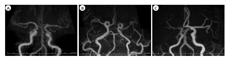
图 1 不同狭窄程度患者的病灶部位的长度以及狭窄程度比较
A: Ⅰ型狭窄; B: Ⅱ型狭窄; C: Ⅲ型狭窄.
Figure 1. Comparison of the length of the lesion site and the degree of stenosis in patients with different degrees of stenosis.
表 1 两种诊断方法对病灶部位的狭窄程度以及病变程度的诊断结果比较
Table 1. Comparison of the diagnostic results of the two diagnostic methods on the degree of stenosis at the lesion site and the degree of lesion (n=72, Mean±SD)
诊断方法 狭窄程度(%) 病变程度(mm) TOF-MRA 59.22±2.41 15.12±2.12 PETRA-MRA 46.63±2.52 12.34±1.97 t 30.637 8.151 P < 0.001 < 0.001 TOF-MRA: 时间飞跃法MR血管成像; PETRA-MRA: 径向采集逐点编码缩短时间MR血管成像.  下载: 导出CSV
表 2 两种诊断方法对病灶部位的图像质量比较
Table 2. Comparison of image quality between the two diagnostic methods on the lesion site (Score, n=72, Mean±SD)
诊断方法 磁敏感伪影评分 血流信号评分 TOF-MRA 2.93±0.61 2.54±0.28 PETRA-MRA 1.49±0.48 1.22±0.34 t 15.742 25.430 P < 0.001 < 0.001
下载: 导出CSV
表 3 不同狭窄程度患者的病灶部位的长度以及狭窄程度比较
Table 3. Comparison of the length of the lesion site and the degree of stenosis in patients with different stenosis degrees (Mean±SD)
狭窄程度分型 狭窄程度(%) 病变程度(mm) Ⅰ型(n=35) 37.52±2.84 10.82±1.68 Ⅱ型(n=21) 47.05±2.78 12.03±1.77 Ⅲ型(n=16) 49.12±1.81 14.21±2.32 F 13.251 14.112 P < 0.001 < 0.001
下载: 导出CSV
表 4 不同狭窄程度患者的磁敏感伪影评分以及血流信号评分比较
Table 4. Comparison of magnetic sensitivity pseudo- film scores and blood flow signal scores of patients with different stenosis degrees (score, Mean±SD)
狭窄程度分型 磁敏感伪影评分 血流信号评分 Ⅰ型(n=35) 1.47±0.32 1.25±0.55 Ⅱ型(n=21) 1.49±0.26 1.23±0.99 Ⅲ型(n=16) 1.52±0.55 1.20±0.73 F 0.858 0.887 P 0.123 0.099
下载: 导出CSV
表 5 不同预后患者的病灶部位的长度以及狭窄程度比较
Table 5. Comparison of the length of the lesion site and the degree of stenosis in patients with different prognosis (Mean±SD)
组别 狭窄程度(%) 病变程度(mm) 预后良好组(n=57) 35.17±2.89 16.38±1.72 预后不良组(n=15) 52.52±2.38 11.52±2.21 t 23.965 7.910 P < 0.001 < 0.001
下载: 导出CSV
表 6 不同预后患者的病灶部位图像质量比较
Table 6. Comparison of image quality of lesion sites in patients with different prognosis (score, Mean±SD)
组别 磁敏感伪影评分 血流信号评分 预后良好组(n=57) 1.47±0.37 1.21±0.25 预后不良组(n=15) 1.49±1.51 1.20±0.36 t 0.051 0.101 P 0.960 0.920
下载: 导出CSV
-
[1] 汝宁, 姚明仁, 白新苹, 等. 脑部磁共振结合静息态fMRI方法在卒中患者认知障碍中的应用[J]. 影像科学与光化学, 2022, 40(1): 188-91. https://www.cnki.com.cn/Article/CJFDTOTAL-GKGH202201038.htm [2] 袁伟壮, 李明利, 冯逢, 等. 高分辨磁共振影像在青年卒中的临床诊断价值[J]. 中风与神经疾病杂志, 2021, 38(11): 984-9. doi: 10.19845/j.cnki.zfysjjbzz.2021.02604 [3] 储小雨, 陈巨罗, 王幼萌, 等. 多模式磁共振成像指导下觉醒型脑卒中再灌注治疗的临床研究[J]. 安徽医科大学学报, 2021, 56(6): 973-6. doi: 10.19405/j.cnki.issn1000-1492.2021.06.027 [4] 顾军, 王觅, 张丹凤, 等. 大脑中动脉粥样硬化性狭窄患者高分辨率磁共振成像血管壁特征与缺血性卒中风险[J]. 国际脑血管病杂志, 2021, 29(6): 401-6. doi: 10.3760/cma.j.issn.1673-4165.2021.06.001 [5] 林志伟, 陈丽芳, 马琪林, 等. 急性缺血性卒中磁共振成像液体衰减反转恢复序列血管高信号征的临床意义分析[J]. 中国脑血管病杂志, 2021, 18(3): 152-7. doi: 10.3969/j.issn.1672-5921.2021.03.002 [6] 吴琼, 任诗媛, 乐赞, 等. 脑机接口综合康复训练对亚急性期脑卒中疗效的静息态功能磁共振研究[J]. 中国康复理论与实践, 2020, 26 (1): 77-84. doi: 10.3969/j.issn.1006-9771.2020.01.014 [7] 李鑫, 郎文娟, 石锴, 等. 联合核磁共振3D CUBE T1高分辨率成像和磁敏感技术评估急性缺血性脑卒中患者的临床预后及其应用价值[J]. 中风与神经疾病杂志, 2020, 37(5): 393-7. https://www.cnki.com.cn/Article/CJFDTOTAL-ZFSJ202005006.htm [8] 冷雯静, 王荔. 脑卒中后抑郁的磁共振波谱成像研究进展[J]. 中华神经医学杂志, 2020, 19(8): 853-8. doi: 10.3760/cma.j.cn115354-20200530-00427 [9] 张春佳, 高正玉, 王强, 等. 脑卒中后肩痛患者肩关节前方撞击综合征的磁共振成像表现[J]. 中华物理医学与康复杂志, 2020, 42(12): 1100-4. doi: 10.3760/cma.j.issn.0254-1424.2020.12.012 [10] 谢晓明, 韩会建, 刘宏亮, 等. 经颅直流电刺激治疗脑卒中后认知功能障碍的静息态功能性磁共振研究[J]. 中华物理医学与康复杂志, 2020, 42(5): 392-6. doi: 10.3760/cma.j.issn.0254-1424.2020.05.002 [11] 赵俊杰, 杨艳红, 郑立峰. 磁共振成像脑小血管病总体负担与卒中后抑郁的相关性[J]. 国际脑血管病杂志, 2020, 28(10): 745-50. doi: 10.3760/cma.j.issn.1673-4165.2020.10.005 [12] 黄旭, 林国辉, 宋建勋. 基底动脉粥样硬化斑块特征与急性缺血性卒中的相关性: 高分辨率磁共振成像研究[J]. 国际脑血管病杂志, 2020, 28(10): 739-44. doi: 10.3760/cma.j.issn.1673-4165.2020.10.004 [13] 林澜, 吕晋浩, 张荣举, 等. 全脑CTP定量分析颅内动脉粥样硬化性狭窄患者血管表面渗透性[J]. 中国医学影像技术, 2020, 36(5): 659-65. https://www.cnki.com.cn/Article/CJFDTOTAL-ZYXX202005006.htm [14] Crespo-Cuevas AM, Canento T, Hernández-Perez M, et al. The Barcelona-Asymptomatic Intracranial Atherosclerosis (AsIA) study: Subclinical cervico-cerebral stenosis and middle cerebral artery pulsatility index as predictors of long-term incident cognitive impairment[J]. Atherosclerosis, 2020, 312: 104-9. doi: 10.1016/j.atherosclerosis.2020.08.025 [15] Kakadia B, Thakkar R, Sanborn E, et al. Nilotinib-associated atherosclerosis presenting as multifocal intracranial Stenosis and acute stroke[J]. J Stroke Cerebrovasc Dis, 2021, 30(8): 105883. doi: 10.1016/j.jstrokecerebrovasdis.2021.105883 [16] You S, Cho Y, Kim B, et al. Synthetic time of flight magnetic resonance angiography generation model based onCycle‐consistent generative adversarial network UsingPETRA-MRAin the patients with treated intracranial aneurysm[J]. Magnetic Resonance Imaging, 2022, 56(5): 1513-28. doi: 10.1002/jmri.28114 [17] Kim JH, Ahn SJ, Park M, et al. Follow-up imaging of clipped intracranial aneurysms with 3-T MRI: comparison between 3D timeof-flight MR angiography and pointwise encoding time reduction with radial acquisition subtraction-based MR angiography[J]. J Neurosurg, 2022, 136(5): 1260-5. doi: 10.3171/2021.7.JNS211197 [18] Norby FL, Alonso A, Rooney MR, et al. Association of ventricular arrhythmias with dementia: the atherosclerosis risk in communities (ARIC) study[J]. Neurology, 2021, 96(6): e926-36. doi: 10.1212/WNL.0000000000011122 [19] Cohen-Cohen S, Lanzino G, Brinjikji W, et al. The role of angioplasty alone in intracranial atherosclerosis: 2-dimensional operative video[J]. Oper Neurosurg (Hagerstown), 2021, 20(5): E350-E351. [20] Chiluka R, Hiluka S, Emani K, et al. AS04 Large Artery Atherosclerosis study of the risk factors for intracranial atherosclerosis disease and its pattern in acute ischemic stroke[J]. Int J Stroke, 2021, 16(S2): 83. [21] You SH, Kim B, Yang KS, et al. Ultrashort echo time magnetic resonance angiography in follow-up of intracranial aneurysms treated with endovascular coiling: comparison of time-of-flight, pointwise encoding time reduction with radial acquisition, and contrast-enhanced magnetic resonance angiography[J]. Neurosurgery, 2021, 88(2): E179-89. [22] Irie R, Suzuki M, Yamamoto M, et al. Assessing blood flow in an intracranial stent: a feasibility study of MR angiography using a silent scan after stent-assisted coil embolization for anterior circulation aneurysms[J]. AJNR Am J Neuroradiol, 2015, 36(5): 967-70. [23] Yang WJ, Zhang YX, Li ZF, et al. Differences in safety and efficacy of endovascular treatment for acute ischemic stroke: a propensity score analysis of intracranial atherosclerosis-related occlusion versus embolism[J]. Clin Neuroradiol, 2021, 31(2): 457-64. [24] 张斐斐, 冉云彩, 朱明, 等. 径向采集逐点编码缩短时间MR血管成像(PETRA-MRA)用于支架成形术治疗大脑中动脉斑块狭窄术后随访[J]. 中国医学影像技术, 2021, 37(9): 1286-90. https://www.cnki.com.cn/Article/CJFDTOTAL-ZYXX202109002.htm [25] 黄茂盛, 伏红超, 梁凯轶, 等. 3.0T MRI可评估缺血性脑卒中患者颈动脉粥样硬化的斑块成分[J]. 分子影像学杂志, 2022, 45(5): 656-60. doi: 10.12122/j.issn.1674-4500.2022.05.05 -

 点击查看大图
计量
- 文章访问数: 150
- HTML全文浏览量: 76
- PDF下载量: 8
- 被引次数: 0