


Application of carotid artery color Doppler ultrasound combined with brain MRI in patients with anterior circulation atherosclerotic cerebral infarction
-
摘要:
目的 探究前循环动脉粥样硬化性脑梗死患者采用颈动脉彩超联合颅脑MRI评估病情严重程度临床价值。 方法 回顾性分析2021年1月~2021年11月医院收治50例前循环动脉粥样硬化性脑梗死患者的资料,依据美国国立卫生研究院脑卒中量表评分,将患者分别纳入轻型脑梗死组(≤7分,n=40)与重型脑梗死组(> 7分,n=10)。患者均接受颈动脉彩超及颅脑MRI检查,分析两者联合在患者病情严重程度上评估价值。 结果 重型脑梗死组患者不稳定斑块比例、狭窄率、搏动指数(PI)、阻力指数(RI)均高于轻型脑梗死组患者,峰值流速(PSV)、舒张末期流速(EDV)低于轻型脑梗死组患者(P < 0.05);重型脑梗死组患者表面弥散系数(ADC)低于轻型脑梗死组患者(P < 0.05),两组患者T1WI信号与T2WI信号检出情况的差异无统计学意义(P > 0.05);Spearman相关性分析显示,ADC、不稳定斑块、PI、RI、PSV、EDV与患者病情严重程度存在相关性(P < 0.05);ROC曲线显示ADC、不稳定斑块、PI、PSV、RI、EDV用于重型脑梗死评估曲线下面积分别为0.638、0.556、0.600、0.608、0.798、0.713,各指标联合曲线下面积值为0.968。 结论 前循环动脉粥样硬化性脑梗死患者病情可以采用颈动脉彩超与颅脑MRI进行评估,两者用于患者病情严重程度评估均有一定价值。 -
关键词:
- 颈动脉彩超 /
- 颅脑MRI /
- 前循环动脉粥样硬化性脑梗死 /
- 病情严重程度
Abstract:Objective To investigate the clinical value of carotid artery color Doppler ultrasound combined with brain MRI in severity evaluation of anterior circulation atherosclerotic cerebral infarction. Methods A retrospective review was conducted on the data of 50 patients with anterior circulation atherosclerotic cerebral infarction who were admitted to the hospital between January and November. The patients were divided into the mild cerebral infarction group (n=40, NIHSS score ≤7) and the severe cerebral infarction group (n=10, NIHSS score > 7) according to the NIHSS score. All patients were examined with carotid artery color Doppler ultrasound and brain MRI. The value of combination of the two methods in severity evaluation was analyzed. Results The proportion of unstable plaques, stenosis rate, pulsatility index (PI) and resistance index (RI) in the severe cerebral infarction group were higher than those in the mild cerebral infarction group. The peak systolic velocity (PSV) and end-diastolic velocity (EDV) were lower than those in the mild cerebral infarction group (P < 0.05). The apparent diffusion coefficient (ADC) of the severe cerebral infarction group was significantly lower than that of the mild cerebral infarction group (P < 0.05). The detection of T1WI signals and T2WI signals showed no statistically significant difference between the groups (P > 0.05). Spearman correlation analysis found that ADC, unstable plaque, PI, RI, PSV, and EDV were correlated with the severity (P < 0.05). ROC curve analysis found that the AUC values of ADC, unstable plaque, PI, PSV, RI, and EDV to evaluate severe cerebral infarction were 0.638, 0.556, 0.600, 0.608, 0.798 and 0.713, respectively. The AUC of joint evaluation with these indicators was 0.968. Conclusion Carotid artery color Doppler ultrasound and brain MRI both can be used for severity evaluation of patients with anterior circulation atherosclerotic cerebral infarction. -

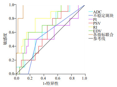
图 1 MRI测定指标、彩色超声参数对重型脑梗死评估价值分析ROC曲线
Figure 1. ROC curves of MRI measurements and color Doppler ultrasound parameters for evaluating severe cerebral infarction.
表 1 两组患者彩色超声参数比较
Table 1. Comparison of color Doppler ultrasound parameters between the two groups (Mean±SD)
组别 不稳定斑块[n(%)] PI PSV(cm/s) RI EDV(cm/s) 轻型脑梗死组(n=40) 4(10.00) 1.08±0.32 62.53±10.29 1.12±0.21 25.34±3.51 重型脑梗死组(n=10) 5(50.00) 1.36±0.45 54.58±9.24 1.34±0.25 23.49±4.08 t/χ2 6.174 2.275 2.198 3.372 2.225 P 0.013 0.027 0.033 <0.001 0.031 PI: 搏动指数; PSV峰值流速; RI: 阻力指数; EDV: 舒张末期流速.  下载: 导出CSV
表 2 两组MRI测定指标比较
Table 2. Comparison of MRI measurements between the two groups [n(%)]
组别 ADC值(μm2/ms, Mean±SD) T1WI信号 T2WI信号 等信号 低信号 等信号 高信号 轻型脑梗死组(n=40) 0.59±0.16 32(80.00) 8(20.00) 10(25.00) 30(75.00) 重型脑梗死组(n=10) 0.50±0.15 8(80.00) 2(20.00) 4(40.00) 6(60.00) t/χ2 3.675 0.195 0.304 P 0.001 0.659 0.581
下载: 导出CSV
表 3 MRI测定指标、彩色超声参数与患者病情严重程度相关性分析
Table 3. Correlation analysis of MRI measurements, color Doppler ultrasound parameters and the patients' condition
参数 r P ADC值 -0.379 0.003 不稳定斑块 0.282 0.030 PI 0.424 0.001 PSV -0.309 0.017 RI 0.552 <0.001 EDV -0.422 0.001
下载: 导出CSV
表 4 MRI测定指标、彩色超声参数对重型脑梗死评估价值分析
Table 4. Value of MRI measurements and color Doppler ultrasound parameters in evaluating severe cerebral infarction
指标 曲线下面积 敏感度(%) 特异性(%) 95% CI P ADC 0.638 90.0 42.5 0.467~0.808 0.182 不稳定斑块 0.556 50.0 70.0 0.367~0.745 0.585 PI 0.600 50.0 82.5 0.368~0.832 0.332 PSV 0.608 80.0 47.5 0.436~0.779 0.297 RI 0.798 80.0 72.5 0.647~0.948 0.004 EDV 0.713 60.0 82.5 0.544~0.881 0.039 各指标联合 0.968 100.0 90.0 0.923~1.000 <0.001
下载: 导出CSV
-
[1] Song XD, Li SX, Zhu M. Plasma miR-409-3p promotes acute cerebral infarction via suppressing CTRP3[J]. Kaohsiung J Med Sci, 2021, 37(4): 324-33. doi: 10.1002/kjm2.12327 [2] Chen LL, Wang WT, Zhang S, et al. Cohort study on the prognosis of acute cerebral infarction in different circulatory systems at 1-year follow-up[J]. BMC Cardiovasc Disord, 2021, 21(1): 521. doi: 10.1186/s12872-021-02291-0 [3] Wang XF, Wang M, Li G, et al. Efficacy of Solitaire AB stent-release angioplasty in acute middle cerebral artery atherosclerosis obliterative cerebral infarction[J]. World J Clin Cases, 2021, 9(19): 5028-36. doi: 10.12998/wjcc.v9.i19.5028 [4] Prabhakaran S, Liebeskind DS, Cotsonis G, et al. Predictors of early infarct recurrence in patients with symptomatic intracranial atherosclerotic disease[J]. Stroke, 2021, 52(6): 1961-6. doi: 10.1161/STROKEAHA.120.032676 [5] 周群, 朱幼玲, 穆艳芳, 等. 前循环血管狭窄与老年患者腔隙或腔隙性脑梗死发生的相关性[J]. 中国临床医学影像杂志, 2019, 30(4): 238-41. https://www.cnki.com.cn/Article/CJFDTOTAL-LYYX201904005.htm [6] 中华医学会神经病学分会, 中华医学会神经病学分会脑血管病学组. 中国各类主要脑血管病诊断要点2019[J]. 中华神经科杂志, 2019, 52(9): 710-5. [7] 马强, 徐达, 李剑. 超声检测老年糖尿病患者颈动脉粥样硬化病变特征及影响因素[J]. 中国老年学杂志, 2021, 41(20): 4375-7. doi: 10.3969/j.issn.1005-9202.2021.20.014 [8] Zhao YY, Lin SB, Chen KL, et al. Ultrasonic characteristics and influencing factors of atherosclerosis in diabetic patients[J]. Am J Transl Res, 2022, 14(5): 3113-20. [9] Xia ZY, Gu ML, Jia XD, et al. Integrated DNA methylation and gene expression analysis identifies SLAMF7 as a key regulator of atherosclerosis[J]. Aging, 2018, 10(6): 1324-37. doi: 10.18632/aging.101470 [10] 童陶然, 周菁菁, 曹昌权. 颈动脉超声定量参数结合MRI对急性脑梗死患者的诊断价值[J]. 中国CT和MRI杂志, 2019, 17(7): 29-31, 49. doi: 10.3969/j.issn.1672-5131.2019.07.009 [11] 谭峥, 耿学斌, 宋玉新, 等. 超声技术评价老年心血管病患者颈动脉粥样硬化的临床价值[J]. 中国老年学杂志, 2021, 41(12): 2467-70. doi: 10.3969/j.issn.1005-9202.2021.12.002 [12] 刘曼, 惠品晶, 丁亚芳, 等. 血管超声评估2型糖尿病患者颅内外动脉粥样硬化性病变的意义[J]. 中国糖尿病杂志, 2019, 27(12): 881-5. https://www.cnki.com.cn/Article/CJFDTOTAL-ZGTL201912001.htm [13] 王明月, 张蕾, 包晶晶, 等. 多普勒超声与超声造影评价颈动脉粥样硬化斑块易损性的临床研究[J]. 中国超声医学杂志, 2021, 37(11): 1215-8. https://www.cnki.com.cn/Article/CJFDTOTAL-ZGCY202111005.htm [14] Tracol C, Vannier S, Hurel C, et al. Predictors of malignant middle cerebral artery infarction after mechanical thrombectomy[J]. Revue Neurol, 2020, 176(7/8): 619-25. [15] Yuan T, Ren GL, Hu XN, et al. Added assessment of middle cerebral artery and atrial fibrillation to FLAIR vascular hyperintensity-DWI mismatch would improve the outcome prediction of acute infarction in patients with acute internal carotid artery occlusion[J]. Neurol Sci, 2019, 40(12): 2617-24. [16] 雷立存, 杜亚强, 刘振宇, 等. 磁共振扩散加权成像对不同时期脑梗死的诊断意义[J]. 河北医科大学学报, 2021, 42(1): 90-4. https://www.cnki.com.cn/Article/CJFDTOTAL-HBYX202101019.htm [17] Su H, Su S, Zhang X, et al. Application of arterial spin labeling and susceptibility weighted imaging in the diagnosis of ischemic cerebrovascular diseases[J]. Int J Clin Exp Pathol, 2020, 13(12): 3052-9. [18] 张骞, 申艳光, 谭丽丽, 等. MRI灌注加权成像参数联合弥散加权成像参数对缺血性脑卒中的诊断价值[J]. 心血管康复医学杂志, 2021, 30(4): 431-5. https://www.cnki.com.cn/Article/CJFDTOTAL-XXGK202104016.htm [19] 袁焕初, 郑晓林, 邹玉坚, 等. 3D SPACE序列及后处理在显示大脑中动脉硬化中的临床价值[J]. 放射学实践, 2020, 35(5): 601-7. https://www.cnki.com.cn/Article/CJFDTOTAL-FSXS202005011.htm [20] 王诺, 朱宣, 张萍, 等. 大脑中动脉粥样硬化斑块的影像学特征[J]. 临床神经病学杂志, 2018, 31(4): 245-8. https://www.cnki.com.cn/Article/CJFDTOTAL-LCSJ201804003.htm [21] 皮年华, 程力, 陈驰, 等. 颈动脉彩超联合头颅MRI在脑梗死病情评估中的价值研究[J]. 中国CT和MRI杂志, 2020, 18(11): 20-2. https://www.cnki.com.cn/Article/CJFDTOTAL-CTMR202011007.htm -

 点击查看大图
计量
- 文章访问数: 123
- HTML全文浏览量: 77
- PDF下载量: 6
- 被引次数: 0