


Correlation between acute pons infarction and characteristic parameters of basilar artery
-
摘要:
目的 采用多模态磁共振技术探讨急性脑桥梗死患者基底动脉迂曲及斑块特征。 方法 搜集35例急性脑桥梗死和40例非急性脑桥梗死患者的临床、实验室及影像资料,所有患者均行多模态磁共振扫描,测量基底动脉迂曲角度及斑块特征(狭窄处管周、管腔及壁面积、参考处管周及管腔面积、斑块负荷、重构模式及重构率),采用单因素分析和多因素二元Logistic回归分析急性脑桥梗死的危险因素。 结果 单因素分析显示斑块负荷、重构模式及重构率组间的差异有统计学意义(P < 0.05),基底动脉迂曲度、狭窄处管周、管腔、壁面积、参考处管周及管腔面积的差异无统计学意义(P > 0.05)。多因素二元Logistic回归分析显示,斑块负荷为急性脑桥梗死的独立保护因素(OR=0.001,95% CI=0.000~0.306),重构模式为急性脑桥梗死的独立危险因素(OR= 2.514,95% CI=1.380~4.580)。 结论 斑块负荷及重构模式可以影响急性脑桥梗死的发生。随着基底动脉斑块负荷的下降,急性脑桥梗死的发病率减低。基底动脉发生正性重构容易导致急性脑桥梗死的发生。 Abstract:Objective To investigate basilar artery tortuosis and plaque characteristics in patients with acute pontine infarction by multimodal magnetic resonance technology. Methods Clinical, laboratory and imaging data of 35 patients with acute pontine infarction and 40 patients with non-acute pontine infarction were collected. The multimodal magnetic resonance scanning was performed to measure the angle of basilar artery tortuation and plaque characteristics (peritubular peritubular area at stenosis, lumen and wall area, reference peritubular and luminal area, plaque load, remodeling mode and remodeling rate). The univariate analysis and multivariate binary logistic regression were used to analyze the risk factors of acute pontine infarction. Results Univariate analysis showed that there were statistically significant differences in plaque load, remodeling mode and remodeling rate between groups (P < 0.05), but no significant differences in basilar artery detour, peritubular periduct at stenosis, lumen, wall area, reference peritubular and luminal area (P > 0.05). Multivariate binary logistic regression analysis showed that plaque load was an independent protective factor for acute pontine infarction (OR=0.001, 95%CI=0.000-0.306) and the remodeling mode was an independent risk factor for acute pontine infarction (OR=2.514, 95% CI=1.380-4.580). Conclusion Plaque load and remodeling patterns can affect the occurrence of acute pontine infarction. With a decrease in basilar plaque load, the incidence of acute pontine infarction decreases. Positive remodeling of the basilar artery can easily lead to acute pontine infarction. -
Key words:
- pons infarction /
- basilar artery /
- high-resolution /
- plaque /
- remodeling
-

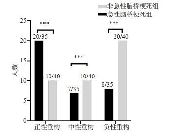
图 1 男,54岁,发现言语不清伴右侧肢体无力6 h来我院就诊
A~B: 2周内分别扫描DWI、MRA、T1VISTA. DWI及ADC图显示急性脑桥梗死; C~D: MRA序列显示正位及侧位BA弯曲角度为163.23°、161.54°; E~G: T1VISTA序列示梗死灶邻近层面基底动脉斑块,测量狭窄处管周和管腔面积分别为17.7 mm2、2.1 mm2, 参考处管周及管腔面积经计算远端及近端面积的平均值分别为14.8 mm2、4.7 mm2.
Figure 1. A54-year-old male patient came to our hospital for treatment with slurred speech accompanied by right limb weakness for 6 h.

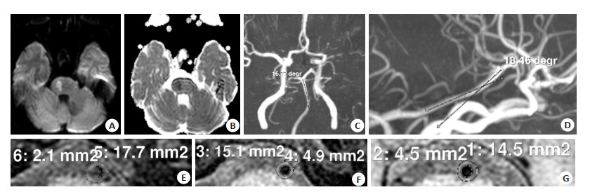
图 2 急性脑桥梗死组和非急性脑桥梗死组动脉重构分布情况
***P < 0.05.
Figure 2. Distribution of arterial remodeling in the acute and non-acute pons infarction groups.
表 1 两组患者一般资料
Table 1. Baseline data of patients in the two groups
项目 急性脑桥梗死组(n=35) 非急性脑桥梗死组(n=40) t/χ2 P 年龄(岁, Mean±SD) 65.89±9.734 65.75±9.644 0.061 0.952 性别(男/女, n) 22/13 21/19 0.819 0.366 糖尿病[n(%)] 13(37.1) 11(27.5) 0.798 0.372 收缩压(mmHg, Mean±SD) 147.34±21.979 151.95±19.779 -0.955 0.347 舒张压(mmHg, Mean±SD) 86.06±11.760 91.08±11.039 -1.905 0.061 吸烟[n(%)] 15(42.9) 21(52.5) 0.695 0.404 饮酒[n(%)] 11(31.4) 16(40.0) 0.595 0.440 高同型半胱氨酸[n(%)] 10(28.6) 11(27.5) 0.011 0.918 高血脂[n(%)] 22(62.9) 23(57.5) 0.223 0.637 高血糖[n(%)] 29(82.9) 29(72.5) 1.142 0.285  下载: 导出CSV
表 2 两组患者BA角度及斑块特征比较
Table 2. Comparison of BAangle and plaque characteristics between the two groups (Mean±SD)
指标 急性脑桥梗死组(n=35) 非急性脑桥梗死组(n=40) t/χ2/Z P MRA正位迂曲度(°) 148.98±16.46 139.86±29.77 1.668 0.100 MRA侧位迂曲度(°) 149.27±13.25 143.08±21.01 1.546 0.127 狭窄处血管面积(mm2) 23.55±8.06 21.74±6.21 1.095 0.277 狭窄处管腔面积(mm2) 4.57±3.10 5.05±2.59 -0.737 0.464 参考处血管面积(mm2) 21.75±6.56 22.49±4.55 -0.578 0.565 参考处管腔面积(mm2) 6.5(4.8, 9.5) 7.2(6.3, 9.2) -1.259 0.208 狭窄层面壁面积(mm2) 18.98±6.38 16.83±5.15 1.621 0.109 斑块负荷 0.81±0.09 0.77±0.09 2.170 0.033 重构模式[n(%)] 8.711 0.013 正性重构 20(57.1) 10(25) 中性重构 7(20) 10(25) 负性重构 8(22.9) 20(50) 重构率(%) 1.09±0.21 0.96±0.16 2.937 0.004
下载: 导出CSV
表 3 急性脑桥梗死的独立因素分析
Table 3. Independent risk factors of acute pontine infarction
因素 回归系数 标准误 Wald 斑块负荷 -7.133 3.035 5.525 重构模式 0.922 0.306 9.078
下载: 导出CSV
-
[1] Qureshi AI, Caplan LR. Intracranial atherosclerosis[J]. Lancet, 2014, 383(9921): 984-98. doi: 10.1016/S0140-6736(13)61088-0 [2] Kang HG, Lee CH, Shin BS, et al. Characteristics of symptomatic basilar artery Stenosis using high-resolution magnetic resonance imaging in ischemic stroke patients[J]. J Atheroscler Thromb, 2021, 28(10): 1063-70. doi: 10.5551/jat.58214 [3] Sui BB, Gao PY. High-resolution vessel wall magnetic resonance imaging of carotid and intracranial vessels[J]. Acta Radiol, 2019, 60 (10): 1329-40. doi: 10.1177/0284185119826538 [4] Lin GH, Song JX, Fu NX, et al. Quantitative and qualitative analysis of atherosclerotic Stenosis in the middle cerebral artery using highresolution magnetic resonance imaging[J]. Can Assoc Radiol J, 2021, 72(4): 783-8. doi: 10.1177/0846537120961312 [5] Sun JL, Liu GQ, Zhang DY, et al. The longitudinal distribution and stability of curved basilar artery plaque: a study based on HR-MRI [J]. JAtheroscler Thromb, 2021, 28(12): 1333-9. doi: 10.5551/jat.62448 [6] 林欢, 王健, 吕志宇, 等. 颅内前后循环缺血性卒中危险因素、卒中机制和梗死模式的对比分析[J]. 天津医药, 2019, 47(2): 179-83. https://www.cnki.com.cn/Article/CJFDTOTAL-TJYZ201902017.htm [7] Yu J, Zhang S, Li ML, et al. Relationship between the geometry patterns of vertebrobasilar artery and atherosclerosis[J]. BMC Neurol, 2018, 18(1): 83. doi: 10.1186/s12883-018-1084-6 [8] Zheng JM, Sun B, Lin RL, et al. Association between the vertebrobasilar artery geometry and basilar artery plaques determined by high-resolution magnetic resonance imaging[J]. BMC Neurosci, 2021, 22(1): 20. doi: 10.1186/s12868-021-00624-5 [9] Zhou L, Yan YF, Du H, et al. Plaque features and vascular geometry in basilar artery atherosclerosis[J]. Medicine, 2020, 99(18): e19742. doi: 10.1097/MD.0000000000019742 [10] 严雪娇, 高洁, 张东升, 等. 症状性基底动脉粥样硬化病人斑块特征与血管几何形态的HR-MRI研究[J]. 国际医学放射学杂志, 2021, 44 (1): 6-12. https://www.cnki.com.cn/Article/CJFDTOTAL-GWLC202101003.htm [11] Teng ZZ, Peng WJ, Zhan Q, et al. An assessment on the incremental value of high-resolution magnetic resonance imaging to identify culprit plaques in atherosclerotic disease of the middle cerebral artery [J]. Eur Radiol, 2016, 26(7): 2206-14. doi: 10.1007/s00330-015-4008-5 [12] Willemink MJ, Coolen BF, Dyvorne H, et al. Ultra-high resolution, 3-dimensional magnetic resonance imaging of the atherosclerotic vessel wall at clinical 7T[J]. PLoS One, 2020, 15(12): e0241779. doi: 10.1371/journal.pone.0241779 [13] 刘潇, 蒋涛, 李敏, 等. 高分辨率MRI评估自发性头颈动脉夹层与缺血性卒中的关系[J]. 中国医学影像技术, 2020, 36(2): 225-8. https://www.cnki.com.cn/Article/CJFDTOTAL-ZYXX202002021.htm [14] Corban MT, Eshtehardi P, Suo J, et al. Combination of plaque burden, wall shear stress, and plaque phenotype has incremental value for prediction of coronary atherosclerotic plaque progression and vulnerability[J]. Atherosclerosis, 2014, 232(2): 271-6. doi: 10.1016/j.atherosclerosis.2013.11.049 [15] Ran YC, Wang YT, Zhu M, et al. Higher plaque burden of middle cerebral artery is associated with recurrent ischemic stroke: a quantitative magnetic resonance imaging study[J]. Stroke, 2020, 51 (2): 659-62. doi: 10.1161/STROKEAHA.119.028405 [16] Ward MR, Pasterkamp G, Yeung AC, et al. Arterial remodeling. Mechanisms and clinical implications[J]. Circulation, 2000, 102 (10): 1186-91. doi: 10.1161/01.CIR.102.10.1186 [17] Qiao Y, Anwar Z, Intrapiromkul J, et al. Patterns and implications of intracranial arterial remodeling in stroke patients[J]. Stroke, 2016, 47 (2): 434-40. doi: 10.1161/STROKEAHA.115.009955 [18] Buijs PC, Krabbe-Hartkamp MJ, Bakker CJ, et al. Effect of age on cerebral blood flow: measurement with ungated two-dimensional phase-contrast MR angiography in 250 adults[J]. Radiology, 1998, 209(3): 667-74. doi: 10.1148/radiology.209.3.9844657 [19] Beausang-Linder M, Bill A. Cerebral circulation in acute arterial hypertension: protective effects of sympathetic nervous activity[J]. Acta Physiol Scand, 1981, 111(2): 193-9. doi: 10.1111/j.1748-1716.1981.tb06724.x [20] Edvinsson L, Owman C, Sjöberg NO. Autonomic nerves, mast cells, and amine receptors in human brain vessels. A histochemical and pharmacological study[J]. Brain Res, 1976, 115(3): 377-93. doi: 10.1016/0006-8993(76)90356-5 [21] Zhang DF, Wu XY, Tang J, et al. Hemodynamics is associated with vessel wall remodeling in patients with middle cerebral artery stenosis[J]. Eur Radiol, 2021, 31(7): 5234-42. [22] Luo JC, Li L, Wang T, et al. Risk factors of new cerebral infarctions after endovascular treatment for basilar artery Stenosis based on high-resolution magnetic resonance imaging[J]. Front Neurol, 2021, 11: 620031. -

 点击查看大图
计量
- 文章访问数: 203
- HTML全文浏览量: 78
- PDF下载量: 5
- 被引次数: 0