


Effect of calcified plaque load on the accuracy of coronary CT angiography in the diagnosis of lumen stenosis
-
摘要:
目的 探究斑块钙化负荷对冠状动脉CT血管造影(CCTA)诊断管腔狭窄准确性的影响。 方法 选取上海市嘉定区中心医院放射科于2019年2月~2022年1月收治的100例患者作为研究对象,所有患者均行冠状动脉造影与CCTA检查,钙化容积与钙化积分以血管段为单位进行记录,按照钙化容积与钙化积分不同将患者分为4组,分析各组血管管腔狭窄程度并探究CCTA判断斑块钙化不同容积与积分的管腔狭窄准确性。 结果 100例患者中入选的钙化冠状动脉段共396个,按照15段进行计算,以管腔狭窄≥50%为有意义狭窄,得到敏感度、特异性、准确率、阳性预测值与阴性预测值分别为99.03%(204/206)、83.16%(158/ 190)、91.41%(362/396)、86.44%(204/236)、98.75%(158/160);当钙化容积≤25 mm2、钙化积分≤80分时,敏感度、特异性、准确率、阳性预测值、阴性预测值均为100%;当196 mm2<钙化容积≤1375 mm2时,敏感度、特异性、准确率、阳性预测值、阴性预测值分别为85.71%、36.36%、75.47%、83.72%、40.00%;当钙化积分>200分时,敏感度、特异性、准确率、阳性预测值、阴性预测值分别为92.86%、75.00%、87.50%、89.66%、91.82%;CCTA诊断不同钙化容积管腔狭窄程度与不同钙化积分管腔狭窄程度的敏感度、特异性、准确率、阳性预测值、阴性预测值具有统计学意义(P<0.05),其中诊断0~25 mm2钙化容积管腔狭窄程度与0~80分钙化积分管腔狭窄程度的敏感度、特异性、准确率、阳性预测值、阴性预测值最高。 结论 不同斑块钙化负荷对CCTA诊断管腔狭窄程度准确性的影响存在差异,应引起临床重视。 Abstract:Objective To investigate the influence of calcified plaque burden on the accuracy of coronary CT angiography (CCTA) in the diagnosis of lumen stenosis. Methods A total of 100 patients who underwent coronary angiography (CAG) and CCTA in the Department of Radiology at Shanghai Jiading District Central Hospital from February 2019 to January 2022 were selected as the research subjects. The calcification volume and calcification score were recorded in vascular segments. The patients were divided into four groups according to the calcification volume and calcification score, and the degree of vascular stenosis in each group was analyzed. The accuracy of CCTA in diagnosing lumen stenoses of different plaque calcification volume and scores was discussed. Results A total of 396 calcified coronary artery segments were selected from the 100 patients. Based on 15 segments, lumen stenosis ≥50% indicated significant stenosis. The sensitivity, specificity, accuracy, positive predictive value and negative predictive value were 99.03% (204/206), 83.16% (158/190), 91.41% (362/396), 86.44% (204/ 236) and 98.75% (158/160), respectively. Under the condition of calcification volume ≤25 mm2 and calcification score≤80 points, the sensitivity, specificity, accuracy, positive predictive value and negative predictive value all were 100%. Under the condition of 196 mm2<calcification volume≤1375 mm2, the sensitivity, specificity, accuracy, positive predictive value and negative predictive value were 85.71%, 36.36%, 75.47%, 83.72% and 40.00%, respectively. When the calcification score was higher than 200, the sensitivity, specificity, accuracy, positive predictive value and negative predictive value were 92.86%, 75.00%, 87.50%, 89.66% and 91.82%, respectively. The sensitivity, specificity, accuracy, positive predictive values and negative predictive values of CCTA to diagnose lumen stenosis of different calcification volumes and different calcification scores were significantly different (P<0.05). The sensitivity, specificity, accuracy, positive predictive value and negative predictive value were the highest to diagnose lumen stenosis of 0-25 mm2 calcification volume and 0-80 calcification score. Conclusion Different calcified plaque loads have different influence on the accuracy of CCTA in the diagnosis of lumen stenosis, which deserves attention in clinical practice. -
Key words:
- coronary CT angiography /
- coronary angiography /
- calcification load /
- coronary stenosis /
- accuracy
-

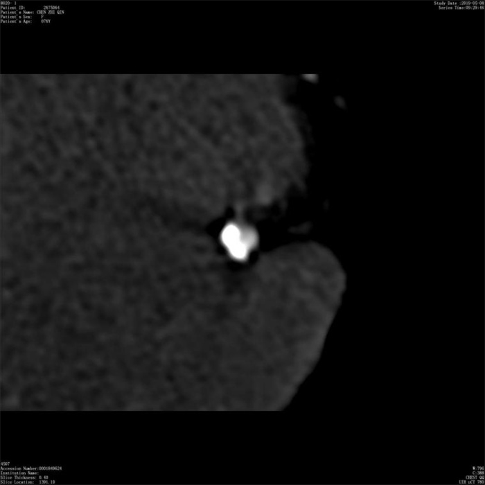
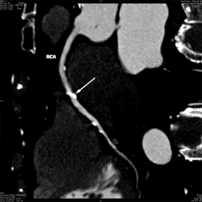
图 1 CCTA曲面重建右冠状动脉多发钙化灶
Figure 1. Multiple calcification of right coronary artery on the curve planar reformation image of CCTA.
表 1 CCTA对不同钙化容积管腔狭窄程度的准确性
Table 1. Accuracy of CCTAin diagnosing the degree of luminal stenosis with different calcification volumes (%)
钙化容积(mm2) 钙化冠状动脉段(n) 敏感度 特异性 准确率 阳性预测值 阴性预测值 0~25 190 100.00 100.00 100.00 100.00 100.00 26~69 84 94.64 96.43 95.24 98.15 90.00 70~196 69 97.96 65.00 88.41 87.27 92.86 197~1375 53 85.71 36.36 75.47 83.72 40.00 χ2 22.19 33.42 46.13 28.46 28.15 P < 0.001 < 0.001 < 0.001 < 0.001 < 0.001  下载: 导出CSV
表 2 CCTA对不同钙化积分管腔狭窄程度的准确性(%)
Table 2. Accuracy of CCTAin diagnosing the degree of luminal stenosis with different calcification scores (%)
钙化积分(分) 钙化冠状动脉段(n) 敏感度 特异性 准确率 阳性预测值 阴性预测值 0~80 168 100.00 100.00 100.00 100.00 100.00 80~200 188 99.19 76.92 91.49 89.05 98.04 >200 40 92.86 75.00 87.50 89.66 91.82 χ2 10.607 13.904 17.385 13.563 11.747 P < 0.005 0.001 < 0.001 0.001 0.003
下载: 导出CSV
-
[1] 谭静, 李静, 张迎花, 等. 冠状动脉早期粥样硬化症患者冠状动脉钙化的影响因素分析[J]. 首都医科大学学报, 2020, 41(6): 978-81. doi: 10.3969/j.issn.1006-7795.2020.06.018 [2] Johnson TW, Räber L, di Mario C, et al. Clinical use of intracoronary imaging. Part 2: acute coronary syndromes, ambiguous coronary angiography findings, and Guiding interventional decision-making: an expert consensus document of the European Association of Percutaneous Cardiovascular Interventions: endorsed by the Chinese Society of Cardiology, the Hong Kong Society of Transcatheter Endocardiovascular Therapeutics (HKSTENT) and the Cardiac Society of Australia and New Zealand[J]. Eur Heart J, 2019, 40(31): 2566-84. doi: 10.1093/eurheartj/ehz332 [3] 官莉, 袁耿彪. 核素心肌灌注显像与冠状动脉造影在冠心病疑诊患者危险度评估中的相性研究[J]. 重庆医学, 2021, 50(15): 2634-8, 2642. doi: 10.3969/j.issn.1671-8348.2021.15.025 [4] 侯波, 焦宁唤, 张拓, 等. 冠状动脉CTA对冠脉临界病变管腔狭窄程度的诊断价值[J]. 中国CT和MRI志, 2021, 19(2): 71-3. https://www.cnki.com.cn/Article/CJFDTOTAL-CTMR202102024.htm [5] 张拓, 王楠, 赵苏丹, 等. 256排颈、冠状动脉联合CT血管造影在冠状动脉狭窄诊断中的价值[J]. 现代科学仪器, 2021(4): 154-7. [6] 陈豫, 魏艳磊, 王泽尉, 等. 冠脉CTA对冠脉临界病变血管狭窄程度的诊断效能评价[J]. 中国CT和MRI杂志, 2020, 18(9): 90-2. doi: 10.3969/j.issn.1672-5131.2020.09.028 [7] 赵亮亮. 探究128层CT血管造影心肌桥-壁冠状动脉图像特征及与冠脉动脉粥样硬化的相关性[J]. 中国CT和MRI杂志, 2021, 19(2): 80-1, 93. doi: 10.3969/j.issn.1672-5131.2021.02.025 [8] 庄德才, 徐大强, 潘姗姗, 等. 混合迭代算法和全模型迭代重建算法在冠状动脉CT血管造影中的应用研究[J]. 中国医学装备, 2022, 19(5): 21-5. https://www.cnki.com.cn/Article/CJFDTOTAL-YXZB202205005.htm [9] 李凘纯, 张雅萍, 李智慧. 冠状动脉CT造影在2型糖尿病合并冠心病患者诊断中的应用价值研究[J]. 中国医学装备, 2021, 18(3): 72-5. doi: 10.3969/J.ISSN.1672-8270.2021.03.018 [10] 李卓, 张磊. CT血管造影和冠状动脉狭窄严重程度在评估病变特异性缺血中的临床价值[J]. 中西医结合心脑血管病杂志, 2021, 19(22): 4012-6. doi: 10.12102/j.issn.1672-1349.2021.22.043 [11] Shwaiki O, Rashwan B, Fink MA, et al. Lower extremity CT angiography in peripheral arterial disease: from the established approach to evolving technical developments[J]. Int J Cardiovasc Imaging, 2021, 37(10): 3101-14. doi: 10.1007/s10554-021-02277-1 [12] 徐延峰, 于淑靖, 董亚鹏, 等. 冠状动脉CT血管造影常规方案团注造影剂对冠状动脉左主干分叉区的影响[J]. 中国医师进修杂志, 2021(11): 1020-5. doi: 10.3760/cma.j.cn115455-20210507-00600 [13] 王晓玲, 方凯, 王冬梅, 等. 肺癌患者肺霉菌感染和肺细菌感染的CT鉴别诊断[J]. 中华医院感染学杂志, 2021, 31(13): 1997-2000. https://www.cnki.com.cn/Article/CJFDTOTAL-ZHYY202113016.htm [14] 师毅冰, 高永广, 张培影, 等. 应用冠状动脉CTA与光学相干断层成像OCT对冠心病的评估[J]. 中国医学计算机成像杂志, 2017, 23(4): 329-32. doi: 10.3969/j.issn.1006-5741.2017.04.009 [15] 王国良, 马光, 滕伟, 等. 冠脉CTA在评估糖尿病患者冠脉临界病变管腔狭窄程度中的应用[J]. 中国CT和MRI杂志, 2018, 16(6): 6-8. https://www.cnki.com.cn/Article/CJFDTOTAL-CTMR201806004.htm [16] 许丽雪, 李芳, 罗南, 等. 钙化斑块对冠状动脉CTA诊断准确性的影响[J]. 中国医学影像学杂志, 2020, 28(10): 741-5. https://www.cnki.com.cn/Article/CJFDTOTAL-ZYYZ202010008.htm [17] 薛秋苍, 徐怡, 孙欣杰, 等. FFRCT联合斑块特征与心肌灌注显像对冠心病患者主要不良心脏事件预测效能的比较[J]. 南京医科大学学报: 自然科学版, 2021, 41(5): 757-62. https://www.cnki.com.cn/Article/CJFDTOTAL-NJYK202105022.htm [18] 陆志锋, 陈晞明, 王世祥. 不同血流储备分数冠状动脉临界病变的血管内超声特征研究[J]. 临床心血管病杂志, 2019, 35(12): 1093-7. https://www.cnki.com.cn/Article/CJFDTOTAL-LCXB201912008.htm [19] White RD, Erdal BS, Demirer M, et al. Artificial intelligence to assist in exclusion of coronary atherosclerosis during CCTA evaluation of chest pain in the emergency department: preparing an application for real-world use[J]. J Digit Imaging, 2021, 34(3): 554-71. [20] 方正, 陈林丽, 郭大静, 等. 校正的管腔内密度变化对冠状动脉支架内再狭窄的诊断价值[J]. 中国医学影像学杂志, 2019, 27(8): 594-8. https://www.cnki.com.cn/Article/CJFDTOTAL-ZYYZ201908010.htm [21] Qiao HY, Tang CX, Schoepf UJ, et al. One-year outcomes of CCTA alone versus machine learning-based FFRCT for coronary artery disease: a single-center, prospective study[J]. Eur Radiol, 2022, 32(8): 5179-88. [22] 张也乐, 曾祥芹, 唐立均, 等. 双源CT冠状动脉CT血管造影对冠状动脉钙化斑块管腔狭窄的诊断价值[J]. 实用放射学杂志, 2018, 34(11): 1697-700. [23] 刘春雨, 谢媛, 苏晓芹, 等. 基于人工智能的冠状动脉CT血管成像检测阻塞性冠状动脉狭窄效能的研究[J]. 国际医学放射学杂志, 2021, 44(5): 516-22. https://www.cnki.com.cn/Article/CJFDTOTAL-GWLC202105008.htm [24] Zhu MM, Gao YJ, Wang J, et al. CCTA-derived strain analysis in detection of regional myocardial dysfunction in coronary artery disease patients with preserved left ventricular ejection fraction: a feasibility study[J]. J Xray Sci Technol, 2022, 30(3): 587-97. [25] 孙欣杰, 徐怡, 朱晓梅, 等. 基于冠状动脉CTA的FFRCT与斑块特征对冠心病患者主要不良心脏事件的预测价值[J]. 中国医学计算机成像杂志, 2021, 27(4): 296-301. https://www.cnki.com.cn/Article/CJFDTOTAL-YJTY202104005.htm -

 点击查看大图
计量
- 文章访问数: 389
- HTML全文浏览量: 154
- PDF下载量: 8
- 被引次数: 0