


Application value of vaginal Doppler ultrasound in the treatment process of polycystic ovary syndrome
-
摘要:
目的分析阴道多普勒超声对多囊卵巢综合征(PCOS)不孕患者治疗过程中卵巢动脉血流动力学、卵泡发育的监测价值。 方法选取2018年8月~2020年8月于医院诊治的267例PCOS不孕患者作为研究对象,所有患者均进行阴道多普勒超声检查,比较治疗前后患者卵巢动脉血流动力学指标:收缩期峰值血流速度、舒张末期血流速度、阻力指数、搏动指数,血清激素水平:促卵泡激素、促黄体生成素、雌二醇,以及卵泡发育(卵泡数量、卵泡大小、卵巢体积)的变化情况。 结果治疗前,PCOS不孕患者超声表现为双侧卵巢增大,卵巢呈现蜂窝状变化,卵泡发育异常,一切面卵泡数量≥12个,直径均 < 10 mm,并且大小不等(直径大多为2~9 mm),呈现栅栏状排列,髓质部回声增强,包膜增厚,卵巢基质内血管清晰,血流丰富,血流信号呈现粗条状,血流频谱呈现高速、低阻状态;治疗后,患者超声表现为卵巢体积大、卵泡数量多,且一侧卵巢内出现优势卵泡,卵巢被膜呈现较强回声,卵巢基质内可见较丰富的血流;与治疗前比较,PCOS不孕患者治疗后卵泡数量增多(P < 0.05),卵泡大小、卵巢体积增大(P < 0.05);与治疗前比较,PCOS不孕患者治疗后卵巢动脉血流动力学指标(即收缩期峰值血流速度、舒张末期血流速度)升高(P < 0.05),阻力指数、搏动指数降低(P < 0.05);与治疗前比较,PCOS不孕患者治疗后促卵泡激素、促黄体生成素以及雌二醇水平升高(P < 0.05)。 结论阴道多普勒超声可对PCOS不孕患者治疗过程中卵巢动脉血流动力学和卵泡发育情况进行监测,对指导治疗PCOS不孕患者具有重要的临床价值。 Abstract:ObjectiveTo analyze the monitoring value of vaginal Doppler ultrasound on ovarian artery hemodynamics and follicular development in infertile patients with polycystic ovary syndrome (PCOS) during treatment process. Methods267 infertile patients with PCOS diagnosed and treated in the hospital from December 2017 to December 2019 were selected as the research subjects. All patients underwent vaginal Doppler ultrasound. And the ovarian artery hemodynamics peak systolic blood flow velocity (PSV), end-diastolic blood flow velocity (EDV), resistance index (RI), pulsatility index (PI)], serum hormones levels [follicle stimulating hormone (FSH), luteinizing hormone (LH), estradiol (E2) and follicular development (follicle number, follicle size, ovarian volume) were compared among the patients before and after treatment. ResultsBefore treatment, the ultrasound manifestations of infertile patients with PCOS showed ovarian enlargement, honeycomb changes of bilateral ovaries and abnormal follicular development. The number of all faceted follicles was ≥12, with diameter less than 10mm in different sizes (mostly of 2-9 mm) and showed a fence-like arrangement, enhanced medulla echo and thickened capsule. And the blood vessels were clear, blood flow was rich and blood flow signal was strip in the ovarian matrix. The blood flow spectrum showed a high-velocity and low-resistance state. After treatment, the ultrasound manifestations showed large ovary volume and a large number of follicles, with dominant follicles present in one side of the ovaries. And ovarian capsule presented a strong echo and there was a richer blood flow in the ovarian matrix. Compared with before treatment, the number of follicles in infertile patients with PCOS increased significantly after treatment (P < 0.05), and the size of follicles and ovarian volume increased significantly (P < 0.05). Compared with before treatment, the ovarian artery hemodynamic indexes of PSV and EDV among infertile patients with PCOS increased significantly after treatment (P < 0.05) while the RI and PI decreased significantly (P < 0.05). Compared with before treatment, the levels of FSH, LH and E2 of infertile patients with PCOS significantly increased after treatment (P < 0.05). ConclusionVaginal Doppler ultrasound can monitor ovarian artery hemodynamics and follicular development in infertile patients with PCOS, and has important clinical value in the guidance treatment of infertile patients with PCOS. -
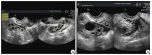
图 1 PCOS不孕患者治疗前后超声表现典型案例
患者女性, 28岁; A:治疗前卵泡主要分布于卵巢包膜下, 且直径均小于1.0 cm; B:治疗后一侧卵巢内出现优势卵泡.
Figure 1. Typical cases of PCOS infertility before and after treatment.
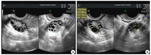
图 2 PCOS不孕患者治疗前后超声表现典型案例
患者女性, 26岁, A:治疗前卵泡呈蜂窝状分布于卵巢内, 且直径均小于1.0 cm; B:治疗后一侧卵巢内出现明显的优势卵泡.
Figure 2. Typical cases of PCOS infertility before and after treatment.
表 1 PCOS不孕患者治疗前后卵泡发育情况比较(Mean±SD,n=267)
Table 1. Comparison of follicular development before and after treatment in patients with PCOS infertility
指标 治疗前 治疗后 t P 卵泡数量(个) 8.46±1.39 15.89±2.75 58.651 < 0.001 卵泡大小(mm) 6.17±1.08 17.80±2.62 102.722 < 0.001 卵巢体积(mL) 7.28±1.15 13.46±2.53 54.882 < 0.001  下载: 导出CSV
表 2 PCOS不孕患者治疗前后卵巢动脉血流动力学比较(Mean±SD,n=267)
Table 2. Comparison of ovarian hemodynamics before and after treatment in patients with PCOS infertility
指标 治疗前 治疗后 t P PSV(cm/s) 20.36±3.44 25.05±4.62 19.016 < 0.001 EDV(cm/s) 7.08±1.32 10.93±1.71 41.524 < 0.001 RI 0.67±0.11 0.54±0.06 24.991 < 0.001 PI 0.97±0.15 0.80±0.12 20.576 < 0.001 PSV: Peak systolic velocity; EDV: End diastolic velocity; RI: Resistance index; PI: Pulsatility index.
下载: 导出CSV
表 3 PCOS不孕患者治疗前后激素水平比较(Mean±SD,n=267)
Table 3. Comparison of hormones levels before and after treatment in patients with PCOS infertility
指标 治疗前 治疗后 t P FSH(U/L) 5.24±0.57 14.37±3.81 68.121 < 0.001 LH(U/L) 6.02±1.28 45.19±11.96 96.683 < 0.001 E2(pmol/L) 148.61±40.45 1169.74±150.62 174.652 < 0.001 FSH: Follicle stimulating hormone; LH: Luteinizing hormone; E2: Estradiol.
下载: 导出CSV
-
[1] Azziz R, Carmina E, Chen ZJ, et al.Polycystic ovary syndrome[J].Nat Rev Dis Primers, 2016, 2:16057. [2] Meier RK.Polycystic ovary syndrome[J].Nurs Clin North Am, 2018, 53(3):407-20. [3] 邹青娥, 于灵, 魏青文, 等.N-乙酰半胱氨酸联合他莫昔芬治疗氯米芬抵抗多囊卵巢综合征的效果[J].分子影像学杂志, 2019, 42(3):401-5. [4] 宋景艳, 孙振高, 王爱娟, 等.消囊育嗣汤联合来曲唑治疗多囊卵巢综合征的效果分析[J].中国妇幼保健, 2019, 34(8):1808-11. [5] Kenigsberg LE, Agarwal C, Sin S, et al.Clinical utility of magnetic resonance imaging and ultrasonography for diagnosis of polycystic ovary syndrome in adolescent girls[J].Fertil Steril, 2015, 104(5):1302-9.e1-4. [6] 戴蓓蓓, 任芸芸, 常才, 等.不同血糖状态的35~40岁PCOS患者超声特征分析[J].中国超声医学杂志, 2019, 35(9):845-9. [7] 中华医学会妇产科学分会内分泌学组及指南专家组.多囊卵巢综合征中国诊疗指南[J].中华妇产科杂志, 2018, 53(1):2-6. [8] Raga F, Caballero O, Bonilla F, et al.The new three-dimensional ultrasound modes allow a better polycystic ovary syndrome ultrasound diagnosis beyond the Rotterdam criteria[J].Donald Sch J Ultrasound Obstet Gynecol, 2015, 9(4):434-45. [9] Rosenfield RL, Ehrmann DA.The pathogenesis of polycystic ovary syndrome (PCOS):the hypothesis of PCOS as functional ovarian hyperandrogenism revisited[J].Endocr Rev, 2016, 37(5):467-520. [10] 李砚, 姚念玲, 王运萍, 等.来曲唑与克罗米芬治疗多囊卵巢综合征的疗效及对促排卵的效果比较[J].海南医学, 2019, 30(15):1965-8. [11] Lee DE, Park SY, Lee SR, et al.Diagnostic usefulness of transrectal ultrasound compared with transvaginal ultrasound assessment in young Korean women with polycystic ovary syndrome[J].J Menopausal Med, 2015, 21(3):149-54. [12] 谢宏基, 王玉莹, 林紫晴, 等.经阴道三维超声检测剖宫产子宫疤痕妊娠的临床意义[J].分子影像学杂志, 2018, 41(3):324-7. [13] Ozdemir O, Sari ME, Kalkan D, et al.Comprasion of ovarian stromal blood flow measured by color Doppler ultrasonography in polycystic ovary syndrome patients and healthy women with ultrasonographic evidence of polycystic[J].Gynecol Endocrinol, 2015, 31(4):322-6. [14] 吕云, 刘素琴, 古咏梅.阴道超声检查多囊卵巢综合征所致不孕症的诊断价值分析[J].山西医药杂志, 2018, 47(1):37-8. [15] Sahu A, Tripathy P, Mohanty J, et al.Doppler analysis of ovarian stromal blood flow changes after treatment with metformin versus ethinyl estradiol-cyproterone acetate in women with polycystic ovarian syndrome:a randomized controlled trial[J].J Gynecol Obstet Hum Reprod, 2019, 48(5):335-9. [16] 饶洪杰, 张露, 柏艳红.经阴道三维容积超声在多囊卵巢综合征中的影像特点及诊断价值评估[J].中国计划生育和妇产科, 2019, 11(6):13-6. [17] 罗俏聪, 刘醒, 余艳.多囊卵巢综合征性不孕症经阴道超声的特征分析[J].沈阳医学院学报, 2017, 19(2):124-6. [18] Mejia RB, Summers KM, Kresowik JD, et al.A randomized controlled trial of combination letrozole and clomiphene citrate or letrozole alone for ovulation induction in women with polycystic ovary syndrome[J].Fertil Steril, 2019, 111(3):571-8.e1. [19] Wu XK, Stener-Victorin E, Kuang HY, et al.Effect of acupuncture and clomiphene in Chinese women with polycystic ovary syndrome:a randomized clinical trial[J].JAMA, 2017, 317(24):2502-14. [20] 丁波, 袁丽, 孙文兵, 等.经阴道超声监测卵泡发育在不孕患者中应用的价值研究[J].中国性科学, 2018, 27(1):92-4. [21] 吴少芬, 周德兴, 林娟, 等.经阴道彩色多普勒超声评价中西医结合治疗多囊卵巢综合征的临床疗效[J].中华中医药学刊, 2019, 37(3):738-41. [22] Garg N, Khaira HK, Kaur M, et al.A comparative study on quantitative assessment of blood flow and vascularization in polycystic ovary syndrome patients and normal women using threedimensional power Doppler ultrasonography[J].J Obstet Gynaecol India, 2018, 68(2):136-41. -


 点击查看大图
计量
- 文章访问数: 850
- HTML全文浏览量: 317
- PDF下载量: 2
- 被引次数: 0