


Comparative study of unilateral dominance and prominent vascular sign after unilateral middle cerebral artery occlusion in M1 segment
-
摘要:
目的探讨单侧大脑中动脉M1段闭塞后,动脉偏侧优势与突出血管征(PVS)之间的相关性,并分析动脉偏侧优势及PVS与患者神经功能缺失程度之间的相关性。 方法收集2018年1月~2020年3月单侧大脑中动脉M1段闭塞的急性缺血性脑卒中患者48例,其中男性27例,女性21例,年龄38~78岁(52.8±7.7岁)。依据三维时间飞跃法磁共振血管成像提示有无患侧大脑前动脉和(或)大脑后动脉偏侧优势分为偏侧优势组(n=26)和对照组(n=22),观察两组患者T2*加权血管成像序列显示的PVS差异。依据T2*加权血管成像序列提示梗死灶周围有无PVS将入组患者分为PVS阳性组(n=27)和阴性组(n=21),分别于患者入院当日及1周后将偏侧优势组和对照组、PVS阴性和PVS阳性组利用美国国立卫生研究院脑卒中评分量表(NIHSS)进行评分,并分析评分差异。 结果48例患者中,偏侧优势组出现PVS为42.31%(11/26),对照组患者出现PVS为72.73%(16/22),对照组高于偏侧优势组(P<0.05);偏侧优势组及PVS阴性组患者入院当日及1周后NIHSS评分均低于对照组及PVS阳性组(P<0.05)。 结论单侧大脑中动脉M1段闭塞后,大脑动脉偏侧优势及PVS阴性提示侧支循环建立及良好的灌注状态,并与患者近期预后密切相关。 Abstract:ObjectiveTo investigate the correlation between arterial hemidominance and prominent vessel sign (PVS) after unilateral middle cerebral artery (MCA) occlusion in M1 segment. Methods48 patients with acute ischemic stroke with unilateral MCA M1 segment occlusion from January 2018 to March 2020 were collected. Among them, there were 27 males and 21 females which the average age was 52.8±7.7 years. All patients were divided into two groups: lateral dominance group (n=26)and control group (n=22) according to the unilateral dominance of ACA and/or PCA revealed by three-dimension time of flight MRA (3D-TOF MRA). The difference of PVS shown by SWAN sequence was observed between the two groups. The patients were divided into PVS positive group(n=27) and negative group (n=21) according to SWAN sequence. On the day of admission and one week after admission, the hemidominance group and the control group, PVS negative and PVS positive groups were scored with the National Institutes of Health Stroke rating scale (NIHSS), and the differences were analyzed. ResultsAmong the 48 patients, the incidence of PVS in the unilateral dominance group was 42.31% (11/26), while that in the control group was 72.73% (16/22), the control group was significantly higher than the lateral dominant group, the difference was statistically significant (P < 0.05). The NIHSS scores of patients in the hemiplegia group and PVS negative group were lower than those in the control group and PVS positive group on the day of admission and one week later, and the difference was statistically significant. ConclusionAfter unilateral MCA M1 segment occlusion, the unilateral dominance of cerebral artery and negative PVS indicate the establishment of collateral circulation and good perfusion state, which is closely related to the short-term prognosis of the patients. -
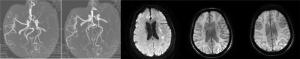
图 1 患者女,66岁,右肢活动不利1 d,入院NIHSS评分为6分,1周后NIHSS评分为3分
A-B: 3D-TOF MRA提示左侧MCAM1段闭塞, 左侧大脑前动脉及大脑后动脉偏侧优势; C: DWI提示左侧脑室旁急性梗死灶; D-E: SWAN提示双侧软脑膜引流静脉及髓纹静脉对称显示, PVS阴性.
Figure 1. Female patient, 66 years old, the right limb movement was disadvantageous for one day. The NIHSS score was 6 when the patient was admitted to hospital and 3 after one week.
表 1 偏侧优势组与对照组PVS检出率比较(%)
Table 1. Comparison of PVS detection rate between the dominant group and the control group
组别 PVS阳性 PVS阴性 偏侧优势组(n=26) 42.31(11/26) 57.69(15/26) 对照组(n=22) 72.73(16/22 27.27(6/22) χ2 4.481 P 0.034 PVS: Prominent vessel sign.  下载: 导出CSV
表 2 偏侧优势与患者入院当日及1周NIHSS评分比较(Mean±SD)
Table 2. Comparison of the score of NIHSS on the day of admission and one week
组别 入院当日 入院1周 偏侧优势组(n=26) 5.36±2.96 3.08±1.65 对照组(n=22) 7.82±3.07 4.96±1.59 t -2.8206 -3.5735 P 0.0071 0.0008
下载: 导出CSV
表 3 PVS与患者入院当日及1周NIHSS评分比较(Mean±SD)
Table 3. Comparison of NIHSS scores of PVS and patients on the day of admission and one week
组别 入院当日 入院1周 PVS阳性组(n=27) 7.68±2.76 5.66±2.41 PVS阴性组(n=21) 5.91±2.96 3.56±1.84 t 2.1416 3.3431 P 0.0376 0.0017 PVS: Prominent vessel sign.
下载: 导出CSV
-
[1] Song JC, Ma ZL, Meng H, et al. Distal hyperintense vessels alleviate Insula infarction in proximal middle cerebral artery occlusion[J]. Int J Neurosci, 2016, 126(11): 1030-5. [2] 闫俊, 常嵘, 李伟旺, 等.大脑中动脉重度狭窄侧枝循环对脑梗死发病机制及NIHSS评分的影响[J].陕西医学杂志, 2017, 46(12): 1636-7. [3] 吕晋浩, 娄昕.脑侧支循环的MRI研究进展[J].中华放射学杂志, 2016, 50(7): 556-8. [4] 刘海峰, 许永生, 陈小莉, 等. 3D-TOF-MRA诊断颅内动脉瘤价值的Meta分析[J].临床放射学杂志, 2017, 36(10): 1396-400. [5] 马永青, 尹喜, 王成伟.磁敏感加权血管成像在指导急性脑梗死溶栓治疗及评估预后中的临床价值[J].实用放射学杂志, 2019, 35(9): 1389-94. [6] 陈越, 周仁兰, 刘林.侧枝循环对急性缺血性卒中预后的影响[J].中国医师杂志, 2019, 21(2): 312-5. [7] Liu LP, Xu AD, Wong LK, et al. Chinese consensus statement on the evaluation and intervention of collateral circulation for ischemic stroke[J]. CNS Neurosci Ther, 2014, 20(3): 202-8. [8] 张丹丹, 宋波, 赵璐, 等.大脑后动脉偏侧优势对同侧大脑中动脉病变卒中严重程度及预后的影响[J].中国卒中杂志, 2017, 12(4): 314-9. [9] 邱晨, 吴敏, 纪学颍, 等.急性卒中患者院前应用NIHSS的准确性分析[J].临床急诊杂志, 2019, 20(6): 480-3. [10] 刘征华.大脑中动脉狭窄或闭塞时的脑血流量变化MRI研究[J].实用放射学杂志, 2019, 35(3): 349-52. [11] 韩楠楠, 常明则, 张格娟, 等.侧支代偿联合NIHSS评分预测大脑中动脉闭塞患者的预后[J].卒中与神经疾病, 2019, 26(1): 60-3. [12] 葛永桂, 郭婷婷, 王玉洁.影响大脑中动脉狭窄后侧支循环建立的因素及其研究进展[J].中华老年心脑血管病杂志, 2019, 21(2): 211-3. [13] 崔勇, 郑智艳, 黄玲, 等.磁共振弥散加权成像和三维时间飞跃法血管成像及三维动脉自选标记在老年人缺血性脑血管病中的应用[J].中华老年医学杂志, 2018, 37(8): 847-50. [14] 周建国, 符大勇, 马先军, 等. ASL对大脑中动脉M1段闭塞后侧支循环建立显示的临床应用[J].实用放射学杂志, 2018, 34(8): 1164-6, 1171. [15] van Seeters T, Hendrikse J, Biessels GJ, et al. Completeness of the circle of Willis and risk of ischemic stroke in patients without cerebrovascular disease[J]. Neuroradiology, 2015, 57(12): 1247-51. [16] 许孝南, 戚玉龙, 李谆晶, 等.侧枝循环对急性缺血性脑卒中静脉溶栓的预后评估[J].卒中与神经疾病, 2017, 24(6): 495-8. [17] 李柏林, 邹育明, 梁春娜, 等.瑞替普酶联合丁苯肽胶囊改善急性进展性大脑中动脉缺血性脑卒中患者的临床预后[J].分子影像学杂志, 2019, 42(3): 414-7. [18] 董天发, 麦慧, 陈展航, 等.慢性脑缺血脑血流动力学的PCMRI研究[J].实用放射学杂志, 2017, 33(6): 625-8. [19] 赵大聪, 鲁广华, 郭江, 等. SWI在超急性大面积脑梗死中的应用价值[J].实用放射学杂志, 2016, 32(4): 514-7. [20] 祁宇, 薛静, 高培毅, 等.磁敏感加权成像突出血管征对急性缺血性卒中缺血半暗带的评估价值[J].国际医学放射学杂志, 2017, 40(6): 651-5. [21] Yu JC, Wang LM, Li ZZ, et al. Related factors of asymmetrical vein sign in acute middle cerebral artery stroke and correlation with clinical outcome[J]. J Stroke Cerebrovasc Dis, 2017, 26(10): 2346- 53. [22] Luo Y, Gong ZY, Zhou YM, et al. Increased susceptibility of asymmetrically prominent cortical veins correlates with misery perfusion in patients with occlusion of the middle cerebral artery[J]. Eur Radiol, 2017, 27(6): 2381-90. [23] 贾亚南, 刘翠翠, 刘俊艳.磁敏感加权成像不对称皮层静脉征与急性缺血性卒中预后的关系研究[J].中国卒中杂志, 2019, 14(7): 639-44. [24] 阿力木·吾甫尔, 韩登峰, 哈力旦·加马力丁, 等.颅内侧枝循环开放程度与颈动脉支架置入术后短期预后的关系[J].中国动脉硬化杂志, 2018, 26(2): 186-9, 193. -

 点击查看大图

计量
- 文章访问数: 605
- HTML全文浏览量: 229
- PDF下载量: 4
- 被引次数: 0