


Significance of X-ray, CT and MRI combined with Weber classification in the differential diagnosis of injury degree of patients with tibiofibular syndesmosis
-
摘要:
目的研究X线、CT与MRI检查联合Weber分型在下胫腓联合损伤患者损伤程度鉴别中的意义。 方法选择2017年10月~2018年10月在我院就诊的75例下胫腓联合损伤患者作为研究对象。Weber分型:A型患者31例,B型患者25例,C型患者19例。右踝骨折患者39例,左踝骨折患者36例,患者的功能损伤情况:Lauge-Hansen分型:旋前外展Ⅰ、Ⅱ、Ⅲ型患者分别为9、5、8例,旋前外旋Ⅰ、Ⅱ、Ⅲ、Ⅳ患者分别为6、7、9、5例,旋后外旋Ⅰ、Ⅱ、Ⅲ、Ⅳ患者分别为9、4、2、3例,旋后内收Ⅰ、Ⅱ患者分别为3、5例。分别对患者开展X线、CT、MRI检查,以MRI检查结果作为金标准,分析X线、CT与MRI检查结果的一致性,分析联合诊断与单独诊断灵敏度之间的差异。 结果通过MRI检查,共发现前韧带损伤患者39例,深韧带损伤患者36例。以此作为金标准,CT诊断结合Weber分型发现浅韧带损伤41例,其中与MRI一致的有35例;发现深韧带损伤34例,与MRI结果一致的有30例。CT诊断结合Weber分型与MRI诊断的一致性较强(P < 0.05);X线结合Weber分型与核磁共振诊断的一致性较强(P < 0.05);通过对患者的不同严重程度的下胫腓联合损伤患者的CT、X线结合Weber分型的联合诊断,其灵敏度高于单独检测,差异有统计学意义(P < 0.05)。 结论X线、CT与MRI检查联合Weber分型在下胫腓联合损伤患者损伤程度的诊断灵敏度显著提升,同时与MRI检查的一致性较高,建议临床推广。 Abstract:ObjectiveTo explore the significance of X-ray, CT and MRI combined with Weber typing in the differential diagnosis of injury degree of patients with tibiofibular syndesmosis. MethodsSeventy-five patients with tibiofibular syndesmosis injury were treated in our hospital from October 2017 to October 2018. Weber classification: 31 patients of type A, 25 patients of type B and 19 patients of type C. There were 39 cases of right ankle fracture and 36 cases of left ankle fracture. According to Lauge-Hansen classification, there were 9, 5 and 8 cases of pronation and abduction type Ⅰ, Ⅱ, and Ⅲ respectively; 6, 7, 9 and 5 cases of pronation and external rotation Ⅰ, Ⅱ, Ⅲ and Ⅳ respectively; 9, 4, 2 and 3 cases of supination and external rotation Ⅰ, Ⅱ, Ⅲ and Ⅳ respectively. There were 3 cases of supinational adduction type Ⅰ and 5 cases of supinational adduction type Ⅱ. X-ray, CT and MRI examination were carried out for the patients respectively. The results of MRI examination were taken as the gold standard. The consistency of X- ray, CT and MRI examination results and the difference between the sensitivity of combined diagnosis and single diagnosis were analyzed. ResultsThirty-nine patients with anterior ligament injury and 36 patients with deep ligament injury were found by MRI. As a gold standard, 41 patients with shallow ligament injury were found by CT diagnosis combined with Weber classification, 35 of which were consistent with MRI, 34 of which were consistent with MRI. The consistency of CT diagnosis combined with Weber typing and MRI diagnosis was stronger (P < 0.05). The consistency of X-ray combined with Weber typing and MRI diagnosis was stronger (P < 0.05). The sensitivity of combined CT and X-ray combined with Weber typing was significantly higher than that of single detection (P < 0.05). ConclusionThe sensitivity of X-ray, CT, examination combined with Weber classification in the diagnosis of the injury degree of tibiofibular syndesmosis is improved. The consistency with MRI is high, so it is recommended to promote the clinical application. -
Key words:
- injury of tibiofibular syndesmosis /
- MRI /
- X-ray /
- CT diagnosis /
- joint diagnosis
-
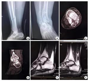
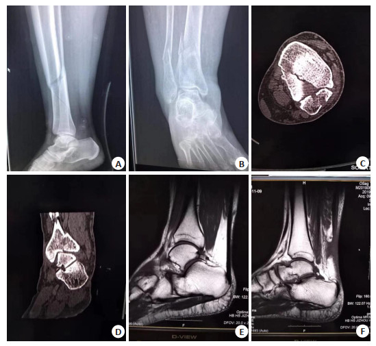
图 1 CT诊断结合Weber分型在下胫腓联合损伤患者中的诊断
A, B:下胫腓联合损伤侧位及正位片; C:下胫腓联合损伤CT横断面; D:下胫腓联合损伤冠状位CT; E, F:下胫腓联合韧带损伤MRI不同矢状位截面
Figure 1. CT diagnosis combined with Weber typing in patients with lower tibiofibular syndesmosis injury
表 1 联合诊断效能分析
Table 1. Effectiveness analysis of joint diagnosis
诊断方法 真阳(n) 假阳(n) 真阴(n) 假阴(n) 准确率(%) 灵敏度(%) 特异度(%) 阳性预测值(%) 阴性预测值(%) 标准误 AUC 95%CI P 浅韧带(n=39) CT 12 6 5 16 43.59 70.59 23.81 42.86 45.45 11.231 0.775 0.520-0.872 0.017 MRI 19 5 11 4 76.92 63.33 73.33 82.61 68.75 9.252 0.603 0.600-0.746 0.000 联合检测 14 11 7 7 53.85 66.67 50.00 66.67 38.89 3.222 0.892 0.607-0.816 0.000 深韧带(n=36) CT 9 5 4 18 36.11 69.23 18.18 33.33 44.44 0.027 0.778 0.762-0.869 0.000 MRI 11 6 7 12 50.00 61.11 36.84 47.83 53.85 1.027 0.792 0.762-0.870 0.000 联合检测 15 7 10 4 69.44 60.00 71.43 78.95 58.82 2.027 0.832 0.760-0.871 0.000  下载: 导出CSV
-
[1] 黄强, 徐向阳, 杨崇林, 等.踝关节镜在诊治Danis-Weber B型踝关节骨折伴下胫腓联合损伤中的意义[J].中华创伤骨科杂志, 2018, 20 (6): 482-6. http://d.old.wanfangdata.com.cn/Periodical/zhcsgkzz201806005 [2] 黄强, 徐向阳, 曹永星, 等.踝关节镜诊断Danis-Weber B型踝关节骨折合并下胫腓联合损伤[J].中华骨科杂志, 2019, 39(11): 660-6. http://d.old.wanfangdata.com.cn/Periodical/zhgkzz98201911002 [3] 王朝亮, 黄素芳, 王仲秋, 等.基于踝关节水平位CT下胫腓联合分离的特点及手术策略[J].中华创伤骨科杂志, 2017, 19(12): 1036-45. http://d.old.wanfangdata.com.cn/Periodical/zhcsgkzz201712006 [4] 于振, 窦强兵, 袁先发, 等. Endobutton钢板与皮质骨螺钉治疗伴踝关节骨折的急性下胫腓联合损伤的比较[J].安徽医药, 2017, 21(7): 1223-7. http://d.old.wanfangdata.com.cn/Periodical/ahyy201707014 [5] 倪斌斌, 王栋梁, 戴力扬.下胫腓联合损伤[J].创伤外科杂志, 2006, 8 (3): 281-3. http://d.old.wanfangdata.com.cn/Periodical/zhcs201502006 [6] 倘艳锋, 杨玉霞, 李红军, 等.经皮骨折断端骨皮质"刺削"并局部注射自体浓缩骨髓液和富血小板血浆混合物治疗骨折延迟愈合[J].中华创伤骨科杂志, 2018, 20(11): 999-1003. http://www.wanfangdata.com.cn/details/detail.do?_type=perio&id=zhcsgkzz201811018 [7] 王耀宗, 张英.悬垂体位撬拨法治疗过伸型胫骨平台骨折[J].中华骨科杂志, 2019, 39(2): 83-9. http://d.old.wanfangdata.com.cn/Periodical/zhgkzz98201902003 [8] 李文菁, 李庭, 孙旭, 等.骨折术后胫腓远端骨性连接的影像学特点及其对踝关节活动度的影响[J].中华医学杂志, 2019, 99(21): 1621-5. http://d.old.wanfangdata.com.cn/Periodical/zhyx201921006 [9] 孙旭, 李庭, 孙志坚, 等.三角韧带加强修补治疗合并三角韧带损伤及下胫腓分离的踝关节骨折[J].中华医学杂志, 2018, 98(39): 3192-6. http://d.old.wanfangdata.com.cn/Periodical/zhyx201839014 [10] 李凡, 徐剑, 李明静, 等.青少年伴三角韧带断裂的踝关节骨折的手术治疗[J].中华小儿外科杂志, 2018, 39(6): 451-5. http://d.old.wanfangdata.com.cn/Periodical/xrwk201806010 [11] 宋虎, 左照光, 孙再杰, 等.腓骨头上入路外侧胫骨平台截骨结合外侧锁定钢板治疗单纯胫骨平台后外侧骨折[J].中华创伤骨科杂志, 2019, 21(2): 166-9. http://d.old.wanfangdata.com.cn/Periodical/zhcsgkzz201902012 [12] Wang L, Zhang YZ, Song ZH, et al. A novel method of using elastic bionic fixation device for distal tibiofibular syndesmosis injury[J]. Int Orthop, 2018, 42(9): 2219-29. http://www.wanfangdata.com.cn/details/detail.do?_type=perio&id=2f01613ee8079afbaa53c645826c0cdd [13] Yuen, Lui TH. Distal tibiofibular syndesmosis: anatomy, bio-mechanics, injury and management[J]. Open Orthop J, 2017, 11: 670-7. https://openorthopaedicsjournal.com/VOLUME/11/PAGE/670/FULLTEXT/ [14] Ye YZ, Zhang LY, Chen Y. Clinical outcomes Tight rope versus traditional screw fixation for the treatment of injury of distal tibiofibular syndesmosis in ankle fracture[J]. Chin J Orthop Traumatol, 2017, 30(5): 441-5. http://www.wanfangdata.com.cn/details/detail.do?_type=perio&id=zggs201705010 [15] Zhang P, Liang Y, He JS, et al. A systematic review of suture-button versus syndesmotic screw in the treatment of distal tibiofibular syndesmosis injury[J]. BMC Musculoskelet Disord, 2017, 18(1): 286-93. http://cn.bing.com/academic/profile?id=b836f799bc14a3db551286f97a3526f7&encoded=0&v=paper_preview&mkt=zh-cn [16] Marschall-Lévesque S, Castellanos-Ryan N, Parent S, et al. Victimization, suicidal ideation, and alcohol use from age 13 to 15 years: support for the self-medication model[J]. J Adolesc Health, 2017, 60(4): 380-7. http://cn.bing.com/academic/profile?id=6b13fe1e02565ad5da582190e602cbcf&encoded=0&v=paper_preview&mkt=zh-cn [17] Tonogai I, Hamada D, Sairyo K. Morphology of the Incisura fibularis at the distal tibiofibular syndesmosis in the Japanese population[J]. J Foot Ankle Surg, 2017, 56(6): 1147-50. http://www.wanfangdata.com.cn/details/detail.do?_type=perio&id=efa513ac504e0d43943c7de071035f94 [18] 孟波.踝关节骨折伴下胫腓联合损伤采用骨锚钉修复40例[J].中国中医骨伤科杂志, 2019, 27(11): 65-7. http://www.wanfangdata.com.cn/details/detail.do?_type=perio&id=zgzygsk201911016 [19] 曾钢, 李春海, 丘雪梅, 等.踝关节镜检在治疗急性踝关节骨折中的作用[J].中华关节外科杂志:电子版, 2018, 12(5): 722-6. http://d.old.wanfangdata.com.cn/Periodical/zhgjwkzz201805022 [20] Agrawal S, Verma N, Perumalla S, et al. Decreasing trend of seroprevalence of hepatic amoebiasis in tertiary care hospital of North India: 2010-2015[J]. J Lab Physicians, 2018, 10(1): 31-3. http://cn.bing.com/academic/profile?id=6f0882ce1184b3e6a866039de631528c&encoded=0&v=paper_preview&mkt=zh-cn [21] Mait AR, Forman JL, Nie BB, et al. Propagation of syndesmotic injuries during forced external rotation in flexed cadaveric ankles[J]. Orthop J Sports Med, 2018, 6(6): 1-14. http://cn.bing.com/academic/profile?id=3b5d08eea410aadc1782c08800ad808e&encoded=0&v=paper_preview&mkt=zh-cn [22] Baldini T, Roberts J, Hao JD, et al. Medial compartment decompression by proximal fibular osteotomy: a biomechanical cadaver study[J]. Orthopedics, 2018, 41(4): e496-e501. http://cn.bing.com/academic/profile?id=c2ff97872b06ca833dd46095e520efce&encoded=0&v=paper_preview&mkt=zh-cn [23] Fitzpatrick E, Goetz JE, Sittapairoj T, et al. Effect of posterior malleolus fracture on syndesmotic reduction: a cadaveric study[J]. J Bone Joint Surg Am, 2018, 100(3): 243-8. http://cn.bing.com/academic/profile?id=05471f895302b2f7bd18cca1df8b6bfb&encoded=0&v=paper_preview&mkt=zh-cn [24] Brown FC, Still E, Koche RP, et al. MEF2C phosphorylation is required for chemotherapy resistance in acute myeloid leukemia[J]. Cancer Discov, 2018, 8(4): 478-97. http://www.wanfangdata.com.cn/details/detail.do?_type=perio&id=2db295a0368fff4f10743e46f1447745 [25] Jeong BO, Baek JH, Song WJ. Ankle arthritis combined with chronic instability of the syndesmosis after ankle fracture with syndesmotic injury: a case report[J]. J Foot Ankle Surg, 2018, 57(5): 1000-4. http://cn.bing.com/academic/profile?id=7f9988a593c5ffad19427c48ece2f228&encoded=0&v=paper_preview&mkt=zh-cn -

 点击查看大图
图(1) / 表(1)
计量
- 文章访问数: 585
- HTML全文浏览量: 226
- PDF下载量: 10
- 被引次数: 0