


Application of multi-row spiral CT in the preoperative assessment of revision surgery after artificial hip replacement
-
摘要:
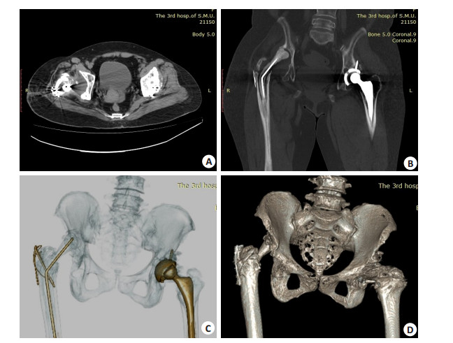
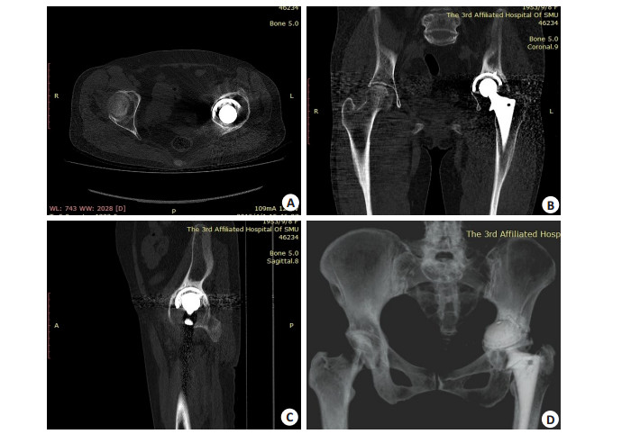
目的探讨多排螺旋CT在人工髋关节翻修术前评估的应用。 方法回顾性分析2014年3月~2019年5月在南方医科大学第三附属医院行人工髋关节翻修术的21例患者资料,其中男性7例,女性14例,中位年龄62岁,共21髋,包括假体松动13髋,假体脱位2髋,假体周围骨折2髋,旷置术后4髋,均于翻修术前行多排螺旋CT检查。由2名放射科医师阅读CT图像,分析假体失败的CT征象及为翻修术手术方案制定提供参考。 结果假体松动13髋,包括髋臼侧假体松动10髋,CT上均可见假体向上向内移位,4髋假体旁见明显透亮线,2髋髋臼内充填大量软组织,2髋髋臼周围有明显的骨质缺损;股骨侧人工假体松动6髋,5髋可见假体移位,2髋可见假体旁明显透亮线,1髋未见明显放射学异常。合并髋臼侧假体内衬磨损1髋,其髋臼侧及股骨侧假体正常间隙消失。假体脱位2髋,表现为股骨头明显向髋关节外上后方移位,髋臼骨质缺损。假体周围骨折2髋,表现为股骨侧假体(股骨柄)末端或中段旁骨质断裂,2髋均合并假体松动。旷置术后4髋,CT上可见存留的假体及间置器、髋臼内填充软组织等,3髋可见髋臼较明显的骨质缺损。 结论多排螺旋CT可检测人工髋关节失败的原因,协助翻修术方案的制定。 Abstract:ObjectiveTo explore the application of multi-row spiral CT in the preoperative assessment of revision surgery after artificial hip replacement. MethodsFrom March 2014 to May 2019, 21 patients (21 hips) of revision surgery after artificial hip replacement in the Third Affilated Hospital of Southern Medical University were included. There were 7 males and 14 female, with median age of 62 years old. The reasons of revision surgery included 13 hips of prosthesis loosening, 2 hips of prosthesis dislocation, 2 hips of periprosthesis fracture, and 4 hips of two-stage exchange. CT examinations were performed to all hips before revision surgery. Two radiologists reviewed the CT images to analyze the radiologic sign of failure of prosthesis and develop a proposal for the revision surgery. ResultsAmong 13 hips of prosthesis loosening, there were 10 hips with artificial acetabulum loosening, which reperent prosthesis shifting upward and inward of all 10 hips, obvious lucent areas of periprosthesis of 4 hips, massive soft tissues filling in the acetabulum of 2 hips, bony defect of the acetabulum of 2 hips. There were 6 hips with artificial femoral loosening, which repesented as prosthesis shifting of 5 hips, obvious lucent areas of periprosthesis of 2 hips, and no definitely abnormal findings of 1 hip. Addtionally, 1 hip with prosthesis loosening coexisted lining abrasion, manifesting as vanish of interval of artificial acetabulum and femoral. There were 2 hips with dislocation of prosthesis, which represented as femoral heads dislocating out of acetabulum laterally, superiorly, posteriorly. There were 2 hips with periprosthesis fractures, which represented as broken bones round the middle segment or the distal of the prosthesis. The both hips were suffered from prosthesis loosening. There were 4 hips of two-stage exchange, which showed residual prosthesis, spacers, soft tissues in the acetabulum on CT images, among which obvious bony defect were found in the acetabulum of 3 hips. ConclusionMulti-row spinal CT could reveal factors of prosthesis failure, and develop a proposal for the revision surgery after artificial hip replacement. -
Key words:
- arthroplasty /
- replacement /
- hip /
- CT /
- postoperative complications
-
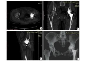
图 1 女,61岁,左人工髋关节置换术后假体松动
CT轴位(A)、多平面重建冠状位(B)、矢状位(C)图像示髋臼侧假体周围不规则透亮区, 髋臼骨质缺失, 容积再现图像(D)示髋臼侧假体向上向内移位, 股骨侧假体亦可见透亮区影
Figure 1. Female, 61 years old, diagnosed prosthesis loosening of left hip
-
[1] Hsiue PP, Chen CJ, Villalpando C, et al. Trends and patient factors associated with technology-assisted total hip arthroplasty in the United States from 2005 to 2014[J]. Arthroplast Today, 2020, 6(1):112-7. doi: 10.1016/j.artd.2019.12.009 [2] Esposito CI, Miller TT, Lipman JD, et al. Biplanar low-dose radiography is accurate for measuring combined anteversion after total hip arthroplasty[J]. HSS J, 2020, 16(1): 23-9. doi: 10.1007/s11420-018-09659-7 [3] Colombi A, Schena D, Castelli CC. Total hip arthroplasty planning [J]. EFORT Open Rev, 2019, 4(11): 626-32. doi: 10.1302/2058-5241.4.180075 [4] Plate JF, Shields JS, Langfitt MK, et al. Utility of radiographs, computed tomography, and three dimensional computed tomography pelvis reconstruction for identification of acetabular defects in residency training[J]. Hip Pelvis, 2017, 29(4): 247-52. doi: 10.5371/hp.2017.29.4.247 [5] DelSole EM, Mercuri JJ. Utility of upright weight-bearing imaging in total hip arthroplasty[J]. Semin Musculoskelet Radiol, 2019, 23 (6): 603-8. doi: 10.1055/s-0039-1697935 [6] 徐海永.术前规范DR片对人工髋关节置换手术的指导研究[D].南宁: 广西医科大学, 2015. [7] 路玉峰, 许鹏, 郭万首, 等.成人正常髋关节旋转中心的X线影像测量研究[J].中华解剖与临床杂志, 2017, 22(2): 99-102. doi: 10.3760/cma.j.issn.2095-7041.2017.02.003 [8] Iwamoto M, Fujii M, Komiyama K, et al. Is lateral acetabular rotation sufficient to correct anterolateral deficiency in periacetabular reorientation osteotomy? A CT-Based simulation study[J]. J Orthop Sci, 2020. DOI: 10.1016/j.jos.2019.12.014. [9] Graulich T, Graeff P, Nicolaides S, et al. Acetabular posterior wall morphology. A CT-based method to distinguish two acetabular posterior wall types[J]. J Orthop, 2020, 20: 160-6. doi: 10.1016/j.jor.2020.01.027 [10] Kobayashi H, Cech A, Kase M, et al. Pre-operative templating in THA. Part Ⅱ: a CT-based strategy to correct architectural hip deformities[J]. Arch Orthop Trauma Surg, 2020, 140(4): 551-62. doi: 10.1007/s00402-020-03341-6 [11] Ghanem M, Zajonz D, Heyde CE, et al. Acetabular defect classification and management: Revision arthroplasty of the acetabular cup based on 3-point fixation[J]. Orthopade, 2020. DOI: 10.1007/s00132-020-03895-8. [12] 张先龙, 蒋垚, 陈云苏.人工髋关节外科学[M].北京:人民军医出版社, 2009. [13] 黄海滨, 袁长深, 寇伯龙.全髋关节翻修术现状[J].重庆医学, 2009, 38 (9): 1116-8. doi: 10.3969/j.issn.1671-8348.2009.09.051 [14] 杨涛, 谢杰, 胡懿郃, 等. 354例Ribbed股骨柄假体置换术的中远期疗效分析[J].中国修复重建外科杂志, 2019, 33(9): 1116-20. http://www.wanfangdata.com.cn/details/detail.do?_type=perio&id=zgxfcjwkzz201909011 [15] 何斌, 章淼锋, 沈跃, 等.人工髋关节置换术后初次翻修的原因分析及翻修术疗效评估[J].中华骨科杂志, 2019, 39(15): 909-17. doi: 10.3760/cma.j.issn.0253-2352.2019.15.001 [16] 蒋梅花, 何川, 冯建民, 等. X线、CT及MRI对髋关节置换术后并发症的诊断价值[J].中国医学计算机成像杂志, 2015, 21(3): 278-82. http://d.old.wanfangdata.com.cn/Periodical/zgyxjsjcx201503018 [17] Patel AR, Sweeney P, Ochenjele G, et al. Radiographically silent loosening of the acetabular component in hip arthroplasty[J]. Am J Orthop, 2015, 44(9): 406-10. http://www.wanfangdata.com.cn/details/detail.do?_type=perio&id=8bb77d4abbfe87238396cff24fe837c5 [18] Bäcker HC, Steurer-Dober I, Beck M, et al. Magnetic resonance imaging (MRI) versus single photon emission computed tomography (SPECT/CT) in painful total hip arthroplasty: a comparative multi- institutional analysis[J]. Br J Radiol, 2020, 93 (1105): 20190738-50. doi: 10.1259/bjr.20190738 [19] Boomsma MF, Slouwerhof I, van Lingen C, et al. CT-based quantification of bone stock in large head metal-on-metal unilateral total hip replacements[J]. Eur J Radiol, 2016, 85(4): 760-3. doi: 10.1016/j.ejrad.2016.01.019 [20] 汪飞, 邱麟, 佘国荣, 等.人工髋关节置换术后并发症影像学表现[J].中国医学影像技术, 2015, 31(7): 1095-9. http://www.wanfangdata.com.cn/details/detail.do?_type=perio&id=zgyxyxjs201507036 [21] Giaretta S, Momoli A, Porcelli G, et al. Diagnosis and management of periprosthetic femoral fractures after hip arthroplasty[J]. Injury, 2019, 50(Suppl 2): S29-33. [22] Rupp M, Kern S, Ismat A, et al. Computed tomography for managing periprosthetic femoral fractures. A retrospective analysis[J]. BMC Musculoskelet Disord, 2019, 20(1): 258-67. doi: 10.1186/s12891-019-2632-y [23] Kwee RM, Broos WA, Brans B, et al. Added value of 18F-FDG PET/ CT in diagnosing infected hip prosthesis[J]. Acta Radiol, 2018, 59 (5): 569-76. doi: 10.1177/0284185117726812 [24] Moon KH, Kang JS, Won MH, et al. The usefulness of threedimensional computed tomography as an assessment of periacetabular osteolysis in revision total hip arthroplasty[J]. Hip Pelvis, 2015, 27(2): 90-7. doi: 10.5371/hp.2015.27.2.90 [25] Salem HS, Marchand KB, Ehiorobo JO, et al. Benefits of CT scanning for the management of hip arthritis and arthroplasty[J]. Surg Technol Int, 2020, 36(5): 1249-58. [26] Tang H, Yang DJ, Guo SJ, et al. Digital tomosynthesis with metal artifact reduction for assessing cementless hip arthroplasty: a diagnostic cohort study of 48 patients[J]. Skeletal Radiol, 2016, 45 (11): 1523-32. doi: 10.1007/s00256-016-2466-8 [27] Horas K, Arnholdt J, Steinert AF, et al. Acetabular defect classification in times of 3D imaging and patient-specific treatment protocols[J]. Orthopade, 2017, 46(2): 168-78. doi: 10.1007/s00132-016-3378-y [28] Greffier J, Pereira F, Hamard A, et al. Effect of tin filter-based spectral shaping CT on image quality and radiation dose for routine use on ultralow-dose CT protocols: a phantom study[J]. Diagn Interv Imaging, 2020. DOI: 10.1016/j.diii.2020.01.002. [29] Trabzonlu TA, Terrazas M, Mozaffary A, et al. Application of iterative metal artifact reduction algorithm to CT urography for patients with hip prostheses[J]. Am J Roentgenol, 2020, 214(1): 137- 43. doi: 10.2214/AJR.19.21748 -

 下载:
下载:
 点击查看大图
图(2)
计量
- 文章访问数: 859
- HTML全文浏览量: 365
- PDF下载量: 6
- 被引次数: 0