

 Find Duplicates
 Check Document
 Submission(new)
 Experts Office
 Editorial Office
2020 Vol. 42, No. 1
Display Method:
2020, 43(1): 1-6.
doi: 10.12122/j.issn.1674-4500.2020.01.01
Abstract:
Functional and molecular imaging can visualize and quantitatively measure not only the change of tissue and organ but also cellular and molecular processes in vivo. As new emerging computer technology, artificial intelligence(AI) is widely applied in the area of medical imaging. The implementation of AI on functional and molecular imaging enables the radiologists to make more efficient and full use of the information and explore the biological nature of images, which have a profound impact on the early detection, effective treatment, prognostic prediction, and pathogenesis exploration of disease. This review briefly summarized the implementation and progress of AI on functional and molecular imaging in image processing, image interpretation, and quality control.
Functional and molecular imaging can visualize and quantitatively measure not only the change of tissue and organ but also cellular and molecular processes in vivo. As new emerging computer technology, artificial intelligence(AI) is widely applied in the area of medical imaging. The implementation of AI on functional and molecular imaging enables the radiologists to make more efficient and full use of the information and explore the biological nature of images, which have a profound impact on the early detection, effective treatment, prognostic prediction, and pathogenesis exploration of disease. This review briefly summarized the implementation and progress of AI on functional and molecular imaging in image processing, image interpretation, and quality control.
2020, 43(1): 7-11.
doi: 10.12122/j.issn.1674-4500.2020.01.02
Abstract:
In China, breast cancer has become the first female malignant tumor, which seriously endangers women’s health. The dense breast is one of the risk factors for breast cancer. It’s crucial to detect and diagnose breast cancer early in the dense breast. On account of the overlap of the tissues and the covering of dense glands, mammography has certain restrictions in the examination of dense breasts. Recently, the functional imaging techniquesenhance the checkout of lesions, such as DCE-MRI, DWI, positron emission computed tomography, and breast specific-gamma imaging. It could reflect the changes of tumor hemodynamics and metabolism. The emerging photoacoustic imaging (PAI) has developed significantly in breast diseases. It provides structural and functional information of biological tissuesand high spatial resolution in deeper tissue imaging. This paper summarizes various examinations including DBT, ultrasonic examination, MRI, PET/CT, BSGI and PAI, prompting clinicians with early diagnosis of dense breast diseases.
In China, breast cancer has become the first female malignant tumor, which seriously endangers women’s health. The dense breast is one of the risk factors for breast cancer. It’s crucial to detect and diagnose breast cancer early in the dense breast. On account of the overlap of the tissues and the covering of dense glands, mammography has certain restrictions in the examination of dense breasts. Recently, the functional imaging techniquesenhance the checkout of lesions, such as DCE-MRI, DWI, positron emission computed tomography, and breast specific-gamma imaging. It could reflect the changes of tumor hemodynamics and metabolism. The emerging photoacoustic imaging (PAI) has developed significantly in breast diseases. It provides structural and functional information of biological tissuesand high spatial resolution in deeper tissue imaging. This paper summarizes various examinations including DBT, ultrasonic examination, MRI, PET/CT, BSGI and PAI, prompting clinicians with early diagnosis of dense breast diseases.
2020, 43(1): 12-15.
doi: 10.12122/j.issn.1674-4500.2020.01.03
Abstract:
At present, functional magnetic resonance imaging (fMRI) has made some progress in the study of the central mechanism of acupuncture (electroacupuncture and hand acupuncture), but different acupuncture parameters and forms of stimulation have different responses in the brain. This paper collects domestic and foreign literatures related to "fMRI and acupuncture". The "stimulus-response" model of acupuncture is taken as the starting point to make a preliminary discussion on the difference of the stimulation amount of electroacupuncture and the central response between electroacupuncture and manual acupuncture.The results show that there are different central response mechanisms for different electroacupuncture parameters in fMRI observation, and multiple parameters should be studied together. Secondly, there are differences between electroacupuncture and hand acupuncture in the afferent pathway, mechanism of action, target, clinical efficacy and other aspects, which also have their own dominant diseases.
At present, functional magnetic resonance imaging (fMRI) has made some progress in the study of the central mechanism of acupuncture (electroacupuncture and hand acupuncture), but different acupuncture parameters and forms of stimulation have different responses in the brain. This paper collects domestic and foreign literatures related to "fMRI and acupuncture". The "stimulus-response" model of acupuncture is taken as the starting point to make a preliminary discussion on the difference of the stimulation amount of electroacupuncture and the central response between electroacupuncture and manual acupuncture.The results show that there are different central response mechanisms for different electroacupuncture parameters in fMRI observation, and multiple parameters should be studied together. Secondly, there are differences between electroacupuncture and hand acupuncture in the afferent pathway, mechanism of action, target, clinical efficacy and other aspects, which also have their own dominant diseases.
2020, 43(1): 16-19.
doi: 10.12122/j.issn.1674-4500.2020.01.04
Abstract:
Malignant proliferation of tumor cells require multiple energy substances, including glucose, fatty acid and protein. Metabolic reprogramming is an important biomarker of cancer which improves the occurrence and development of malignant tumors. The Warburg effect is a key metabolic hallmark of cancer, which significantly increased glucose consumption and provided energy for tumor metabolism. Using this characteristic, 18F-FDG imaging can accurately reflect tumor biological characteristics, location, progress and response to treatment. Molecular imaging such as PET has been widely used in cancer diagnosis and prognosis after treatment. In addition, fatty acid metabolites also regulate the post-translational modifications of protein and provides lipid signal molecule as well as membrane phospholipid to resist the effect of chemotherapeutic drugs. Thus, taking advantage of fatty acid synthesis in tumors, radiolabeled short-chain fatty acid can used for the diagnosis of cancer. This review focuses on the research of fatty acid metabolism and molecular imaging in cancer.
Malignant proliferation of tumor cells require multiple energy substances, including glucose, fatty acid and protein. Metabolic reprogramming is an important biomarker of cancer which improves the occurrence and development of malignant tumors. The Warburg effect is a key metabolic hallmark of cancer, which significantly increased glucose consumption and provided energy for tumor metabolism. Using this characteristic, 18F-FDG imaging can accurately reflect tumor biological characteristics, location, progress and response to treatment. Molecular imaging such as PET has been widely used in cancer diagnosis and prognosis after treatment. In addition, fatty acid metabolites also regulate the post-translational modifications of protein and provides lipid signal molecule as well as membrane phospholipid to resist the effect of chemotherapeutic drugs. Thus, taking advantage of fatty acid synthesis in tumors, radiolabeled short-chain fatty acid can used for the diagnosis of cancer. This review focuses on the research of fatty acid metabolism and molecular imaging in cancer.
2020, 43(1): 20-24.
doi: 10.12122/j.issn.1674-4500.2020.01.05
Abstract:
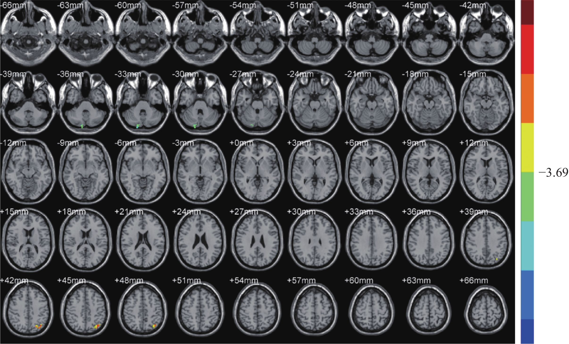
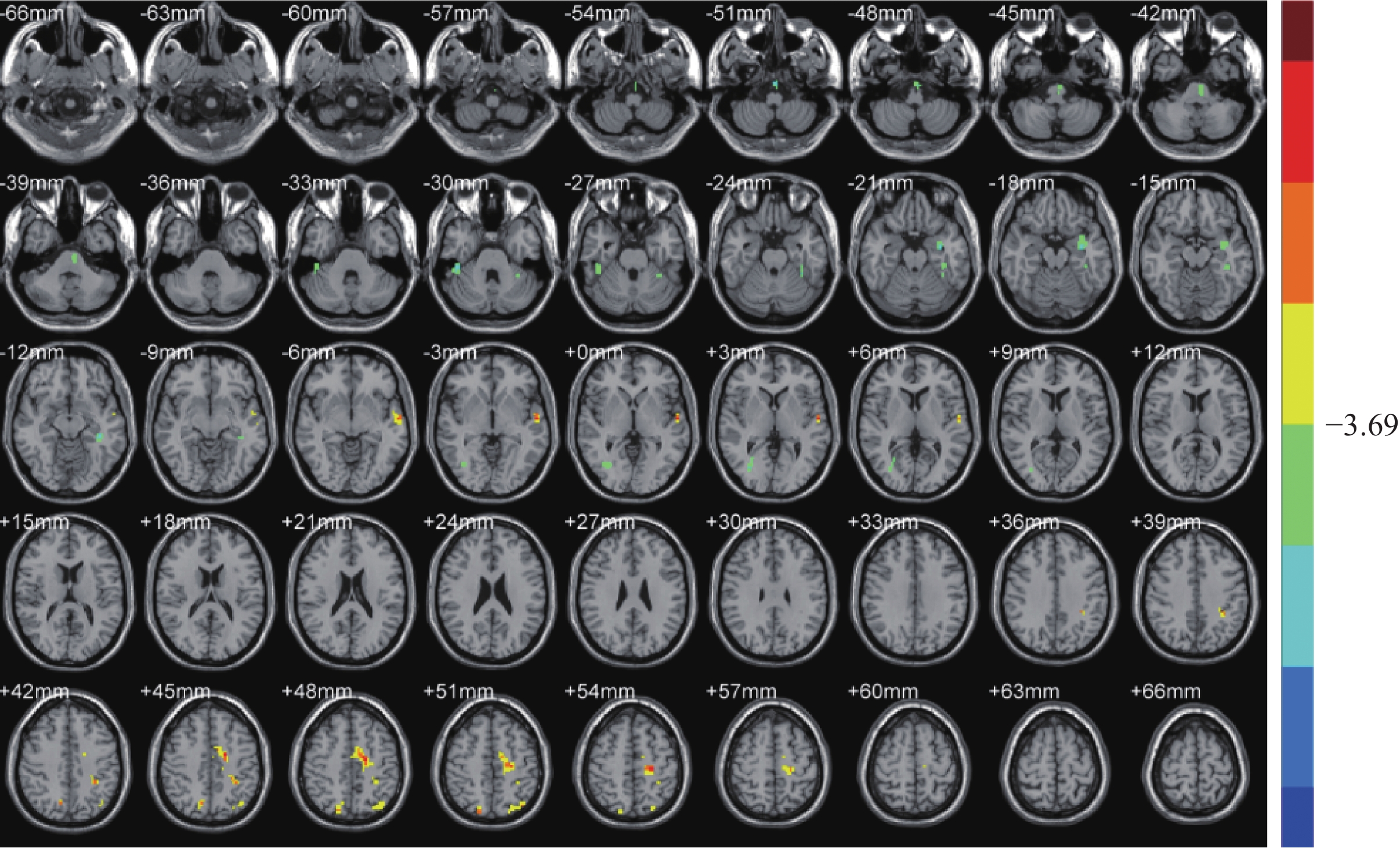
ObjectiveTo investigate the activation of brain regions after international standard scalp acupuncture on healthy middle-aged and elderly women using resting state brain functional imaging (Rs-fMRI). MethodsWe recruited 11 healthy middle-aged and elderly female volunteers who had voluntarily signed informed consent from January 2017 to May 2017, with the age from 60 to 63 years old (average 56.18±3.82). They had voluntarily signed informed consent were recruited to receive scalp acupuncture stimulation at MS5, the left MS6, and the left MS7. Rs-fMRI data of subjects were obtained by Siemens 3.0 t superconducting MRI before and after acupuncture. It was based on Matlab 2012a platform, using DPABI、SPM12 and REST1.8 software for data analysis and mapping with fractional amplitude low-frequency fluctuation (fALFF) and regional homogeneity (ReHo) as an outcome indicator. ResultsFinally, the changes of local brain activity after the international standard scalp acupuncture intervention in ten subjects were as follows: fALFF decreased at the right posterior cerebellar lobe, cerebellopontine angle, uvula and declive (T= −6.576 1), and increased at the left angular gyrus, precuneus and BA7 (T= 6.219 3). ReHo decreased at mainly the left spindle gyrus (T= −5.609 5), and the land the left pontine (T= −6.431 6), and mainly the superior temporal gyrus (BA38) and para hippocampus (T= −6.442 4), and decreased at Cortical cortex (T=−5.520 1) and right fusiform gyrus (T= −6.477 6) as well, but increased at the bilateral anterior wedge, BA7, and angular gyrus (T=4.822 4 and 5.606 4), the left middle temporal superior and superior temporal gyrus (BA21, BA22) are increased (T= 6.745 8), and increased at mainly the left top parietal lobe and central posterior gyrus (T= 8.089 1) are increased, and the left marginal lobe, cingulate gyrus (BA24), auxiliary sports area, central anterior gyrus (BA4), central posterior gyrus (BA3) (T= 6.714 1) are increased too. ConclusionBrain function in healthy middle-aged and elderly women shows specific changes in brain areas related to sensory, motor and speech functions after the first intervention with the international standard scalp acupuncture.
2020, 43(1): 25-30.
doi: 10.12122/j.issn.1674-4500.2020.01.06
Abstract:
ObjectiveTo explore the gender differences in endovenous radio-frequency ablation(RFA) of great saphenous varicosis vein (GSVV) and evaluate the application ande experience of color Doppler flow imaging(CDFI). MethodsWe included 72 patients with GSVV (96 extremities) in vascular surgery department, and 70 patients (94 extrenities) underwent endovenous RFA from March 2018 to June 2019, in a third-class hospital of Wuhan. CDFI examination was performed before, during and after operation. Then results were analyzed to assess the importance and experience of the CDFI. ResultsAmong 72 patients with GSVV, 70 patients were performed to endovenous RFA for GSVV without high ligation and they all received satisfactory recover after the operation with none suffering from severe complications such as deep venous thrombosis or pulmonary embolism.One month after surgery, all of GSVV following RFA were reviewed. Symptoms of affected extremities such as edema,pain,local acid bilges and numbness were obviously relieved or disappeared.No significant difference was observed in age,surgical extremity, grade of CEAP, time and velocity of reflux of affected extremities between male and female groups (P>0.05).The difference of the diameter of left GSVV between two groups was not significant (P>0.05). The difference in the diameter of right GSVV between two groups was significant (P<0.05). ConclusionCDFI can figure out whether a patient suffers from GSVV and help to choose the optimal treatment.It also guides the ongoing operation during the intraoperative time and assesses therapeutic effect after operation.Short-term effect of endovenous RFA without high ligation of GSVV is satisfactory,with none encountering sever complications.The diameter of right GSVV of female patients is narrower than that of male patients.


2020, 43(1): 31-35.
doi: 10.12122/j.issn.1674-4500.2020.01.07
Abstract:
Breast cancer is one of the most common malignant tumors among women throughout the world. Its incidence is increasing year by year, which is a serious threat to women's health. Lymph node metastasis is an important prognostic indicator in breast cancer. The traditional methods of imaging still have great challenges in the preoperative evaluation of lymph node metastasis status in breast cancer patients. As a new high-throughput feature extraction technology, radiomics can extract deep information of images and use it to establish clinical diagnosis, prognosis and prediction models. Radiomics has been widely used and studied in clinical diagnosis and therapy. At present, radiomics based on MRI, ultrasound and mammography have gradually been applied to the prediction of breast cancer lymph node metastasis, and has become a hot topic in academic research. This article reviews the definition and workflow of radiomics, then reviews the research progress of radiomics in breast cancer lymph node metastasis.
Breast cancer is one of the most common malignant tumors among women throughout the world. Its incidence is increasing year by year, which is a serious threat to women's health. Lymph node metastasis is an important prognostic indicator in breast cancer. The traditional methods of imaging still have great challenges in the preoperative evaluation of lymph node metastasis status in breast cancer patients. As a new high-throughput feature extraction technology, radiomics can extract deep information of images and use it to establish clinical diagnosis, prognosis and prediction models. Radiomics has been widely used and studied in clinical diagnosis and therapy. At present, radiomics based on MRI, ultrasound and mammography have gradually been applied to the prediction of breast cancer lymph node metastasis, and has become a hot topic in academic research. This article reviews the definition and workflow of radiomics, then reviews the research progress of radiomics in breast cancer lymph node metastasis.
2020, 43(1): 36-40.
doi: 10.12122/j.issn.1674-4500.2020.01.08
Abstract:
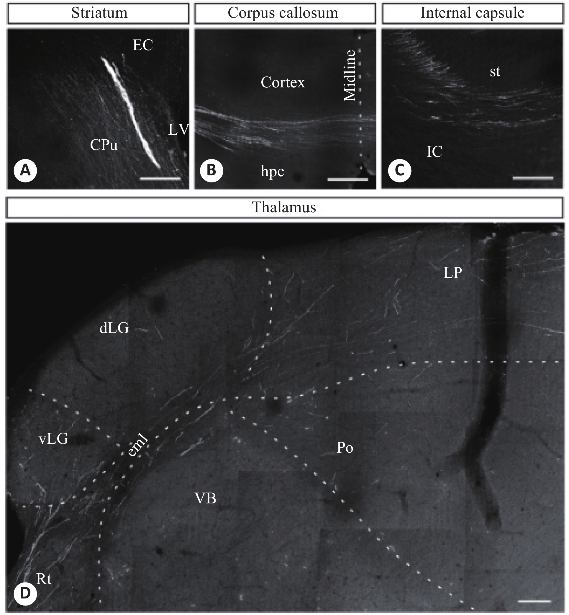
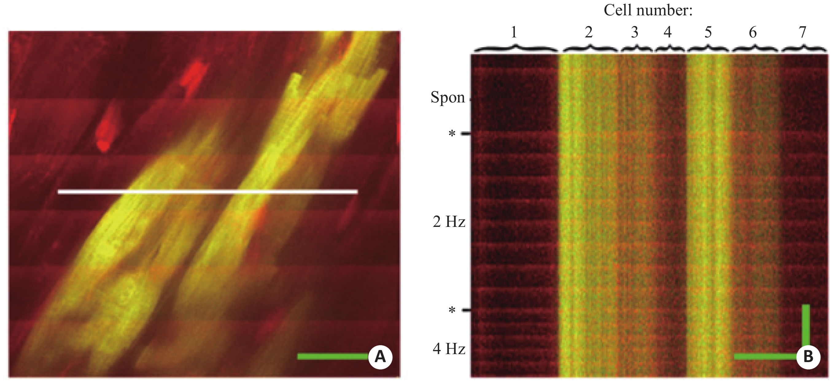
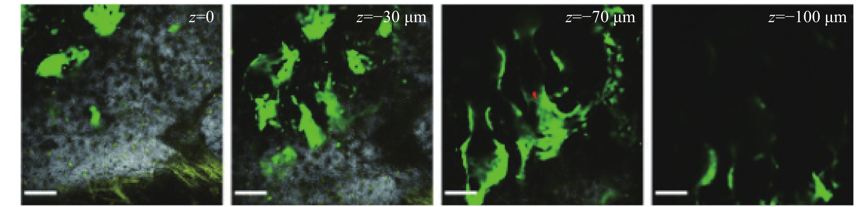
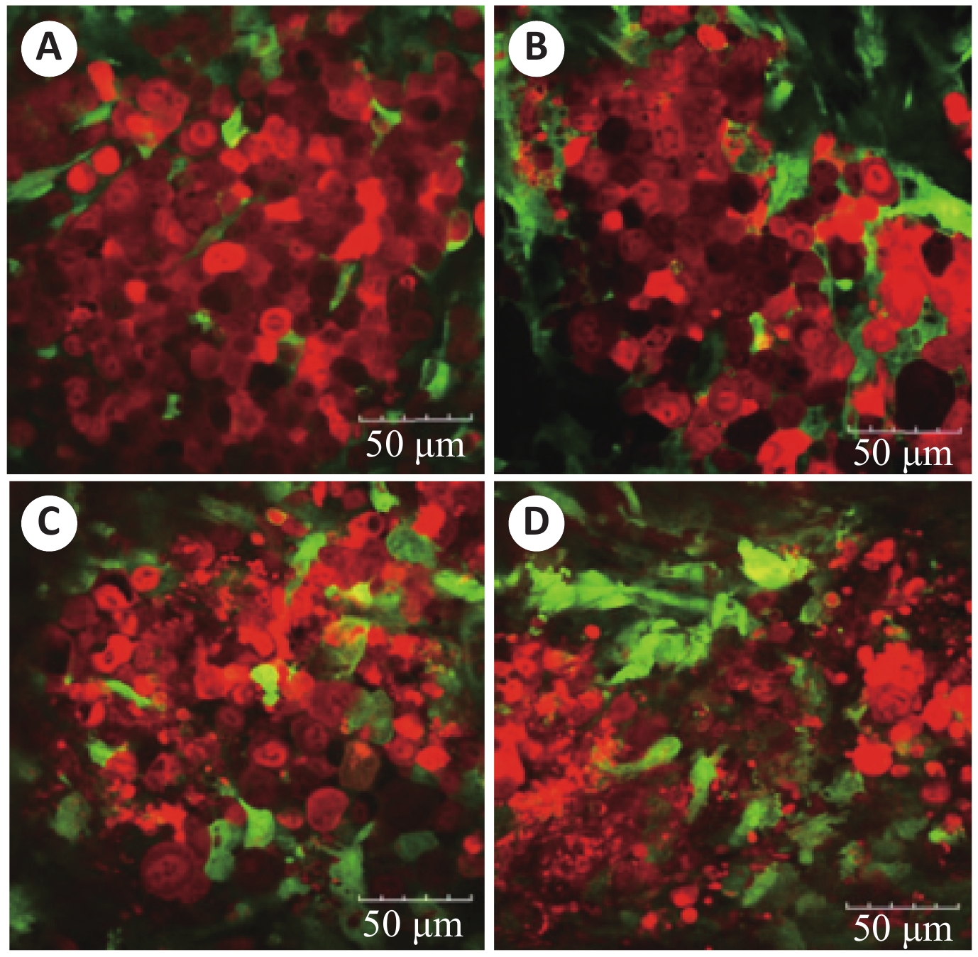
Two-photon imaging technique realizes in vivo 3D deep-tissue imaging. It is an important tool of in vivo imaging and applied in research on in vivo and in situ biological tissue broadly. Research model established by cell transplantation helps study of process and mechanism in the real microenvironment. Using two-photon imaging, morphological observation and functional evaluation about donor cells is realized in living tissue, eliminating differences in vitro tissue culture models. Research of disease models are promoted by utilizing two-photon imaging. This technique is an important tool for research on disease and therapy pathphysiologic events. Cell transplants are used to establish research models of central nervous system, myocardium, bone marrow and anticancer drug. This article reviews the application of research on these regions with two-photon imaging technique, and discusses the development of this technique.
Two-photon imaging technique realizes in vivo 3D deep-tissue imaging. It is an important tool of in vivo imaging and applied in research on in vivo and in situ biological tissue broadly. Research model established by cell transplantation helps study of process and mechanism in the real microenvironment. Using two-photon imaging, morphological observation and functional evaluation about donor cells is realized in living tissue, eliminating differences in vitro tissue culture models. Research of disease models are promoted by utilizing two-photon imaging. This technique is an important tool for research on disease and therapy pathphysiologic events. Cell transplants are used to establish research models of central nervous system, myocardium, bone marrow and anticancer drug. This article reviews the application of research on these regions with two-photon imaging technique, and discusses the development of this technique.
2020, 43(1): 41-44.
doi: 10.12122/j.issn.1674-4500.2020.01.09
Abstract:
The incidence of oral and maxillofacial tumors was increased year by year. Surgery is still an important treatment at present. The surgical plan and postoperative prognosis depend on the size of cancer, the extent of invasion,whether accompanied by lymph node metastasis and distant metastasis. Imaging examination plays a more and more important role in the preoperative evaluation in patient with oral and maxillofacial tumors. PET/MR is the most advanced molecular imaging equipment in the world, the perfect combination of structure and functional imaging plays an indispensable role in the diagnosis and treatment of tumors, but its clinical application value in oral and maxillofacial tumors is rarely reported. The purpose of this review is to explore the application value of PET/MR in oral and maxillofacial tumors through clinical cases combined with relevant literature at home and abroad.
The incidence of oral and maxillofacial tumors was increased year by year. Surgery is still an important treatment at present. The surgical plan and postoperative prognosis depend on the size of cancer, the extent of invasion,whether accompanied by lymph node metastasis and distant metastasis. Imaging examination plays a more and more important role in the preoperative evaluation in patient with oral and maxillofacial tumors. PET/MR is the most advanced molecular imaging equipment in the world, the perfect combination of structure and functional imaging plays an indispensable role in the diagnosis and treatment of tumors, but its clinical application value in oral and maxillofacial tumors is rarely reported. The purpose of this review is to explore the application value of PET/MR in oral and maxillofacial tumors through clinical cases combined with relevant literature at home and abroad.
2020, 43(1): 45-48.
doi: 10.12122/j.issn.1674-4500.2020.01.10
Abstract:
High-resolution magnetic resonance vascular wall imaging (HR-VWI) is a magnetic resonance imaging technology with ultra-high resolution. By suppressing blood flow signals, it can clearly present fibrous caps, lipid cores, intramural hematomas and lumen stenosis of atherosclerotic plaques. HR-VWI provides important imaging information and has great value for clinical diagnosis and treatment in atherosclerotic cardiovascular and cerebrovascular diseases. This article reviews the imaging forming principle of HR-VWI and its clinical application with scientific research in intracranial atherosclerotic diseases.
High-resolution magnetic resonance vascular wall imaging (HR-VWI) is a magnetic resonance imaging technology with ultra-high resolution. By suppressing blood flow signals, it can clearly present fibrous caps, lipid cores, intramural hematomas and lumen stenosis of atherosclerotic plaques. HR-VWI provides important imaging information and has great value for clinical diagnosis and treatment in atherosclerotic cardiovascular and cerebrovascular diseases. This article reviews the imaging forming principle of HR-VWI and its clinical application with scientific research in intracranial atherosclerotic diseases.
2020, 43(1): 49-52.
doi: 10.12122/j.issn.1674-4500.2020.01.11
Abstract:
Thyroid cancer is one of the most common endocrine system malignancies, in which differentiated thyroid cancer accounts for more than 90% of the incidence of thyroid cancer, with a good prognosis. However, the 5-year relative survival rate of thyroid cancer patients in China is far from that in some developed countries. Early and accurate detection of recurrent disease and appropriate treatment strategies can improve the prognosis of patients with recurrent disease. Therefore, early detection of recurrence and metastasis of these patients is essential. With the transition of diagnostic technology from system to molecular level, multi-modal molecular imaging is increasingly important. PET can provide functional information of tumor cells, while CT and MRI can provide anatomical information of tumors.The combination of functional imaging technology and anatomical imaging technology can achieve complementary advantages and is of great significance for the diagnosis of disease recurrence and metastasis. Two major directions of combined PET fusion imaging are PET-CT and PET-MRI. In recent years, PET-CT has more advantages than conventional imaging in the efficacy and prognosis evaluation of DTC diagnosis staging, and PET-MRI is another excellent multi-mode imaging technology after PET-CT. Due to its high-resolution soft tissue and multi-sequence and multi-parameter imaging characteristics, PET-MRI is increasingly important. Therefore, the clinical application and future prospect of PET-CT and PET-MRI in postoperative recurrence/metastasis of differentiated thyroid cancer is reviewed.
Thyroid cancer is one of the most common endocrine system malignancies, in which differentiated thyroid cancer accounts for more than 90% of the incidence of thyroid cancer, with a good prognosis. However, the 5-year relative survival rate of thyroid cancer patients in China is far from that in some developed countries. Early and accurate detection of recurrent disease and appropriate treatment strategies can improve the prognosis of patients with recurrent disease. Therefore, early detection of recurrence and metastasis of these patients is essential. With the transition of diagnostic technology from system to molecular level, multi-modal molecular imaging is increasingly important. PET can provide functional information of tumor cells, while CT and MRI can provide anatomical information of tumors.The combination of functional imaging technology and anatomical imaging technology can achieve complementary advantages and is of great significance for the diagnosis of disease recurrence and metastasis. Two major directions of combined PET fusion imaging are PET-CT and PET-MRI. In recent years, PET-CT has more advantages than conventional imaging in the efficacy and prognosis evaluation of DTC diagnosis staging, and PET-MRI is another excellent multi-mode imaging technology after PET-CT. Due to its high-resolution soft tissue and multi-sequence and multi-parameter imaging characteristics, PET-MRI is increasingly important. Therefore, the clinical application and future prospect of PET-CT and PET-MRI in postoperative recurrence/metastasis of differentiated thyroid cancer is reviewed.
2020, 43(1): 53-58.
doi: 10.12122/j.issn.1674-4500.2020.01.12
Abstract:
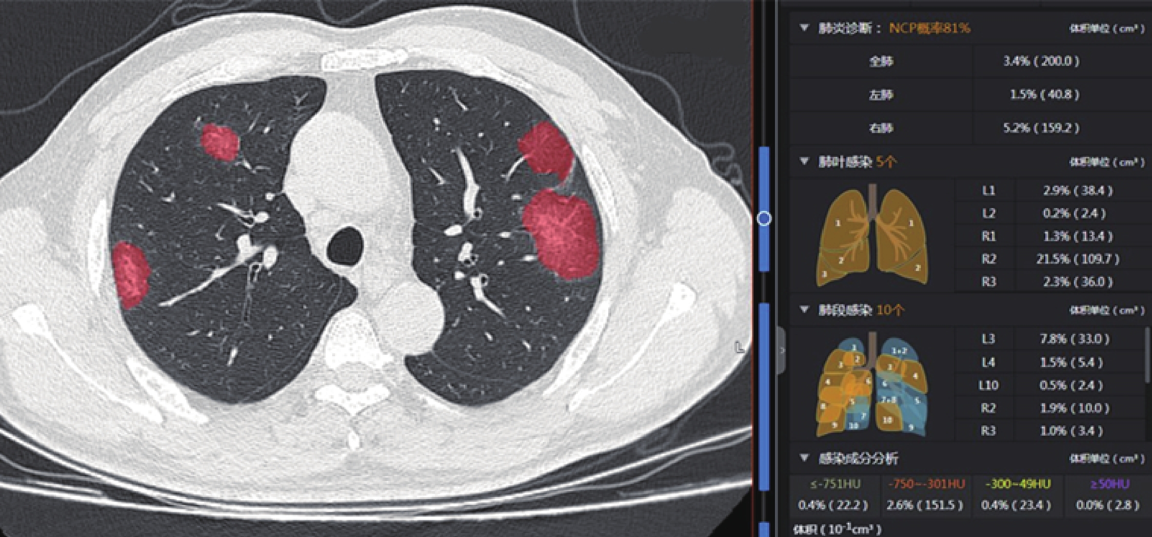
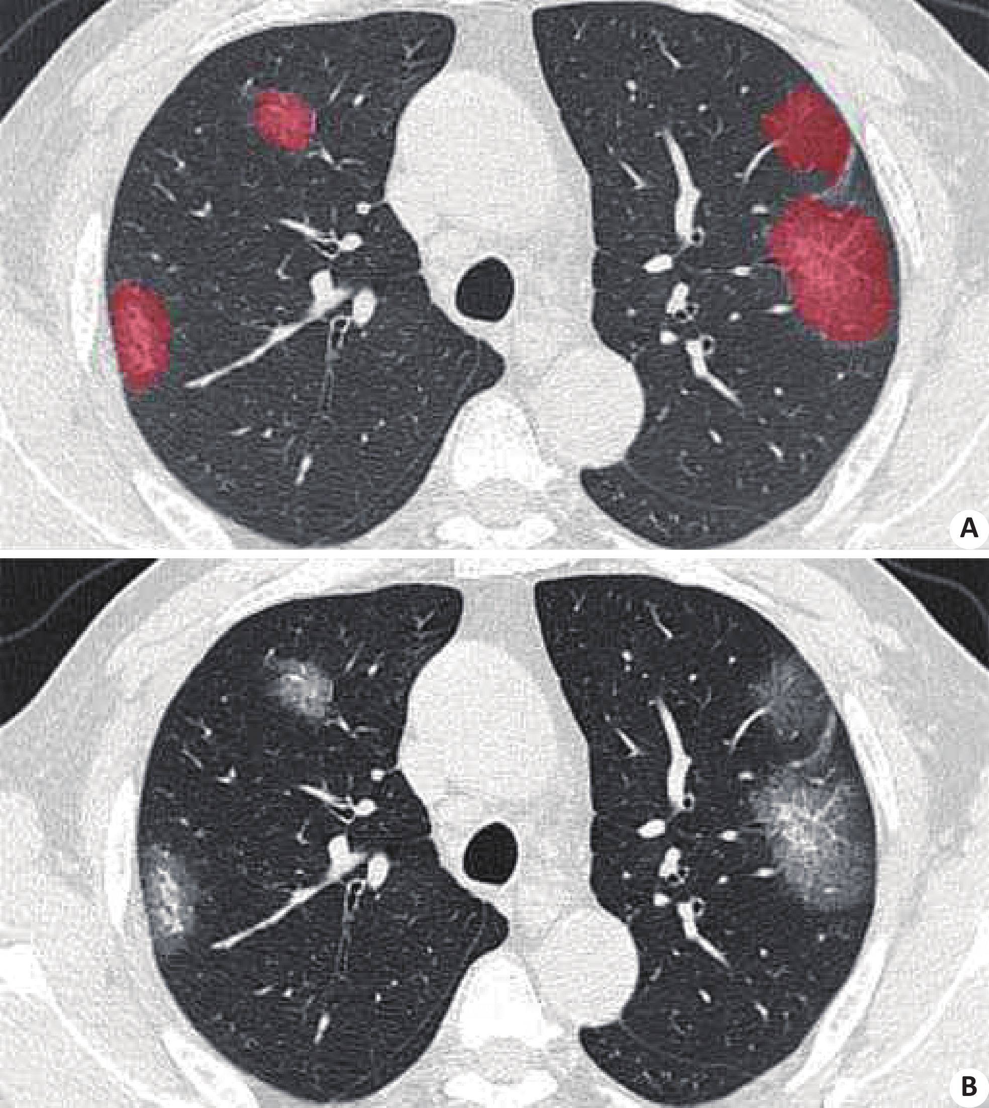
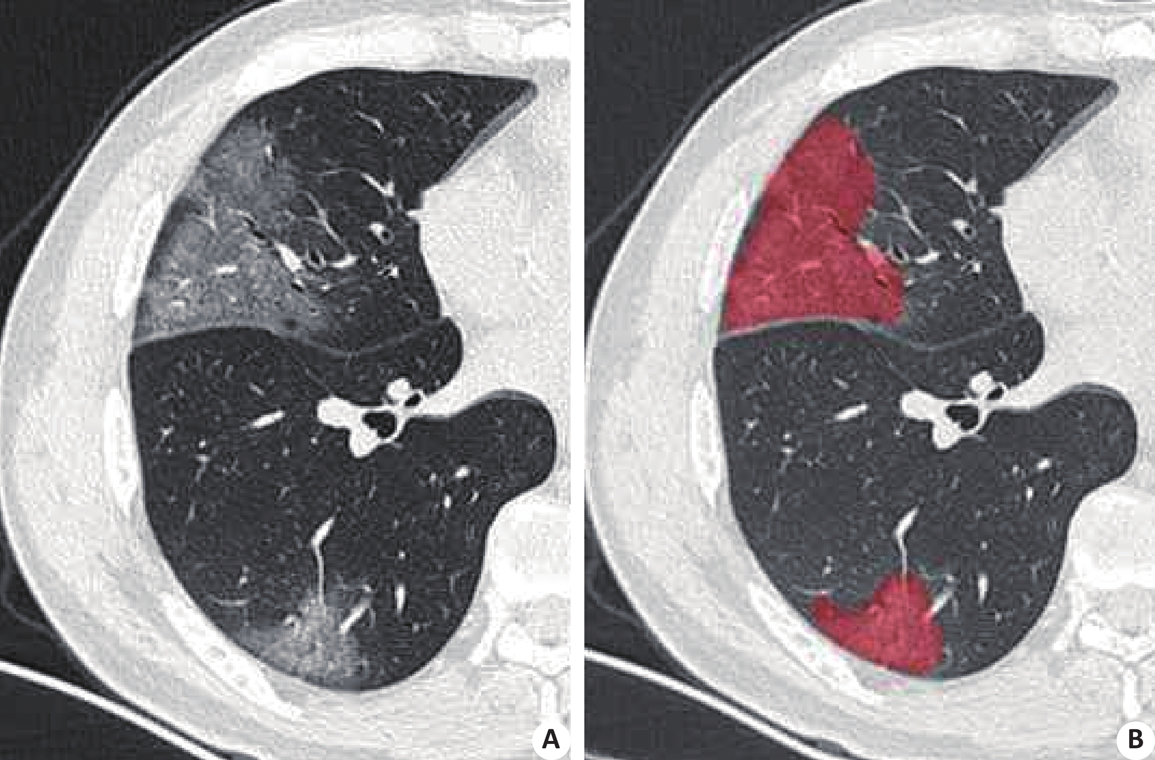
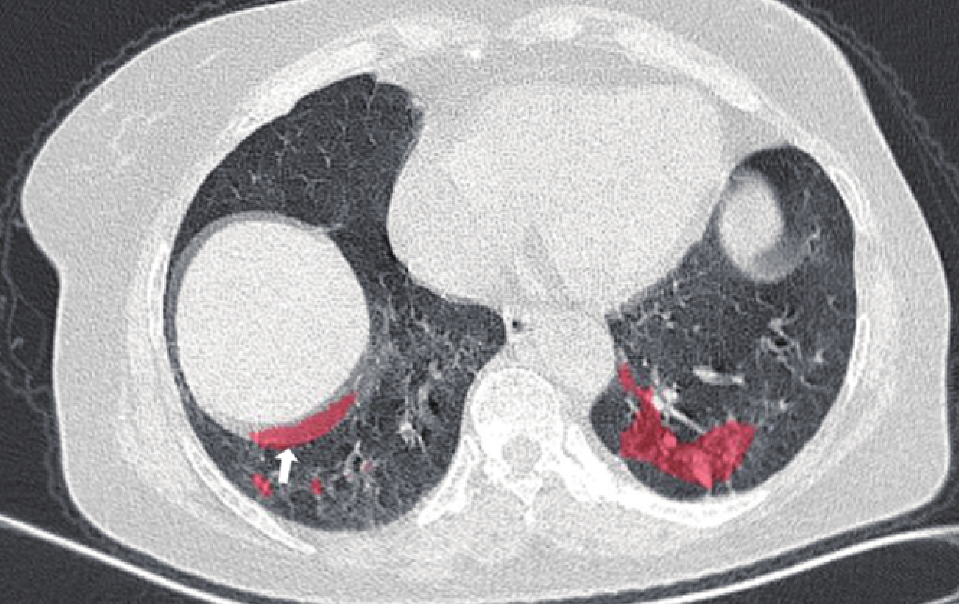
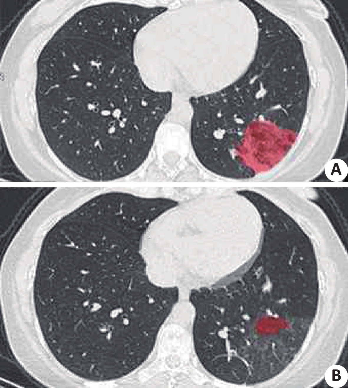
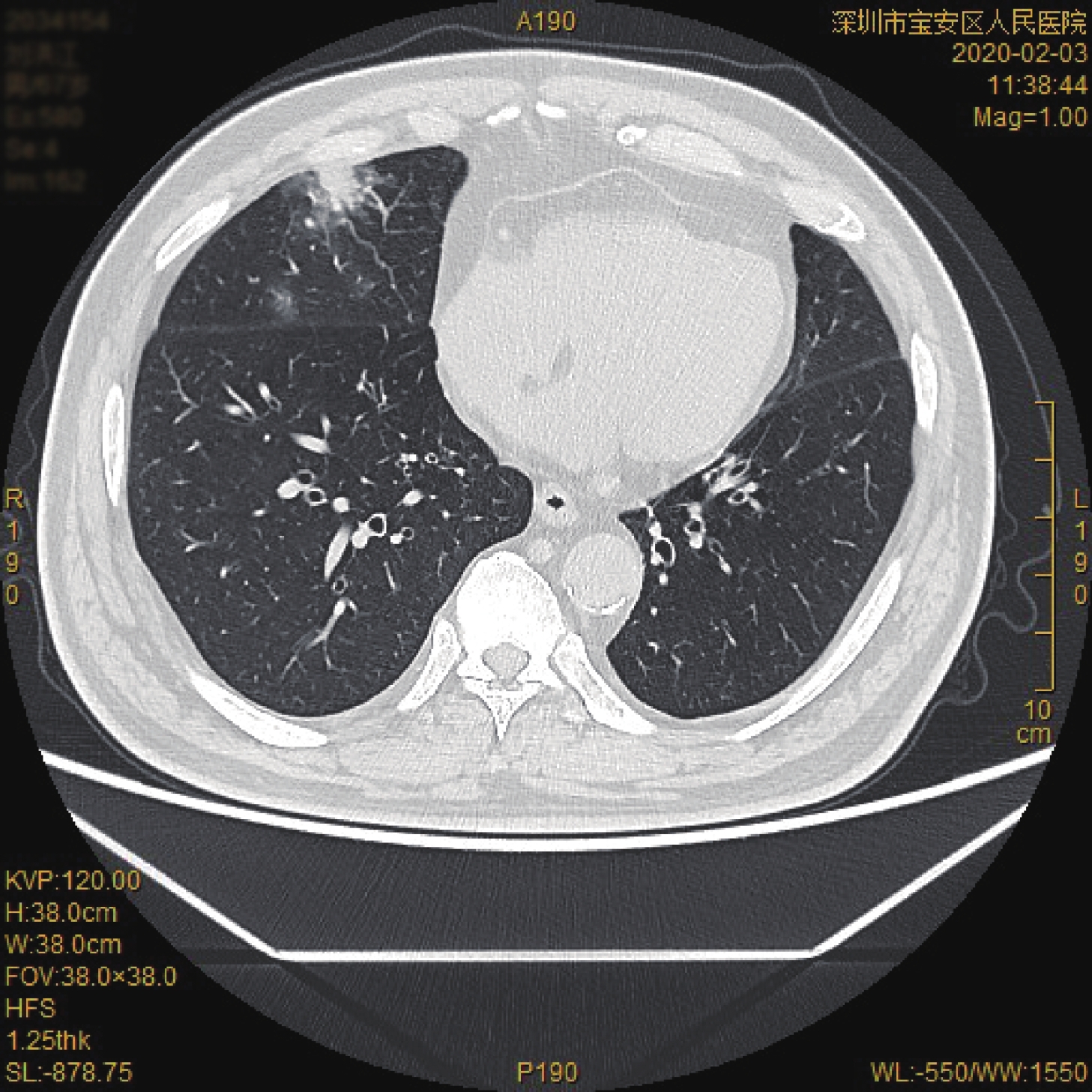
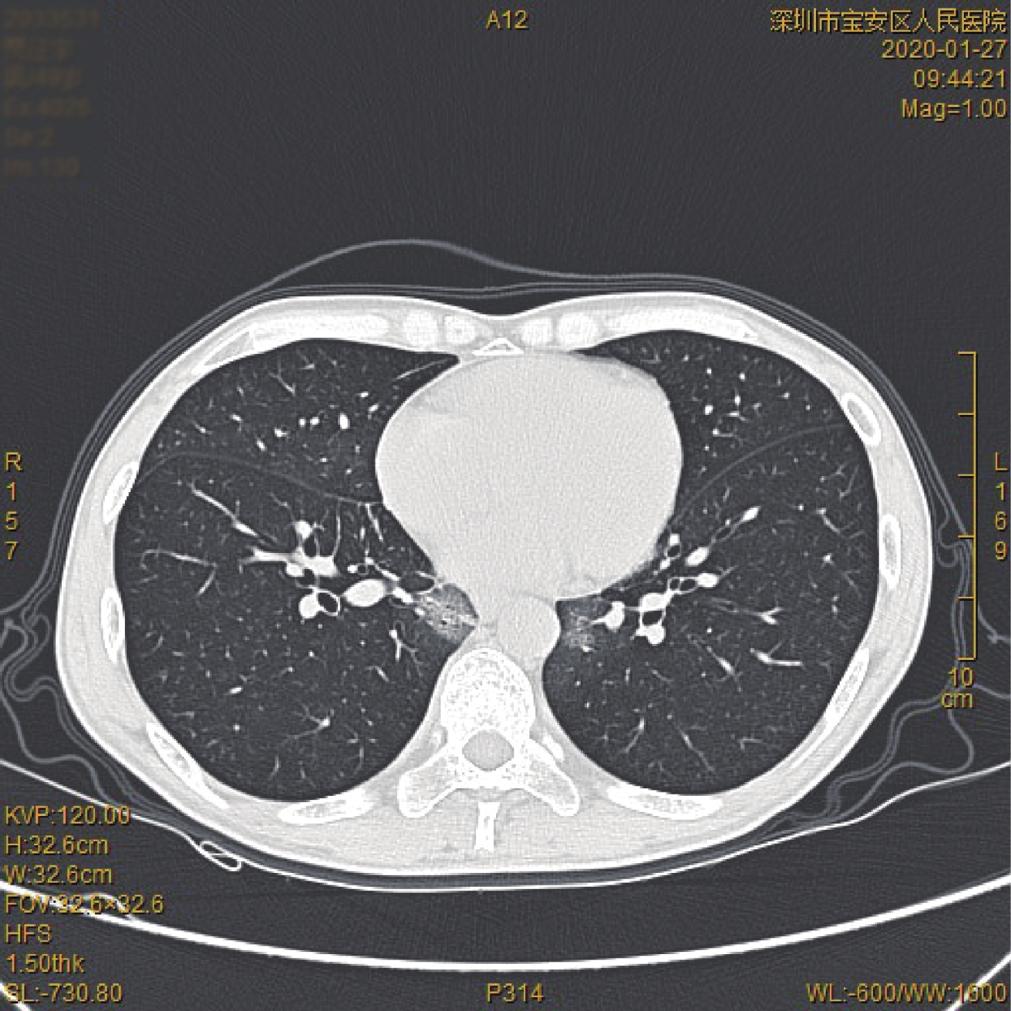
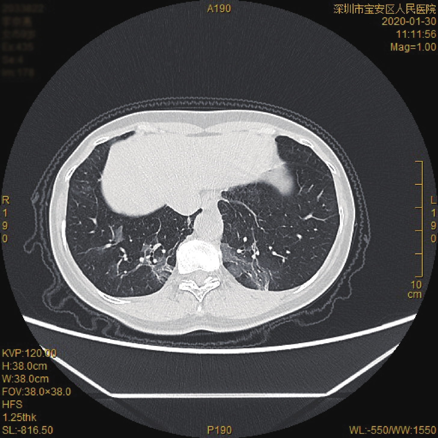
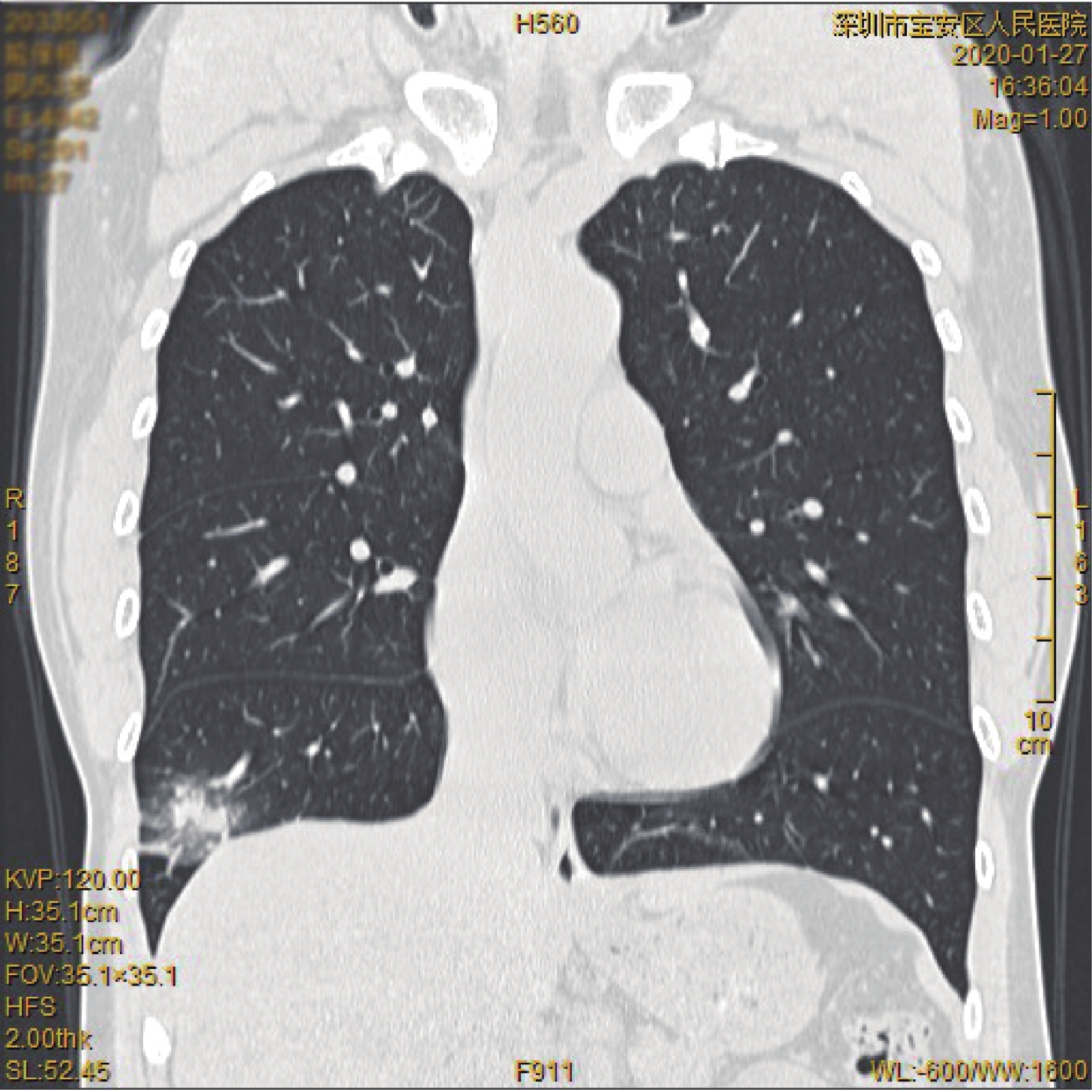
ObjectiveTo explore the application value of artificial intelligence-assisted CT in the screening of the novel coronavirus pneumonia and the monitoring. MethodsCT imaging data of 27 patients with novel coronavirus pneumonia were collected, including 14 males and 13 females with the age from 28 to 85 years old (average 48.9±14.3). The imagings were loaded to the "uAI novel coronavirus pneumonia Intelligent Assisted Analysis System" based on deep learning models. Then the software automatically identified and labeled the pneumonia lesions in batches, and automatically calculated the total volume of lesions, the volume of internal ground glass shadow and the volume of consolidation area. After that, the PACS system was used to manually review the diagnosis of lesions identified by the artificially assisted diagnosis software, record the false positive or false negative situation in the software recognition area, and manually repair a few false positive or false negative images. ResultsArtificial intelligence-assisted diagnosis software automatically identified and labeled the pneumonia lesions. It calculated the total volume of the patient's lesions, the volume of the internal ground glass shadow and the volume of the consolidation area. The results of manual reexamination showed that the range of lesions labeled by the artificial intelligence -assisted diagnosis software was more consistent with that observed by the naked eye. There were no false-positive or false-negative cases in 20 clinical general-type patients. Among the severe and critical patients, 3 patients showed false positive manifestations of local software-labeled lesions, and the difference between patients in different clinical types was significant (P<0.05). The follow-up function provided by the artificial intelligence-assisted diagnosis software visually showed the comparison of the changes in the range and density of the two lesions by the forms of pictures and charts. The manual review diagnosis showed that 2 patients presented false negative manifestations in the local lesion labeled area, and 3 patients presented false positive manifestations, and the difference between patients in different clinical types was significant (P<0.05). ConclusionArtificial intelligence-assisted CT can effectively identify the lesions of novel coronavirus pneumonia and provide detail information about the lesions. In terms of the assessment of patients' condition, the changes in the lesion range and internal density differences can be visually shown by pictures and charts, which provide objective data support for clinical evaluation and improve the work efficiency of imaging physicians.

2020, 43(1): 59-63.
doi: 10.12122/j.issn.1674-4500.2020.01.13
Abstract:
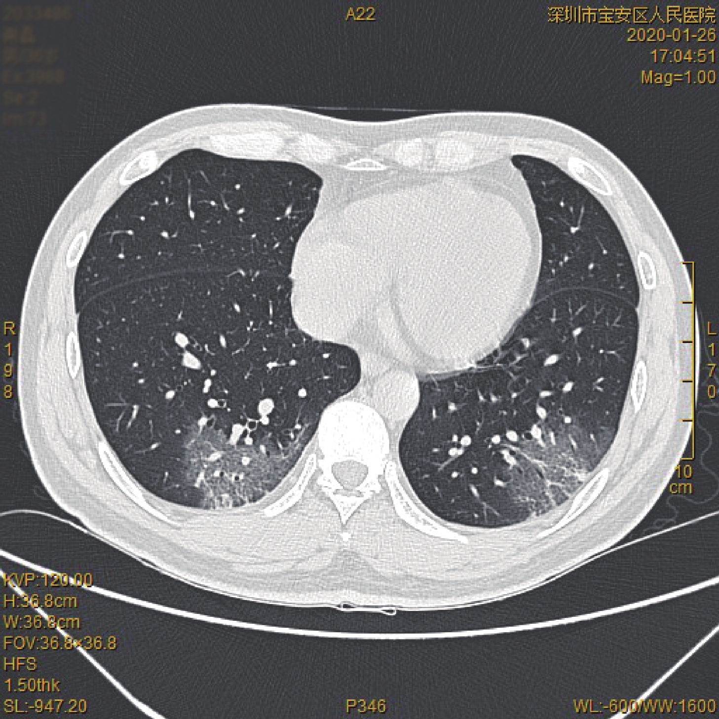
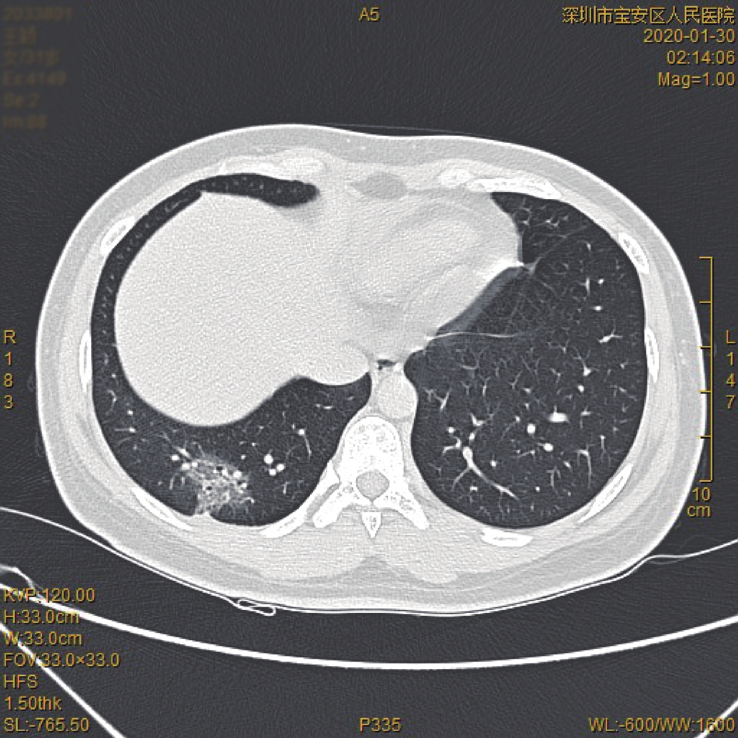
ObjectiveTo investigate the clinical characteristics and CT features of novel coronavirus pneumonia (COVID-19). MethodsThe clinical and CT data of 13 cases of COVID-19 diagnosed by nucleic acid test in our hospital were retrospectively analyzed. The CT features focused on the density, number, distribution, location and morphology of the lesions, as well as pleural thickening, pleural effusion, mediastinal lymph node enlargement or other accompanying signs. ResultsThere were 9 males and 4 females, aged 31-67 years, with an average age of 49±12 years. Common clinical symptoms included fever (8/13), dry cough or cough (3/13), accompanied by diarrhea, nausea and vomiting in 1 case (1/13). Lymphocyte count in peripheral blood decreased in 2 cases (2/13) and CRP increased in 5 cases (5/13). 10 cases (10/12) have multiple lesions, 11 cases (11/12) involve the dorsal and posterior basal segment of the lower lobe bilateral lung. Peripheral distribution (located in subpleural or subpleural area of interlobular fissure) was dominant in 12 cases (12/12), and distribution along the bronchial tree in 10 cases (10/12), There were 10 cases (10/12) with mass and patchy morphology, 9 cases (9/12) with grid shadow. The density was mainly ground-glass opacity lesions (11/12 cases), thickening of the adjacent bronchial bundle was observed in 9 cases (9/12), Pleural thickening or traction deformation (including interlobular fissure distortion) was observed in 8 cases (8/12). ConclusionThe clinical features of COVID-19 are fever and increased CRP. The imaging features is peripheral GGO, and HRCT of the chest can detect the pulmonary changes of COVID-19 patients at an early stage.
2020, 43(1): 64-69.
doi: 10.12122/j.issn.1674-4500.2020.01.14
Abstract:
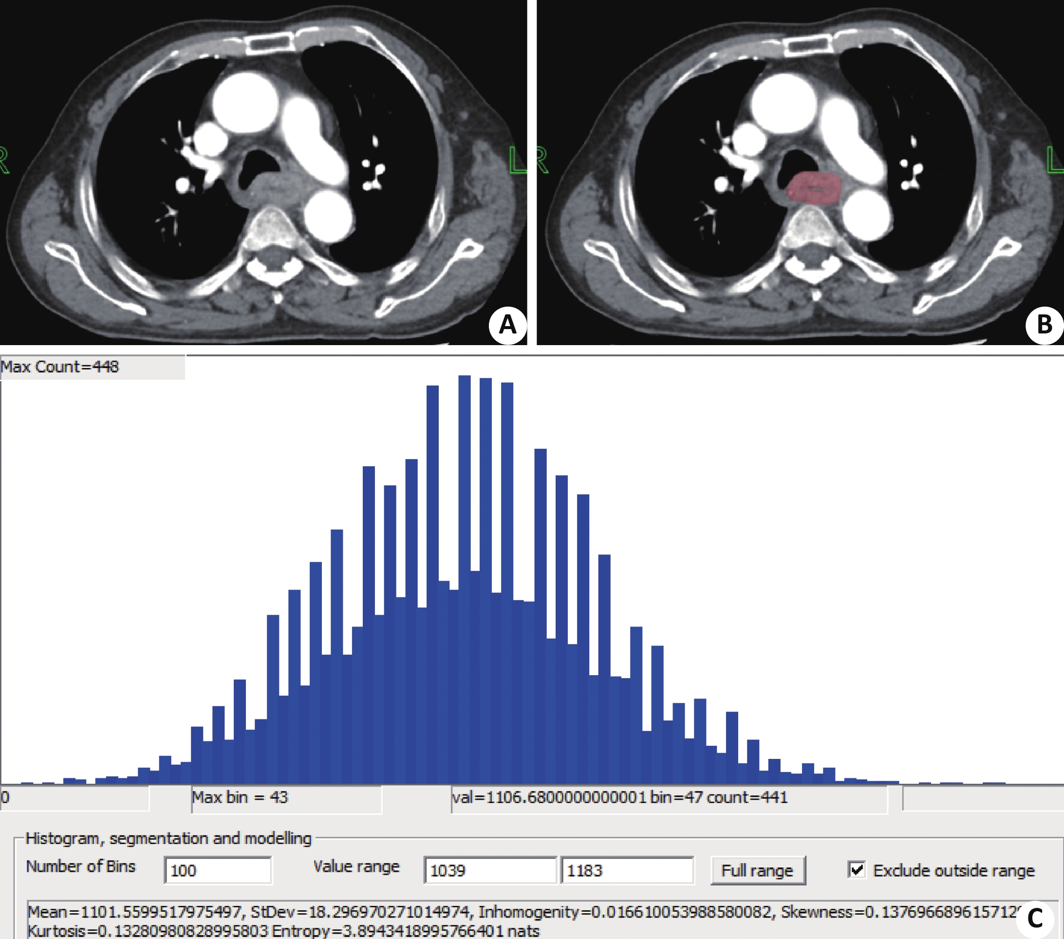
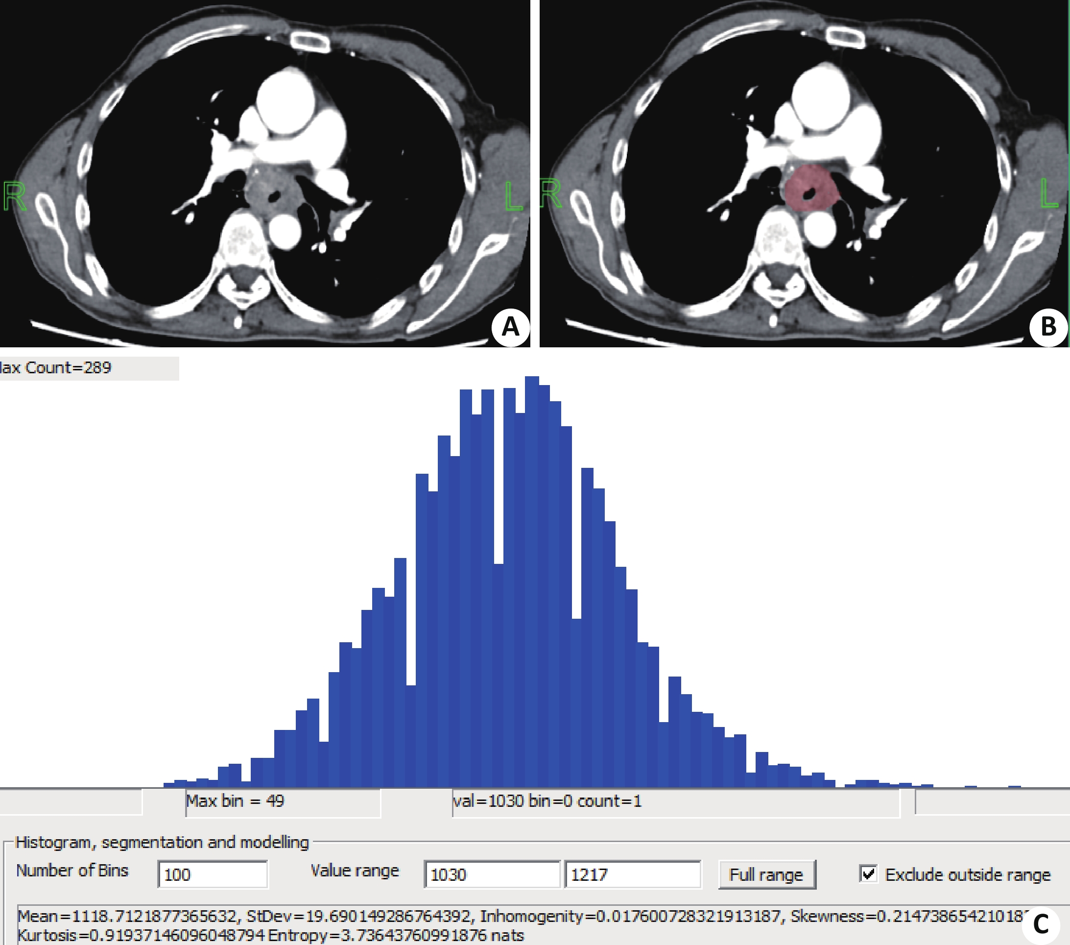
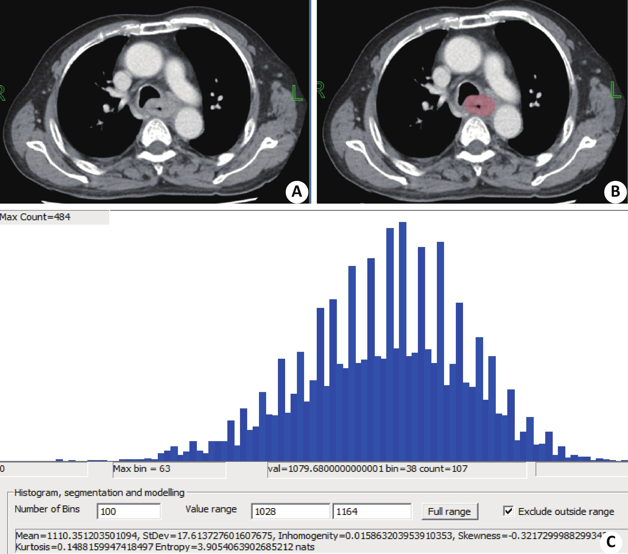
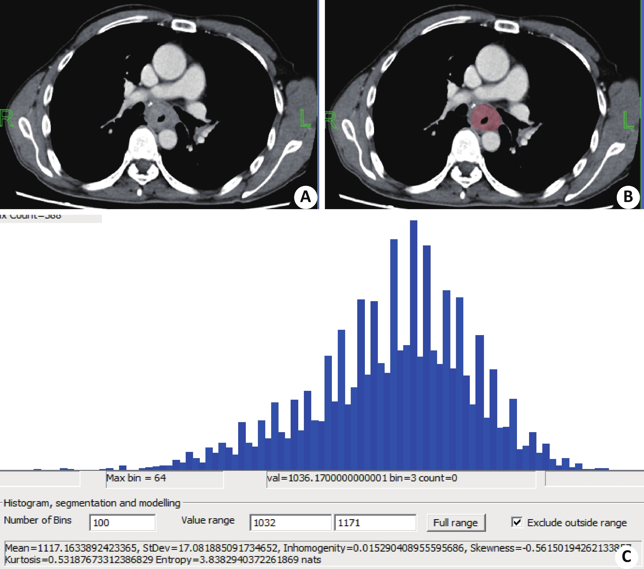
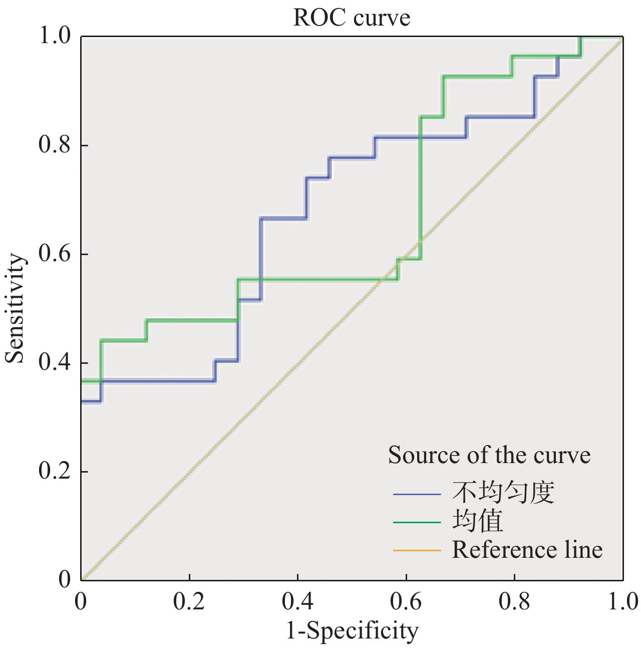
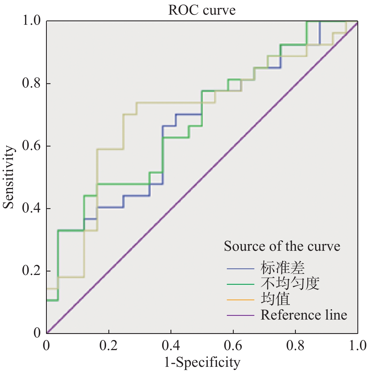
ObjectiveTo explore the feasibility of texture analysis of enhanced CT images to predict the metastasis of lymphonodus in patients with esophageal squamous cell carcinoma. MethodsThe patients of esophageal squamous cell carcinoma in our hospital were selected. According to the pathological results, the patients were divided into lymphonodus metastasis group and non metastasis group. 51 patients with esophageal squamous cell carcinoma were included, 27 in lymphonodus metastasis group (25 males and 2 females with an average age of 63.4±6.5 years old) and 24 in non metastasis group (20 males and 4 females with an average age of 63.5±9.4 years old). The chest CT enhancement were performed before treatment. The enhanced CT images (arterial phase and venous phase) were imported into the Firevoxel software for texture analysis. The texture parameters were recorded including mean, median, standard deviation, heterogeneity, skewness, kurtosis and entropy. The differences of texture parameters between the two groups were analyzed. The value of CT texture parameters in identifying lymphonodus metastasis was analyzed by ROC curve. ResultsThere were significant differences in heterogeneity and entropy between the two groups in arterial phase (P< 0.05). There were significant differences in standard deviation, heterogeneity and entropy between the two groups in venous phase (P< 0.05). In arterial phase, the optimal cut-off values for the diagnosis of lymph node metastasis of esophageal cancer were heterogeneity and entropy and the areas under the curve were 0.685 and 0.674, respectively. In venous phase, the standard deviation, inhomogeneity and entropy had significant significance in distinguishing lymph node metastasis of esophageal cancer. Among them, the area under the curve of entropy was the largest (0.704), the best critical value was 3.842, the sensitivity was 70.4%, and the specificity was 75%. It had a certain accuracy for the diagnosis of lymph node metastasis of esophageal cancer. ConclusionThe texture analysis of enhanced CT images (especially the venous phase) is helpful to identify lymph node metastasis of esophageal cancer and provides help for noninvasive evaluation of lymph node metastasis before surgery.
2020, 43(1): 70-75.
doi: 10.12122/j.issn.1674-4500.2020.01.15
Abstract:
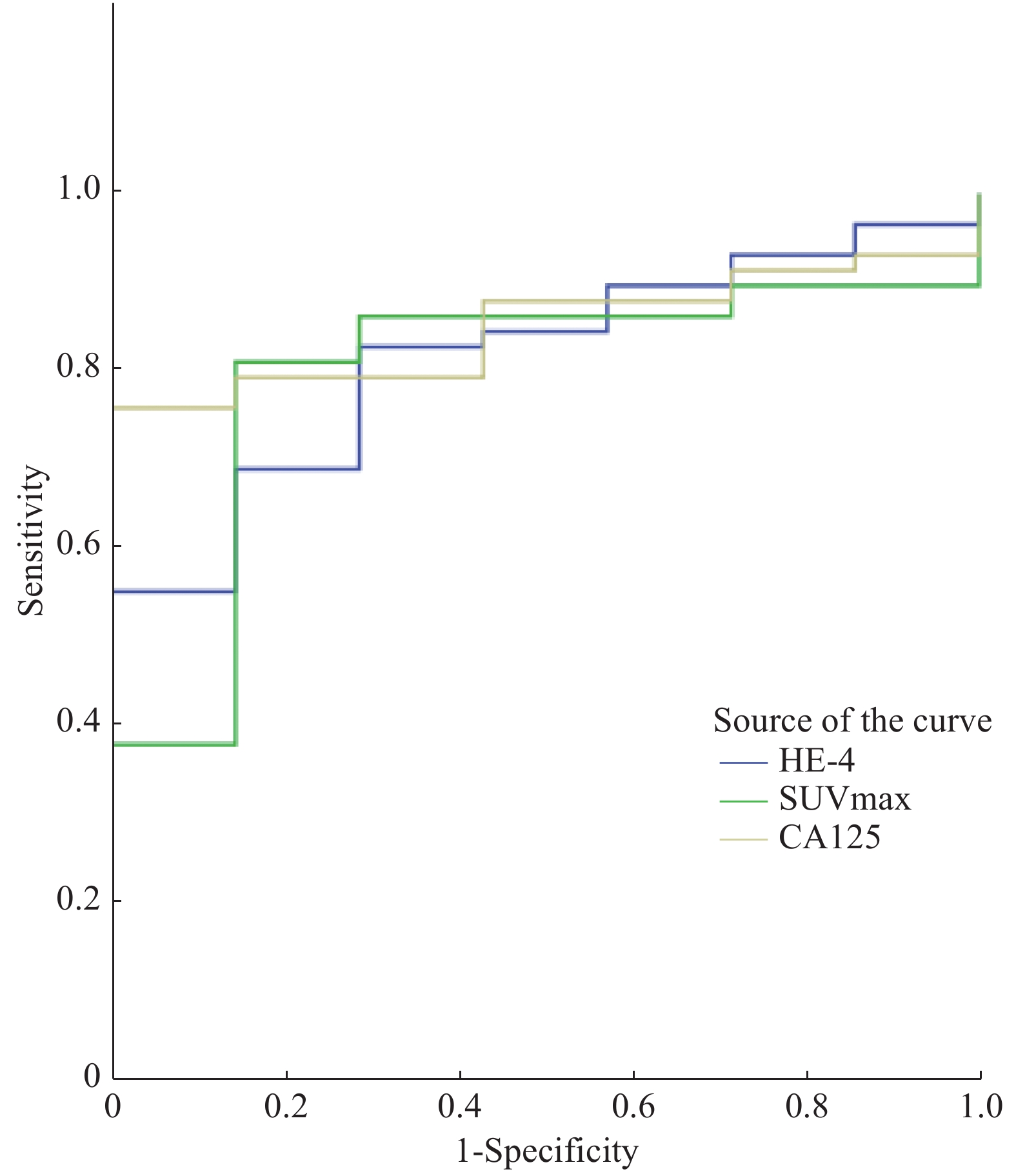
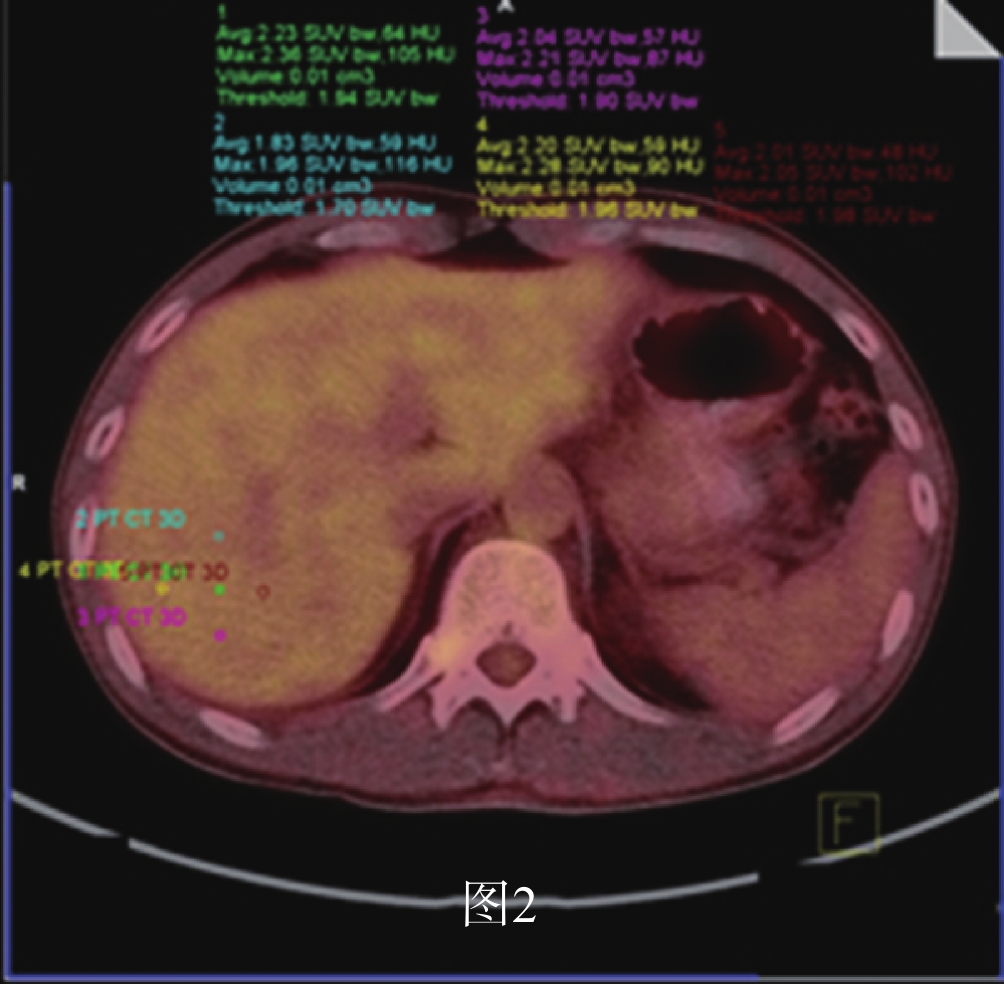
ObjectiveTo investigate the diagnostic value of 18F-fluorodeoxyglucose PET/CT imaging combined with serum carbohydrate antigen 125 (CA125) and human epididymal protein 4 (HE4) in the diagnosis of postoperative recurrence / metastasis in patients with epithelial ovarian cancer. MethodsFrom July 2018 to September 2019, 65 patients with epithelial ovarian cancer who had detected serum CA125 and HE4 before 18F-FDG PET/CT imaging were retrospectively analyzed. The patients were divided into 18F-FDG PET/CT imaging diagnosis group and 18F-FDG PET/CT+CA125+HE4 diagnosis group and followed up according to the follow-up criteria. The diagnostic results of each group were compared with the follow-up results. ResultsThe sensitivity, specificity, positive predictive value, negative predictive value and consistent rate of 18F-FDG PET/CT imaging in evaluating postoperative recurrence / metastasis of patients with epithelial ovarian cancer were 96.22%, 66.7%, 92.73%, 80.0%, 90.77%, respectively. The sensitivity, specificity, positive predictive value, negative predictive value and consistent rate of 18F-FDG PET/CT+CA125+HE4 in evaluating postoperative recurrence / metastasis of patients with epithelial ovarian cancer were 98.1%, 66.7%, 92.9%, 88.9% and 92.3%, respectively. Receiver operating characteristic of CA125 and HE4 subjects in recurrence / metastasis group showed that the critical value were 20.65 U/mL and 45.5 pmol/L respectively. The sensitivity, specificity, positive predictive value, negative predictive value and consistent rate of 18F-FDG PET/CT+CA125critical value + HE4critical value for evaluating postoperative recurrence / metastasis of epithelial ovarian cancer patients were 98.1%, 75.0%, 94.6%, 90.0%, 93.9%, respectively. Conclusion18F-FDG PET/CT imaging has an advantage in evaluating postoperative recurrence / metastasis in patients with epithelial ovarian cancer. 18F-FDG PET/CT+CA125+HE4 detection has a high diagnostic value in evaluating postoperative recurrence / metastasis in patients with epithelial ovarian cancer. The combined detection of the three is better than that of single detection and combined detection of both. The detection of 18F-FDG PET/CT+CA125critical value + HE4critical value has a higher diagnostic value in evaluating postoperative recurrence/metastasis of patients with epithelial ovarian cancer. During the clinical follow-up, it was found that serum CA125≥20.65 U/mL and serum HE4≥45.5 pmol/L. We should be vigilant when it continues to increase, and 18F-FDG PET/CT imaging should be selected to improve the detection rate of recurrence/metastasis of epithelial ovarian cancer and to detect recurrence/metastasis lesions in the early stage.
2020, 43(1): 76-81.
doi: 10.12122/j.issn.1674-4500.2020.01.16
Abstract:
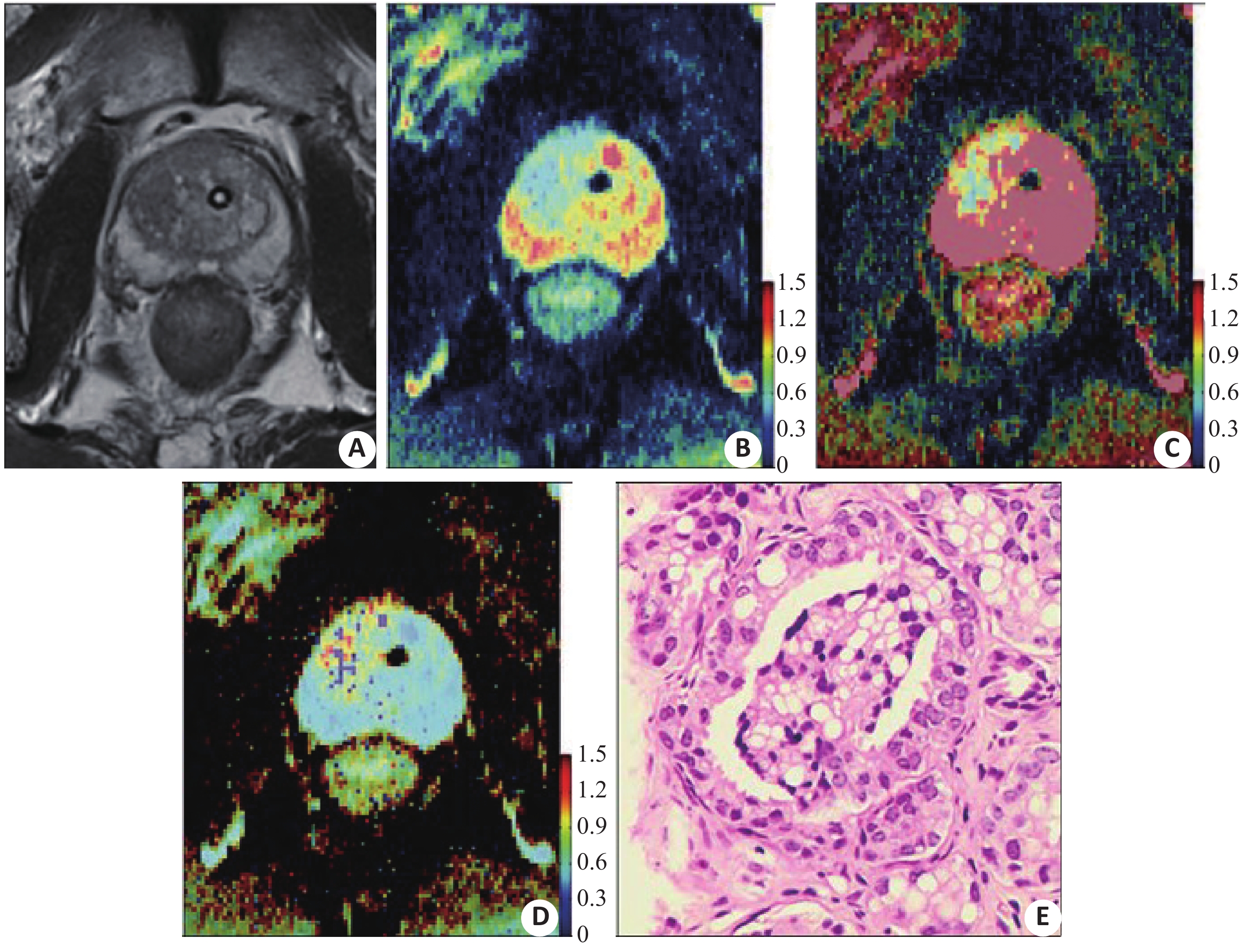
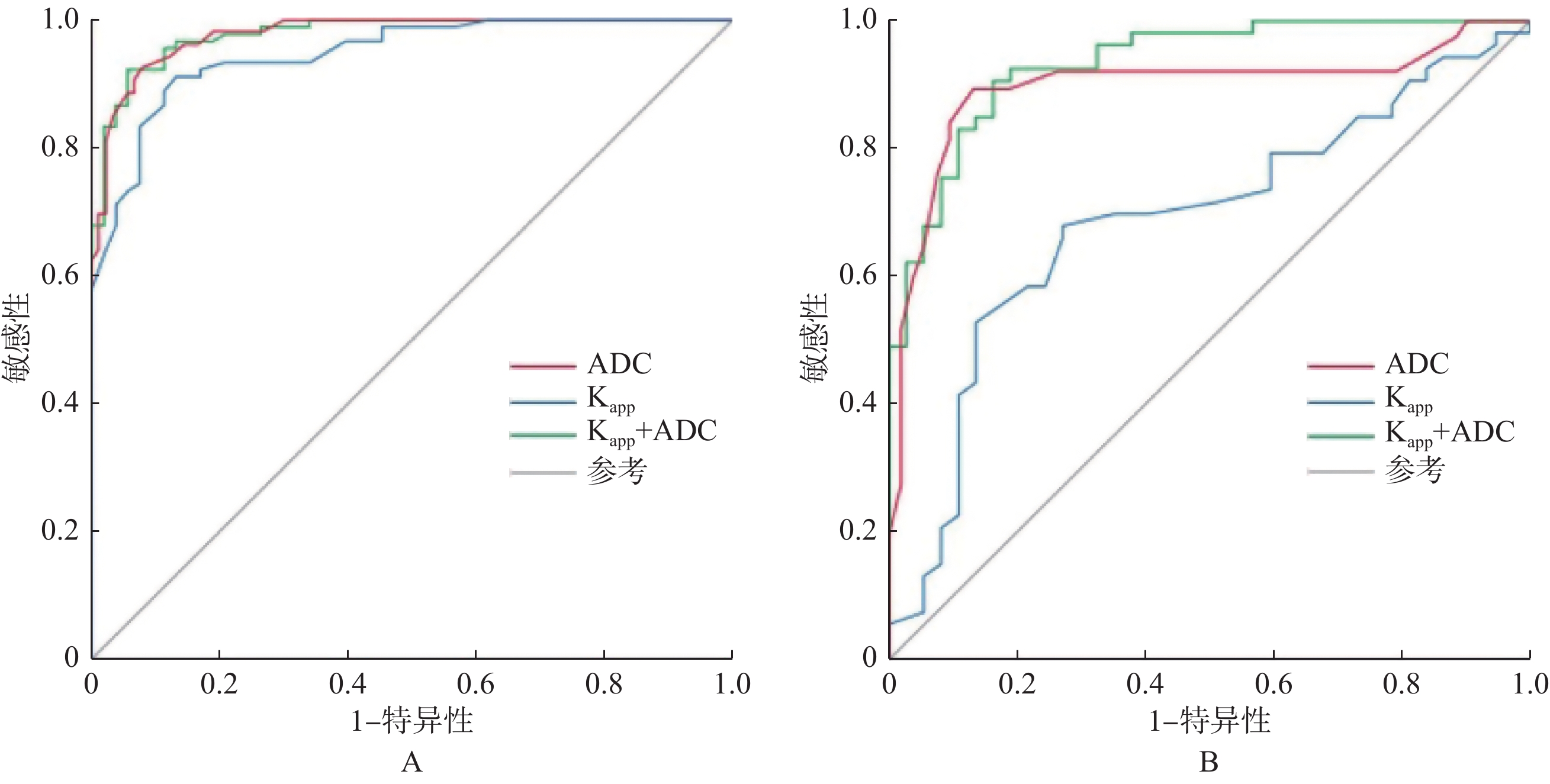
ObjectiveTo investigate the value of diffusion kurtosis imaging (DKI) combined with DWI in diagnosis and assessment of aggressiveness of prostate cancer (PCa). MethodsWe retrospectively analyzed 80 patients from May 2015 to June 2019 underwent DKI and MR images and confirmed by prostate biopsy, including 49 patients with PCa and 31 patients with benign prostatic hyperplasia (BPH). The patients with PCa were divided into a low-grade PCa group (Gleason score≤3+3) and intermediate- and high-grade PCa group (GS≥3+4). DKI-derived parameter (apparent kurtosis coefficient, Kapp) and a DWI-derived parameter (apparent diffusion coefficient, ADC) were obtained. The differences of Kapp and ADC between PCa and BPH, the correlation between DKI and DWI parameters and Gleason score were compared. ROC curves were drawn to evaluate the diagnostic efficacy. Results65 ROIs in 49 patients with PCa were drawn, including ROIs in 26 low-grade tumors and ROIs in 39 intermediate- and high-grade tumors. PCa and intermediate- and high-grade PCa had significantly lower ADC values and higher Kapp values than BPH and low-grade PCa (all P<0.01). The AUCs of Kapp were significantly lower than the AUCs of ADC in the diagnosis (0.947 vs 0.978, P<0.001) and grading (0.689 vs 0.894, P=0.008) of PCa. The AUCs of the combination of the two metrics were significantly higher than the AUCs of Kapp for the diagnosis (0.979 vs 0.947, P=0.013) and grading (0.934 vs 0.689, P<0.001) of PCa, and they were higher than the AUCs of ADC without significance between groups (P>0.05). The combination of the two metrics significantly increased the specificity in grading of PCa compared with Kapp alone (0.838 vs 0.730, P=0.035). ConclusionBoth ADC and Kapp can be used as quantitative parameters in detection and assessment of aggressiveness of PCa. The combination of DKI and DWI has no significant superiority to DWI alone in detection and assessment of the aggressiveness of PCa.
2020, 43(1): 82-87.
doi: 10.12122/j.issn.1674-4500.2020.01.17
Abstract:
ObjectiveTo explore the effect of Eadyn on norepinephrine application. MethodsThe study was a prospective and observative cohort study. A total of 68 patients with severe pneumonia admitted to intensive care unit (ICU) of Foshan Second People's Hospital were enrolled from June 2018 to June 2019, including 38 males and 30 females with the age from 27 to 77 years old (average 58.60±8.72). All the patients were treated by mechanical ventilation and planned to use noradrenaline. Based on whether Eadyn was used as as an indicator for the use of norepinephrine, the patients were divided into the monitoring group (32 cases) and the control group (36 cases). The patients in the monitoring group were monitored continuously by pulse indicating continuous cardiac output (PiCCO). According to the real-time eadyn which calculated by the hemodynamic indexes, norepinephrine was adjusted at any time.Patients in the control group were monitored for invasive blood pressure.The ealy goal directed therapy(EGDT) was carried out with the balanced salt solution. The indexes were compared after treatment. ResultsAfter 12 hours of resuscitating and vasopressor treatment, the severity scores were significantly lower in the monitoring group than in the control group (P<0.05). At the beginning of 24 hours, the indexes of respiratory and circulatory function in the monitoring group were better than those in the control group. The CVP and BNP in the monitoring group were lower than those in the control group (P<0.05). Compared with the lung CT images after 96 hours, the exudation in the control group increased significantly(P<0.05). The time of mechanical ventilation in the monitoring group were 9.47±2.7 days, and that in the control group were 14.42±2.9 days. The length of stay in ICU in the monitoring group were 11.31±3.2 days, and that in the control group were 20±2.9 days. From the statistics of 28 days, 4 cases died in the monitoring group with a mortality rate of 12.5%, and 8 cases died in the control group with a mortality rate of 22.22%. The difference was significant (P<0.05). The prognosis of the monitoring group was better than that of the control group. ConclusionUsing Eadyn as an indicator for norepinephrine application is beneficial to severe pneumonia shock patients.

2020, 43(1): 88-93.
doi: 10.12122/j.issn.1674-4500.2020.01.18
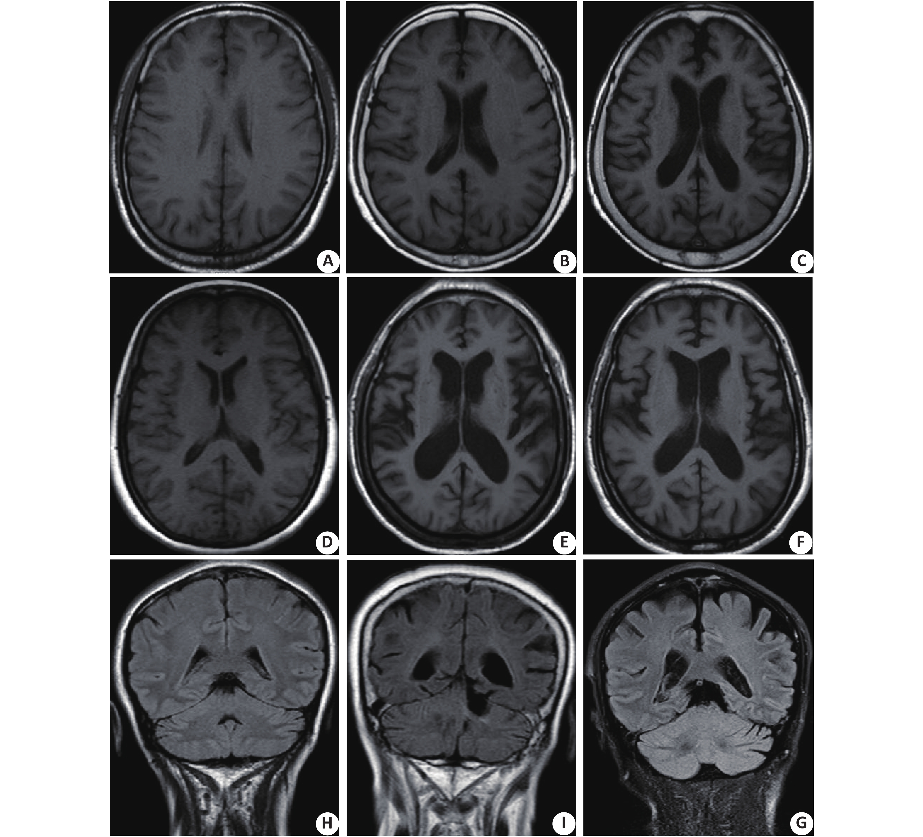
Abstract:
ObjectiveTo explore the clinical and imaginal characteristics of PD patients with probably RBD(P-RBD) and explore the influencing factors. MethodsA total of 99 PD patients were eligible and enrolled in the study, 88 of them had complete brain MRI data. By the RBDQ-HK scale, patients were divided into the P-RBD group (RBDQ-HK score≥18) with 33 patients and the NP-RBD group (RBDQ-HK score <18) with 66 patients. All patients were assessed with the comprehensive Parkinson's Disease Rating Scale (UPDRS) and Hoehn. Yahr Scale. The motor function, depression, anxiety, cognitive function and MRI data of the patients were assessed. Logistic regression was used to analyze the correlation between P-RBD and its influencing factors. ResultsThe incidence of P-RBD in Parkinson's disease patients was 33.3% (33/99). Compared with NP-RBD group, P-RBD group had earlier onset age, higher male proportion, longer disease duration, lower use rate of antidepressants, more on-off phenomenon, more treatment of complications (P<0.05). However, the differences of the degree of leukopathy, global atrophy, frontal lobe atrophy, parietal lobe atrophy and temporal lobe atrophy between the two groups were not significant (P>0.05). Multivariate logistic regression analysis showed that men, earlier onset age and antidepressants were the significant related factors of P-RBD(P<0.05). ConclusionsP-RBD is common in Parkinson’s disease patients. Men and earlier onset age are risk factors for P-RBD in PD patients. The antidepressants can decrease the risk of P-RBD in PD patients.
2020, 43(1): 94-98.
doi: 10.12122/j.issn.1674-4500.2020.01.19
Abstract:
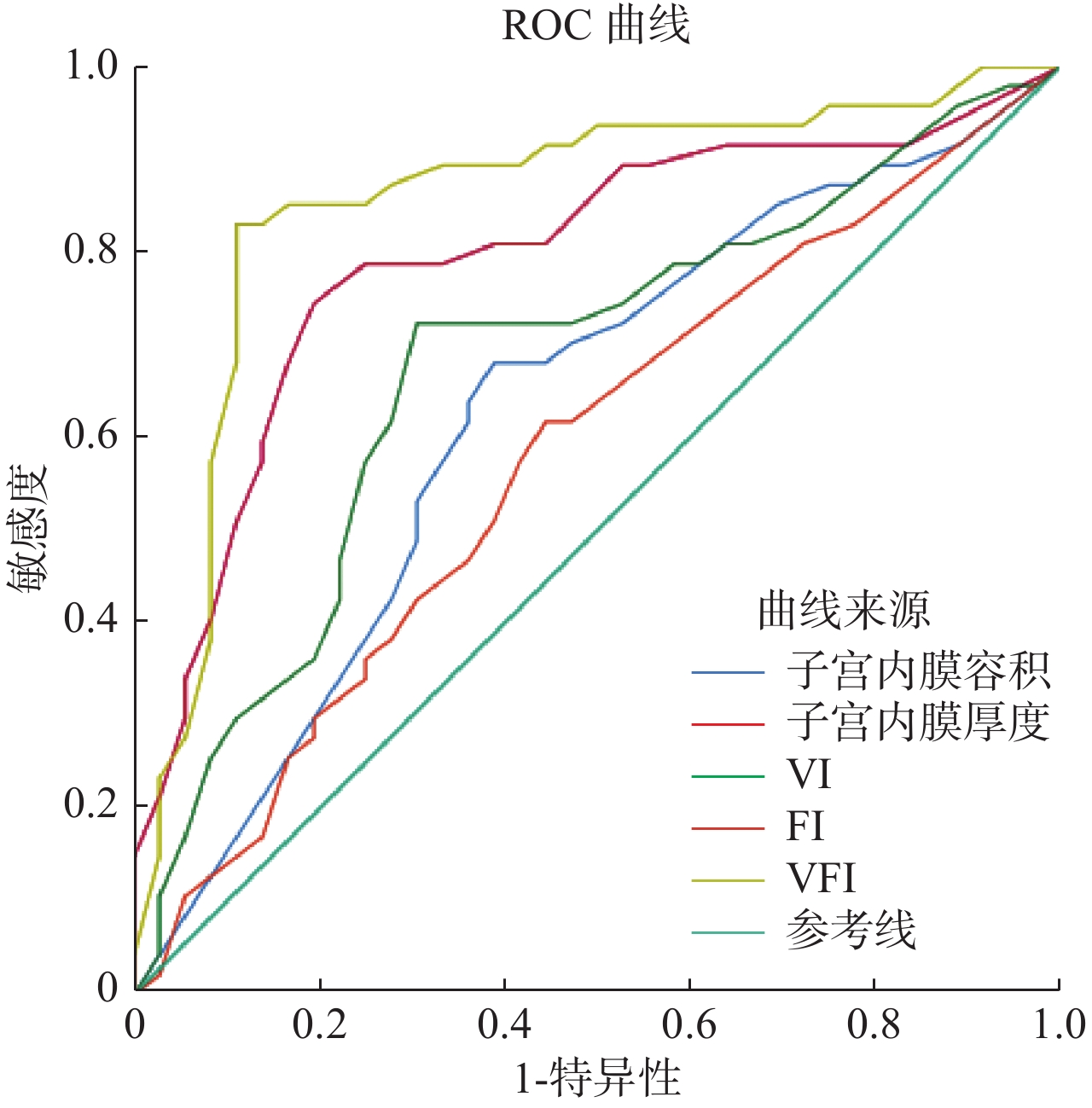
ObjectiveTo investigate the value of three-dimensional contrast-enhanced ultrasound (3D-CEUS) in the diagnosis of severe intrauterine adhesions (SIUA) after transcervical resection of adhensions (TCRA). MethodsThe clinical data of 83 patients with IUA after TCRA were retrospectively analyzed. All patients underwent hysteroscopy, transvaginal two-dimensional ultrasonography and 3D-CEUS. The endometrial thickness, volume, vascular index (VI), flow index (FI), vascularization flow index (VFI) were analyzed. Based on the results of hysteroscopy, the value of 3D-CEUS in diagnosing SIUA after TCRA was analyzed. ResultsThirty-six cases of SIUA were confirmed after TCRA. The three-dimensional imaging of SIUA showed abnormal uterine cavity shape, narrow, irregular margin and discontinuous endometrial echo. The accuracy rate of IUA classification of TCRA by transvaginal three-dimensional ultrasound was 90.36%, which was in good agreement with hysteroscopy (kappa=0.795, P<0.05). The endometrial thickness, volume, VI, FI, VFI index of SIUA patients were lower than those of moderate and mild groups (P<0.05). Dual Logistic regression analysis showed that the thickness, volume, VI, FI and VFI of endometrium were significantly correlated with SIUA after TCRA (P<0.05). The results of ROC analysis showed that endometrial thickness and VFI had a higher efficiency in identifying SIUA, with AUC of 0.794 and 0.856, sensitivity and specificity of 80.56%, 74.47%, 88.89% and 82.98%, respectively. ConclusionTransvaginal three-dimensional ultrasound can clearly show the degree and extent of intrauterine adhesions after TCRA. It provide a reliable reference for IUA classification. The measurement of endometrial parameters by 3D-CEUS can be used as a quantitative index for SIUA diagnosis.

2020, 43(1): 99-102.
doi: 10.12122/j.issn.1674-4500.2020.01.20
Abstract:
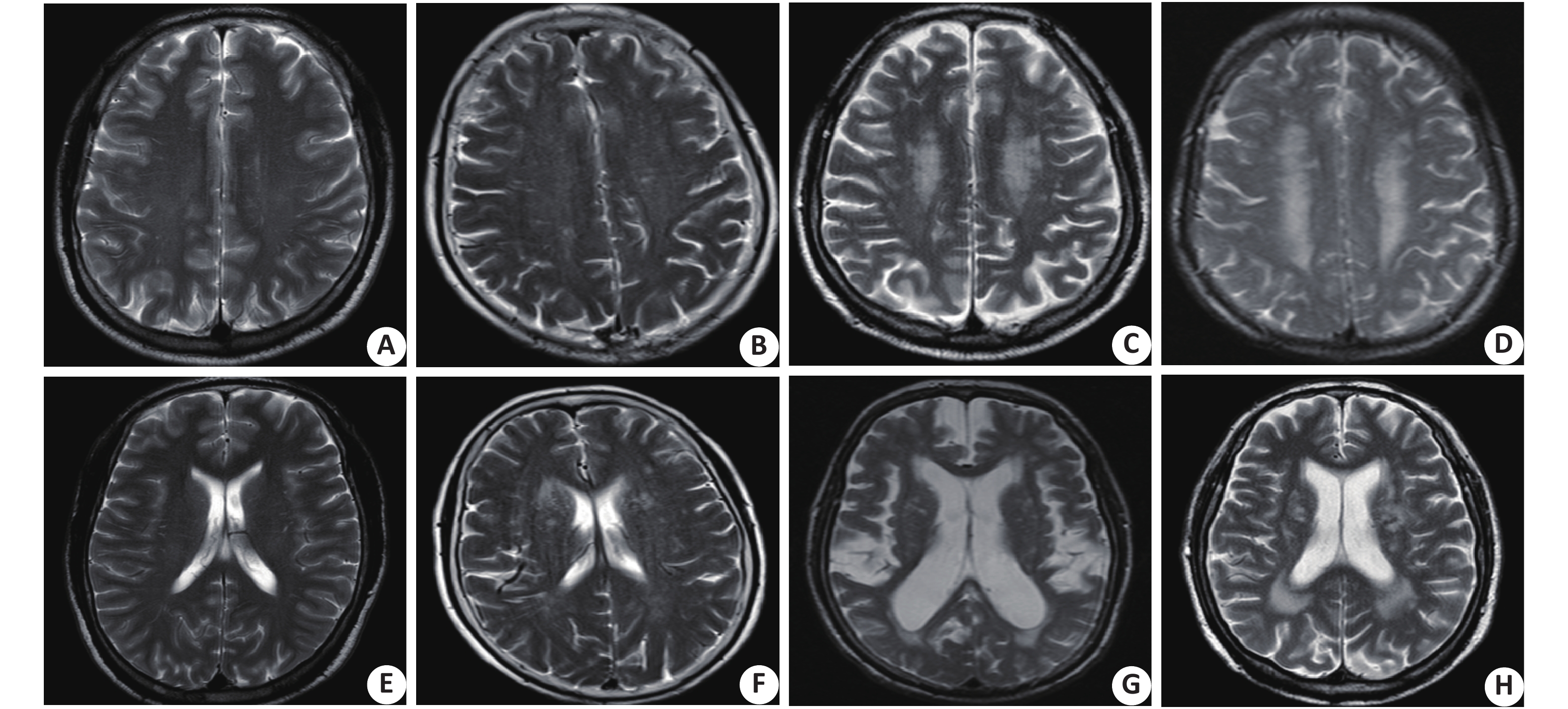
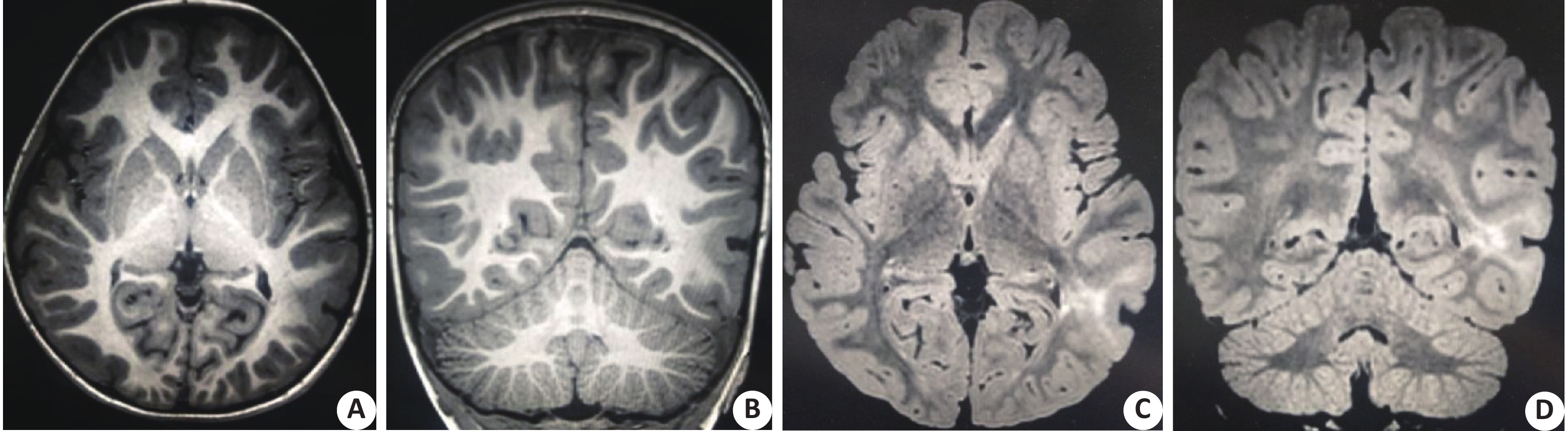
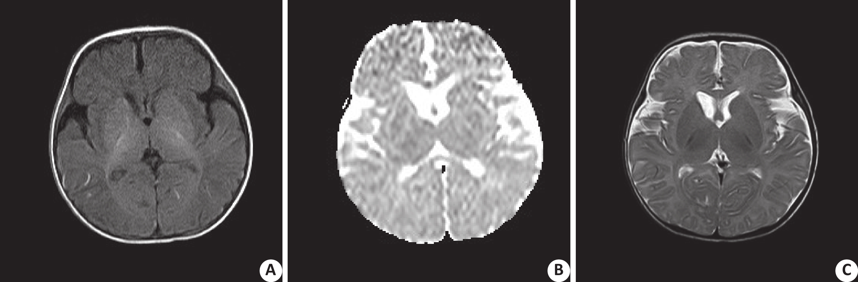
ObjectiveTo explore simplex focal cortical dysplasia (FCD) imaging characteristics with children by 3D high resolution magnetic resonance imaging. MethodsWe retrospectively analyzed MRI data of 57 children (33 males, 24 females, 6 months to 11.5 years old ) with simple FCD confirmed by pathology from March 2016 to October 2018 in Qilu Children's Hospital of Shandong University. The main MRI positive signs and the frequency of various signs in different types of FCD and subtypes were analyzed. The differences in the detection rates of various signs in different types of FCD and subtypes were compared. ResultsThe main MRI signs of type Ⅰ FCD were focal gray-white matter blurring (mild) (n=9, 39.13%), focal cortex thinning (n=9, 39.13%), segmental cerebral lobe atrophy, and local white matter volume reduction (n=8, 34.78%). The detection rate of the three signs was significantly higher than that of FCD Ⅱ(P<0.05). The main MRI signs of type Ⅱ were blurred gray matter boundary (obvious) (n=29, 85.29%), focal cortex thickening (n=25, 73.53%), transmantle sign (n=16, 47.06%), abnormal signal in gray-white matter(n=17, 50.00%; n=24 cases, 70.59%), abnormal sulci and morphology (n=13, 38.24%). The detection rate of the above signs was significantly higher than that of children with type I(P<0.05). The detection rate of focal gray-white matter in FCDⅡb type was blurred (more obvious). The focal cortical thickening, transmantle sign (like a cone signal extending from the subcortex to the ventricle), and abnormal white matter signal detection rates(100.00%, 95.00%, 80.00%, 90.00% respectively)were significantly higher than that in FCD Ⅱa (P < 0.05). Ther difference in the detection rate of MRI signs of type FCD Ⅰwas not significant (P>0.05). ConclusionHigh-resolution imaging could determine the type of FCD before surgery. It has a discriminating value for each subtype of FCD Ⅱ, which is significant in guiding the formulation of its preoperative plan.
2020, 43(1): 103-107.
doi: 10.12122/j.issn.1674-4500.2020.01.21
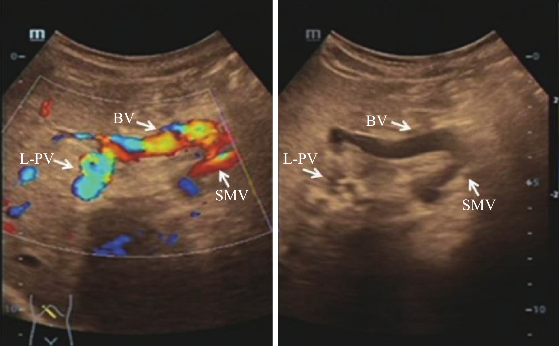
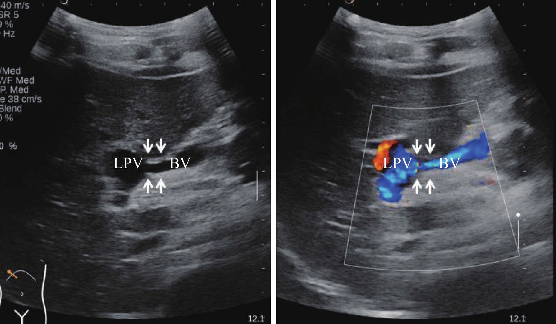
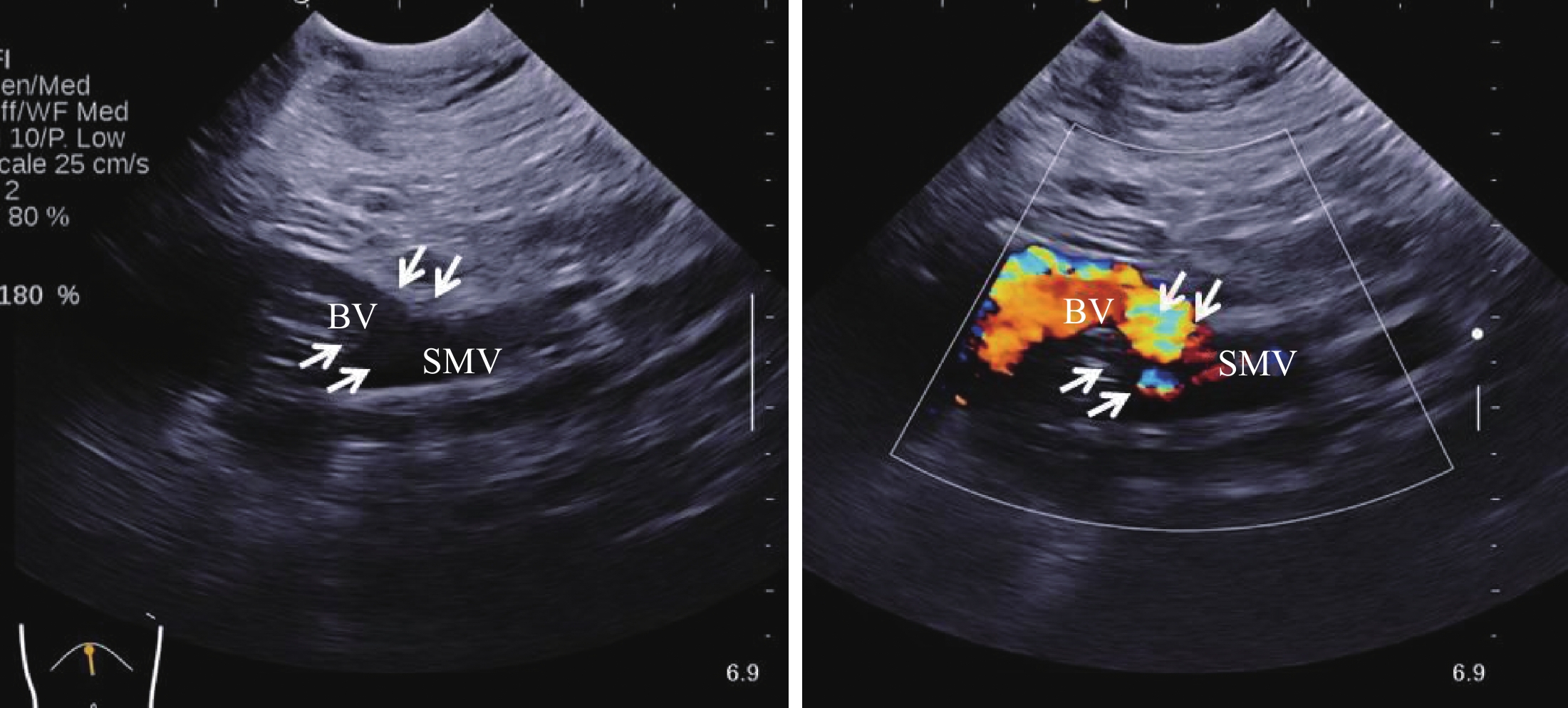
Abstract:
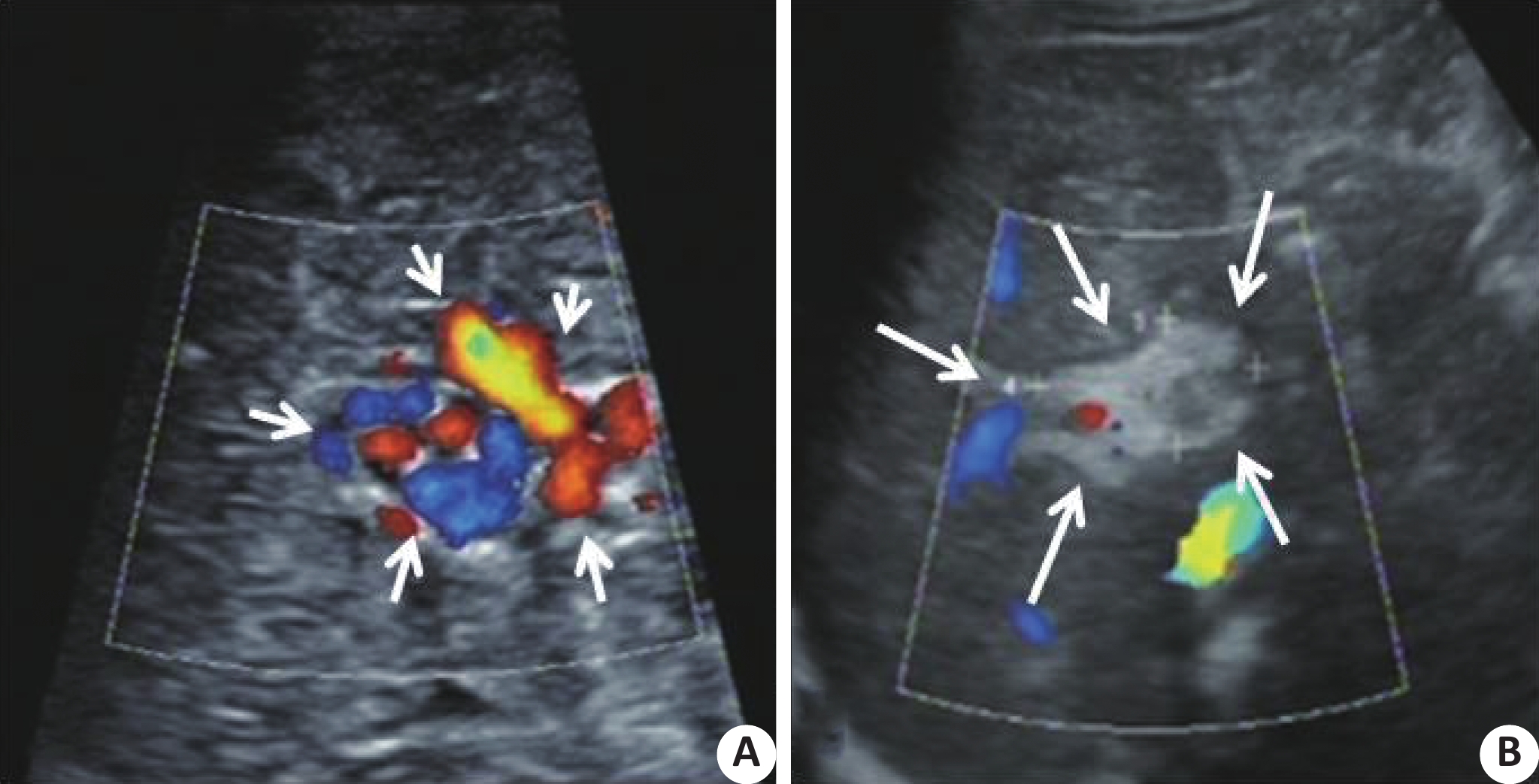
ObjectiveTo evaluate the value of ultrasonography in evaluating the efficacy of left portal vein shunt (Rex shunt) in children with cavernous transformation of portal vein combined with portal hypertension. MethodsFrom October 2014 to January 2016, 10 children underwent Rex shunt surgery due to cavernous transformation of portal vein combined with portal hypertension in our hospital were selected. The diameters of the liver and spleen were measured by two-dimensional ultrasound before and after surgery, and the size changes were calculated. The left portal vein, postoperative bypass vein and anastomotic diameter before and after surgery were measured. Doppler ultrasound was used to evaluate the flow patency of the left portal vein, bypass vein and anastomotic stoma. ResultsThe blood flow signal in the area of cavernous transformation of portal vein after Rex shunt surgery was significantly decreased in 10 cases. The oblique diameter of the right lobe of the liver, the upper and lower diameter of the left lobe of the liver, and the area of the spleen were 98.5±3.5 mm, 53.3±2.7 mm, and 42.9±5.9 cm2 before the operation, respectively. The oblique diameter of the right lobe of the liver (109.3±1.9 mm) one week after the operation and the oblique diameter of the right lobe of the liver (108.7±1.0 mm), upper and lower diameter of the left lobe (64.0±2.5 mm), and the area of the spleen (28.5±3.6 cm2) one year after the operation had significant statistical significance compared with that before the operation (P < 0.05). After the operation, the anastomotic stoma of the left portal vein and the left portal vein was clearly displayed by ultrasound, the display rate was 100%, and the display rate of the anastomotic stoma of the superior mesenteric vein was 70%. The diameter of the left portal vein before operation was 2.58±0.34 mm, and the diameter of the left portal vein after operation was 5.33±0.61 mm (6 months after operation) and 6.90±0.95 mm (1 year after operation), both of which were statistically significant (P < 0.05). At 1 week, 3 months, 6 months and 1 year after surgery, the diameter of the relevant vessels was as follows: bypass vein: 5.96±0.80 mm, 6.90±0.68 mm, 7.41±0.56 mm; Anastomosis of the left portal vein: 2.77±0.37 mm, 2.71±0.36 mm, 3.53±0.32 mm; Anastomosis of the superior mesenteric vein: 3.26±0.16 mm、3.40±0.17 mm、3.63±0.11 mm. ConclusionUltrasound can evaluate the size of liver and spleen, left portal vein before and after Rex shunt surgery, postoperative bypass vein and anastomotic diameter, and postoperative blood flow status of the left portal vein, bypass vein and anastomotic stoma, providing reliable indicators for the evaluation of surgical effect.
2020, 43(1): 108-111.
doi: 10.12122/j.issn.1674-4500.2020.01.22
Abstract:
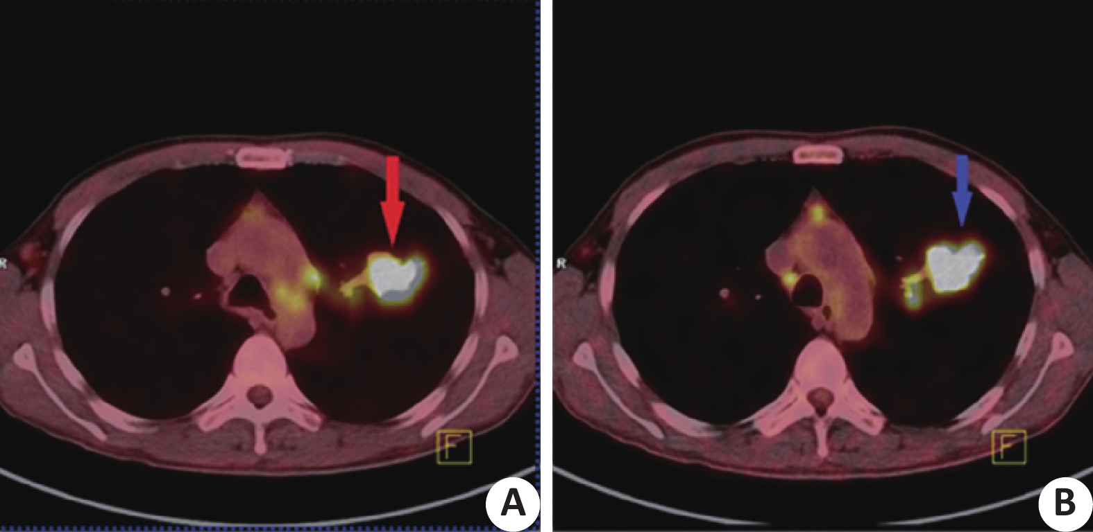
objectiveTo compare the effects of breath-holding scanning with ultra HD reconstruction in improving pulmonary PET/CT detection of respiratory motion artifacts. MethodsThe inclusion criteria were established: clear space-occupying lesions could be seen in the CT images of the patients, the PET had a matching high uptake focus, and the patients held their breath well. According to the above inclusion conditions, 60 patients examined in PET/CT center from 2019.1-2019.6 were selected. the images obtained by routine collection were used as the control group, breath-holding scanning of single bed lung with ultra HD weighted images was used as the observation group. compared with Image fusion quality, average Standard Uptake Value, 40% of Metabolic Tumor Volume, target(high uptake foci) background(SUVMax of hepatic blood pool) ratio (T/BMax)between the control group and the observation group, try to explaination the results based on the imaging principle. ResultsIn the control group, 17 cases had good fusion, accounting for 28.33%. In the observation group, there were 58 cases with good fusion, accounting for 96.67%. The SUVavg observation group was 8.28±2.45 and the control group was 6.84±2.58, which were significantly higher than the control group (χ2= 10.50, P<0.05). MTV40% was 5.61±4.40 in the observation group and 7.70±5.39 in the control group, which were significantly lower than the control group (χ2=5.37, P<0.05), 6.29±2.39 in the T/Bmax observation group and 4.87±1.78 in the control group, which were significantly higher than the control group (χ2=13.71, P<0.05). ConclusionHold-breath scanning with ultra HD reconstruction make imagings has a better fusion; SUVavg, MTV40%, T/B max measurements were less affected by partial volume effect and moving boundary expansion effect. In addition, the application of more accurate PSF in the reconstruction process makes the quantitative indicators more accurate, It is worthy to use PET/CT for reference in clinical PET/CT collection of patients with pulmonary diseases.
2020, 43(1): 112-116.
doi: 10.12122/j.issn.1674-4500.2020.01.23
Abstract:
ObjectiveTo evaluate the value of ultrasound-guided thoracic paravertebral nerve block in thoracotomy for patients with pulmonary tuberculosis. MethodsNinety cases of tuberculosis patients with ASA grade I ~ II, aged 18 ~ 60, who underwent elective thoracotomy were randomly divided into the group of general anesthesia alone (group G), the group of general anesthesia combined with ultrasound-guided thoracic paravertebral nerve block (group P) and the group of general anesthesia combined with epidural block (group E), with 30 cases in each group.Patients in group P were subjected to single paravertebral nerve block under ultrasound guidance before anesthesia induction. The patients in group E were subjected to anaesthesia induction with anterior thoracic epidural puncture and indwelling epidural catheter.All the patients in 3 groups were treated with intravenous analgesia by static aspiration combined with general anesthesia. We recorded MAP and HR of patients entering the operating room (T0), before induction of intubation (T1), before skin cutting (T2), 5 min after skin cutting (T3), after extubation (T4) and 2 h after surgery (T5). Intraoperative sufentanil dosage, operative time and number of dopamine users were recorded. VAS score and the number of compressions of the analgesic pump were recorded at 2, 6, 12, 24, 48, 72 h after the operation. ResultsCompared with group G, MAP of group P decreased at time points T3 and T4 (P<0.05), and HR decreased at time points T3, T4 and T5 (P<0.05). MAP and HR in group E decreased at time points T1, T2, T3, T4 and T5 compared with that in group G and group P (P<0.05). The intraoperative sufentanil dosage in group P and group E were lower than that in group G (P<0.05).The number of dopamine users in group E was more than that in group G and group P (P<0.05), while the number of dopamine users in group P was more than that in group G (P<0.05). In the quiet and cough condition, the score of group P was lower than that of group G at 2, 6 and 12 h after surgery (P<0.05). The score of group E was lower than that of group G at 2, 6 h after surgery (P<0.05). The number of postoperative analgesic pump presses in group P and group E were less than that in group G (P<0.05). ConclusionUltrasound-guided thoracic paravertebral nerve block operation has a high success rate, definite analgesic effect and stable perioperative hemodynamics. It can reduce opioid dosage during thoracotomy in patients with tuberculosis and enhance the early postoperative analgesic effect. It can be safely and effectively applied to thoracotomy anesthesia in patients with tuberculosis.
2020, 43(1): 117-121.
doi: 10.12122/j.issn.1674-4500.2020.01.24
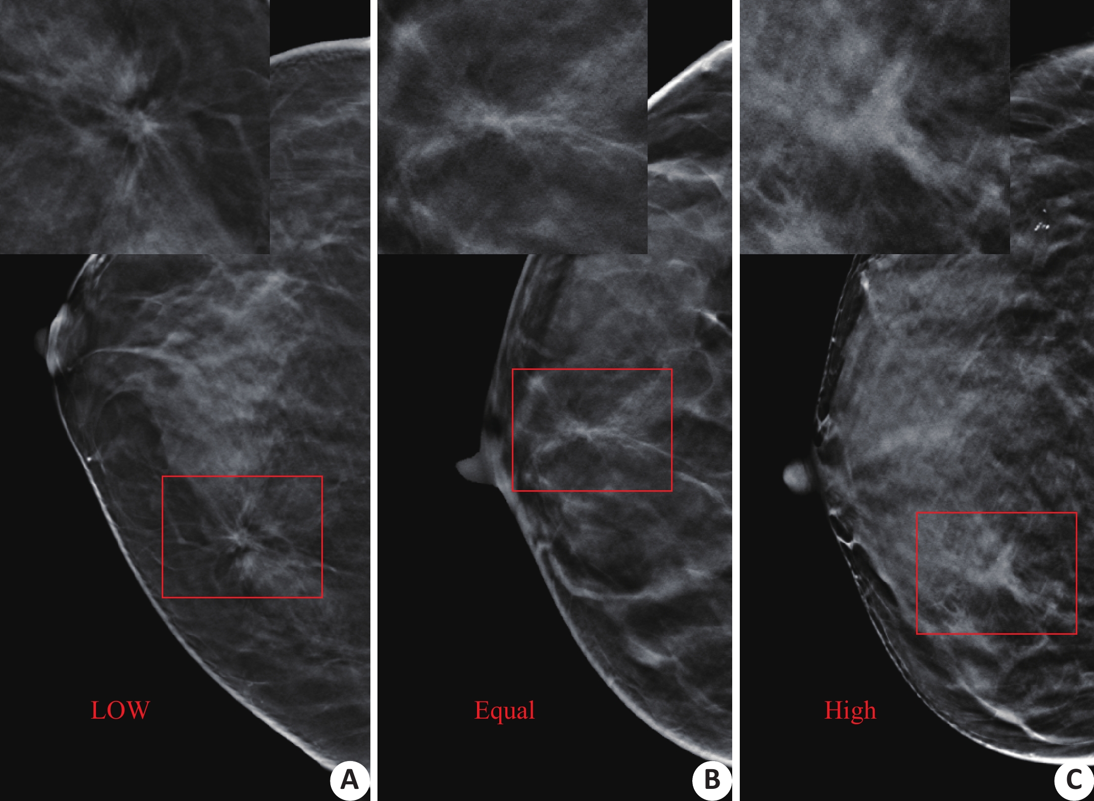
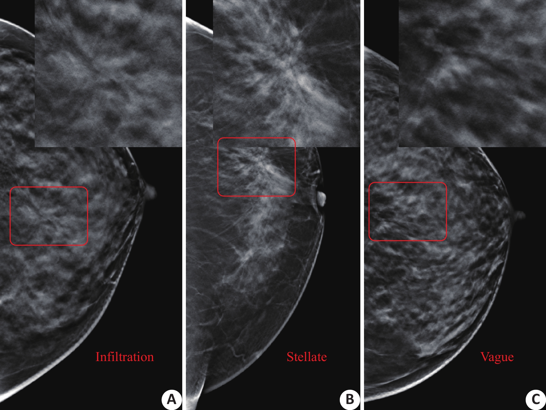
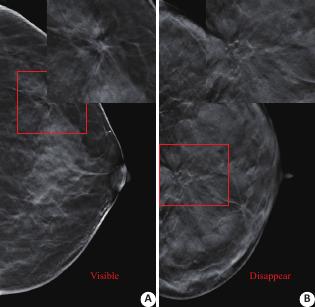
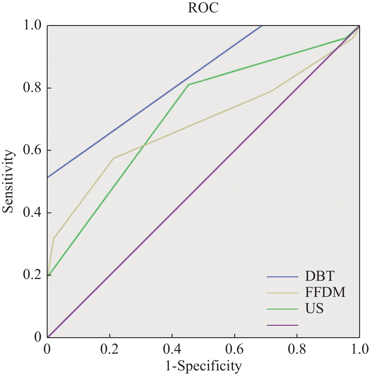
Abstract:
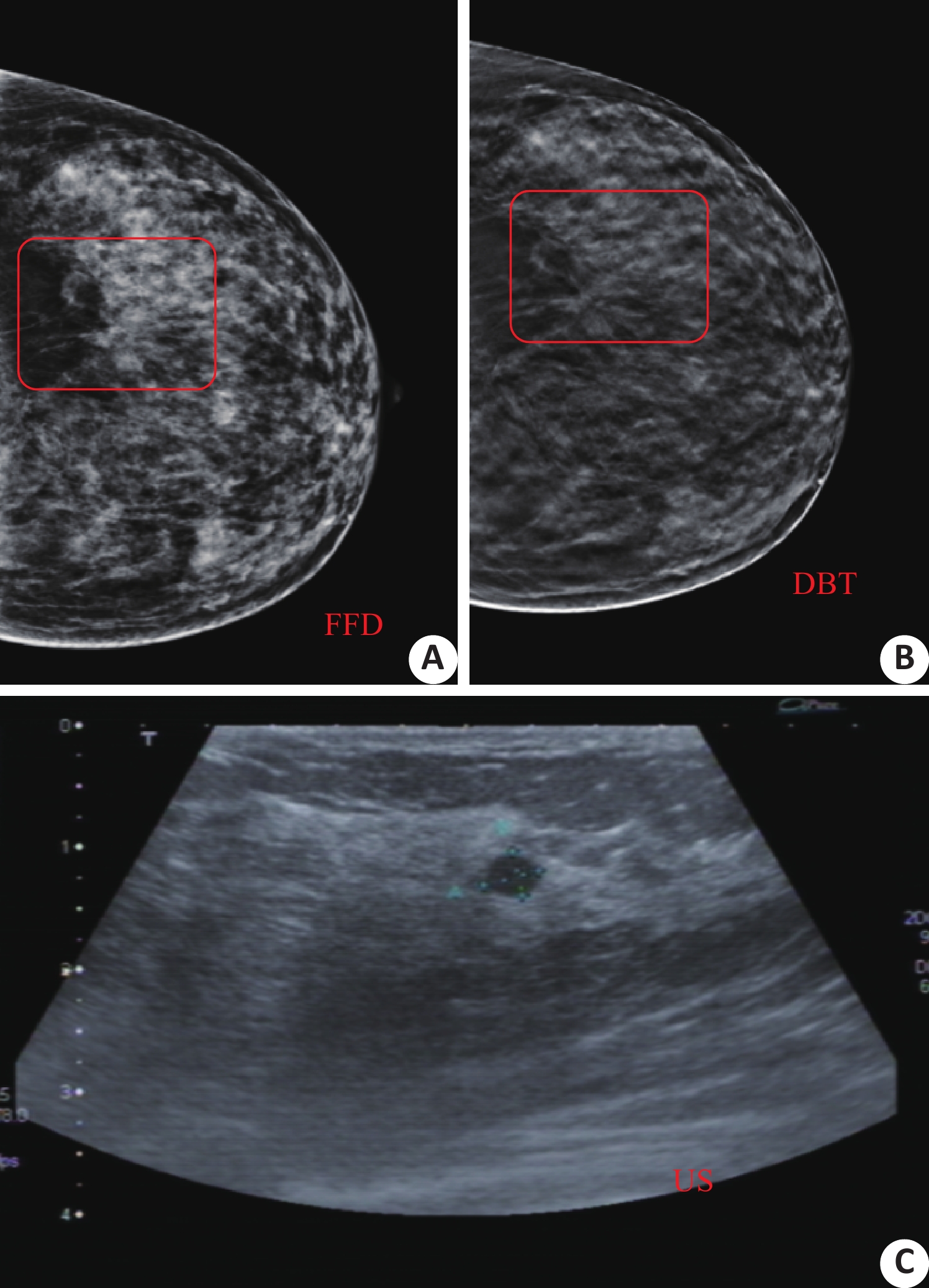
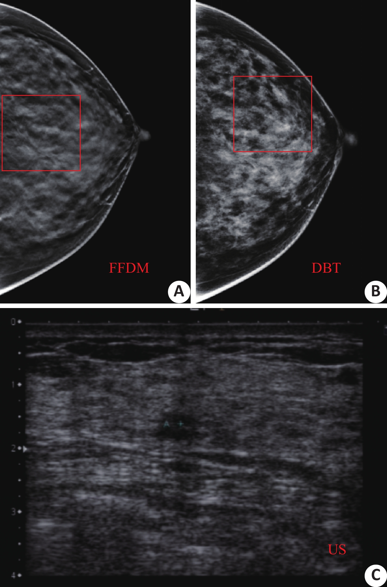
ObjectiveTo study the comparative study of mammography (full digital mammography FFDM, digital mammography DBT) and ultrasonography on benign and malignant structural distortion and its X-ray sign analysis. MethodsCollecting 51 patients, with the age from 20 to 70 years old (average 44.84±8.738) , who underwent FFDM, DBT and color Doppler ultrasound examinations from June 2013 to December 2018 and found signs of structural distortion and pathological findings. According to the mammography report and data system, the diagnostic diagnosis and the analysis of the signs of structural distortion of the three imaging methods were compared. ResultsFisher's precise algorithm was used to analyze the X-ray signs of structural distortion, which had significant difference between benign and malignant (P<0.05). The diagnostic efficiency of three methods in breast diseases was analyzed by ROC curve. The study found that the area under the curve were greater than 0.5, is a valuable diagnostic sensitivity. ConclusionCompared with FFDM,and US, DBT can better observe the structural distortion and improve the diagnostic efficacy.
2020, 43(1): 122-125.
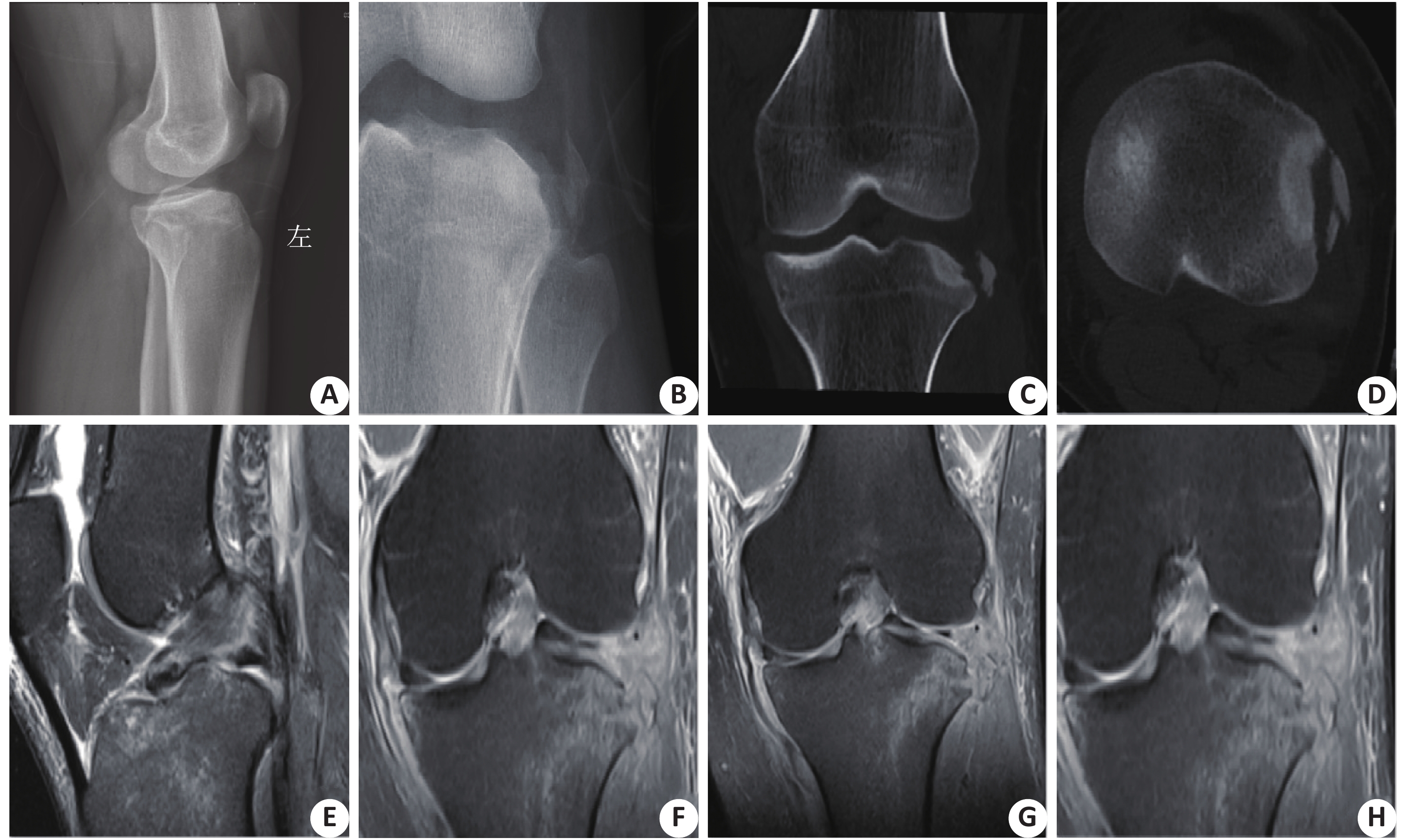
doi: 10.12122/j.issn.1674-4500.2020.01.25
Abstract:
ObjectiveTo explore the diagnostic value of X-ray and CT combined with MRI in tibial plateau fracture. MethodsThe clinical data of 144 patients (144 sides) with tibial plateau fractures in our hospital from March 2017 to March 2019 were retrospectively analyzed, including 80 males and 64 females with the age from 20 to 78 years old (average 52.35±10.49). All patients were examined by CT and MRI after routine X-ray examination. The coincidence rate of X-ray diagnosis, X-ray combined with CT and three combined diagnoses and the coincidence rate of fracture classification diagnosis were compared. The role of imaging diagnosis methods in soft tissue diagnosis was discussed. ResultsThe difference of the coincidence rate between X-ray+CT and MRI in the diagnosis of tibial plateau fractures was not significant (P>0.05). But the coincidence rate was significantly higher than that of X-ray alone (P<0.05). The difference of the diagnosis of tibial plateau fractures between the two methods and the three methods was not significant (P>0.05). The coincidence rate of the combination of the two and the three in the diagnosis of type I, type II and type III fractures was higher than that of the X-ray diagnosis alone(P<0.05). There were 60 cases of soft tissue injury diagnosed by MRI, but no diagnosis was made by X-ray and CT. ConclusionThe coincidence rate of X-ray combined with CT in the diagnosis and classification of tibial plateau fracture is high, but further combined with MRI examination can make a judgment of soft tissue injury.
2020, 43(1): 126-129.
doi: 10.12122/j.issn.1674-4500.2020.01.26
Abstract:
ObjectiveTo explore the value of dual-source CT in the evaluation of central neurocytoma. MethodsA total of 110 cases of primary neuronal tumor treated in our hospital from March 2017 to February 2019 were examined by dual source CT and conventional plain scan. There were 59 males and 51 females with the age from 27 to 81 (average 51.34±4.71) included. ResultsThe coincidence rate of double source CT images was significantly higher than that of conventional plain scan (P<0.05). The detection rate of calcified foci by dual source CT was significantly higher than that of conventional plain scan (P<0.05). The difference between the CT value of the virtual plain scan image and the CT value of the conventional plain scan image was not significant (P>0.05). The results of the two methods were consistent (Cronbach α=0.91). The detection rate of double source CT was significantly higher than that of control group (P<0.05). ConclusionCompared with conventional plain scan, dual source CT can improve the detection rate and image quality of neuroma.
2020, 43(1): 130-133.
doi: 10.12122/j.issn.1674-4500.2020.01.27
Abstract:
ObjectiveTo evaluate the clinical value of 3D ASL in the assessment of collateral circulation after unilateral internal carotid artery (ICA) occlusion. MethodsFrom January 2018 to February 2020, 22 patients with unilateral ICA occlusion and no other moderate or severe stenosis of intracranial artery were studied by 3D-TOF MRA In ASL sequence perfusion imaging, including 13 males and 9 females with the age from 35 to 76 (average 52.2±15.5). The functool software was used to automatically generate the pseudocolor image of cerebral blood flow (CBF) from the original data. The areas of interest (ROI=200±20 mm2) were selected from the blood supply area of ICA on the occluded side and the frontal lobe, parietal lobe, paraventricular white matter area and basal ganglia area of the mirror image area. The CBF values of the time delay (PLD) between the responsible ICA blood supply area and the mirror image area were compared. ResultsThere were 12 cases of ICA occlusion on the left side and 10 cases of ICA occlusion on the right side. The CBF value in the blood supply area of ICA occlusion was significantly lower than that in the mirror area in 3D ASL (PLD: 1 525 ms). The difference between the two groups was significant (P<0.05). When PLD was 2 525 ms, the CBF value in the blood supply area of ICA occlusion was slightly lower than that in the mirror area, but the difference was not significant (P>0.05). Conclusion3D ASL imaging technology can be used to evaluate the establishment of collateral circulation and perfusion state after unilateral ICA occlusion, which is of great value for the selection of treatment plan and the prediction of clinical prognosis.

2020, 43(1): 134-139.
doi: 10.12122/j.issn.1674-4500.2020.01.28
Abstract:
ObjectiveTo investigate the clinical effect and safety of dexmedetomidine combined with sufentanil and propofol in painless gastroscopy for obese patients. MethodsA total of 136 obese patients who were to undergo painless gastroscopy were randomly divided into group D (with dexmedetomidine combined with sufentanil and propofol as the anesthetic regimen) and group C (with sufentanil combined with propofol as the anesthetic regimen), with 68 cases each. There were 37 males and 31 females with an average age of 44.6±8.5 in observation group, 39 males and 29 females with an average age of 43.2±6.8 in control group. The anesthetic effect, induction time, examination time, waking time, directional force recovery time and propofol dosage were compared between the two groups. The changes of mean arterial pressure (MAP), heart rate (HR) and respiratory rate (RR) in two groups were observed before the induction of anesthesia (T0), at the time of endoscopic induction (T1), from the endoscopic body to the level of epiglottis (T2), at the end of operation (T3), and 5 min after operation (T4). The stress indicators such as cortisol (Cor) and norepinephrine (NA) at T0 and T4 were measured. The perioperative adverse reactions were recorded. The satisfaction of endoscopists, anesthesiologists and patients were investigated. ResultsThe excellent and good rate of anesthesia in group D was higher than that in group C (92.65% vs 72.06%, P<0.05).The induction time, awakening time and directional force recovery time of group D were shorter than that of group C (P<0.05), and the dosage of propofol was lower than that of group C (P<0.05).The changes of MAP, HR and RR in group C at T1, T2 and T3 were significantly greater than those in group D (P<0.05). Compared with T0, Cor and NA levels in the two groups increased in T4, but the degree of increase in group D was lower than that in group C (P<0.05). The incidences of body movement, cough, respiratory depression and hypotension in group D were lower than that in group C (P<0.05).The satisfaction of endoscopists, anesthesiologists and patients in group D was higher than that in group C. ConclusionFor painless gastroscopy in obese patients, the dexmedetomidine combined with sufentanil and propofol have good anesthetic effect, stable intraoperative vital signs and few adverse reactions.
2020, 43(1): 140-143.
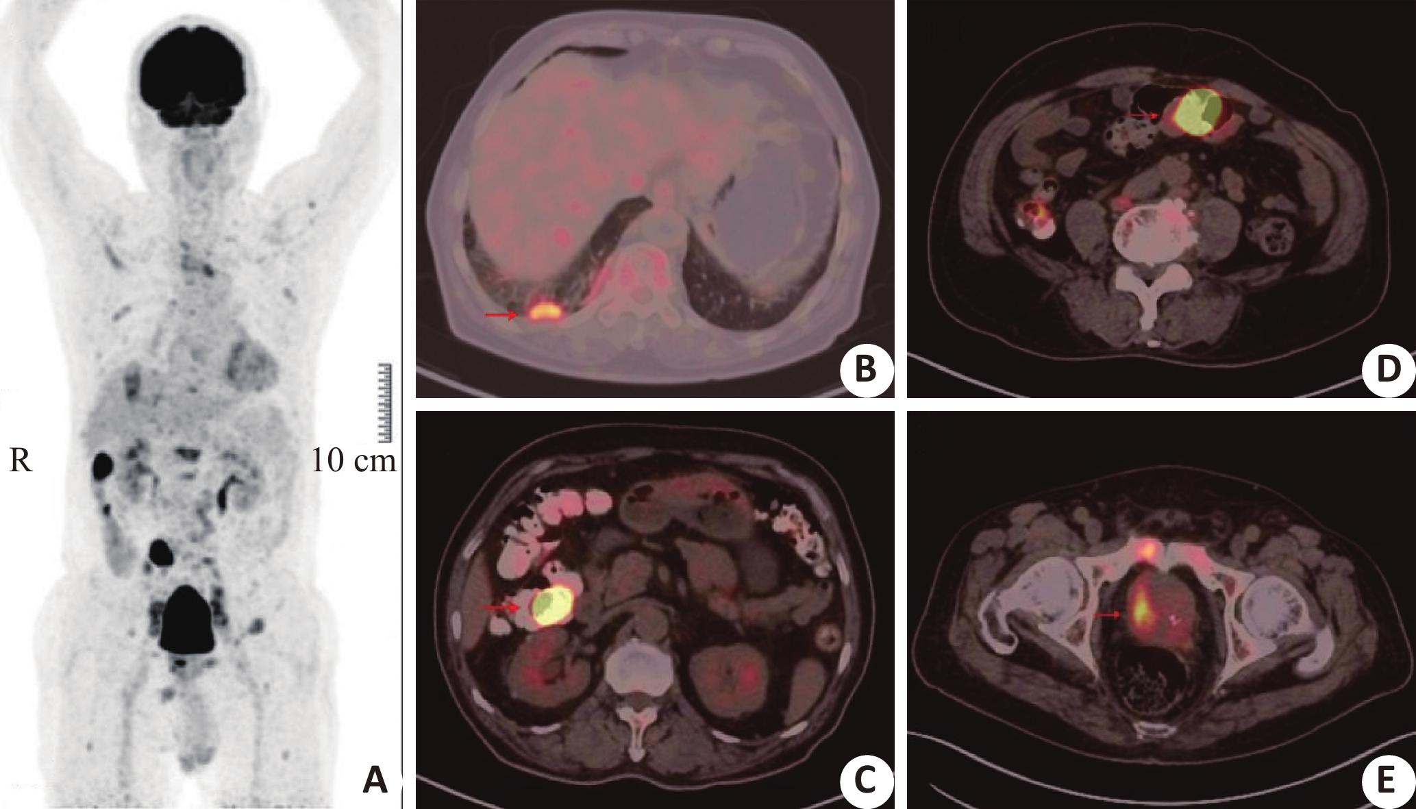
doi: 10.12122/j.issn.1674-4500.2020.01.29
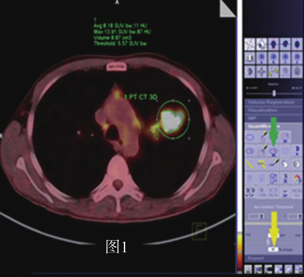
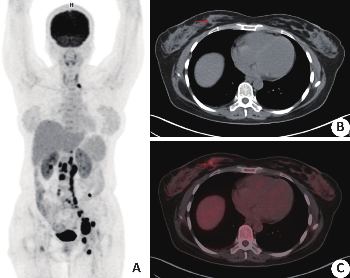
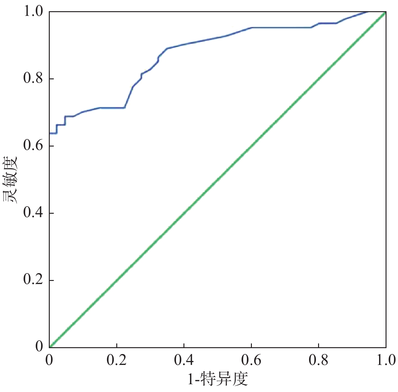
Abstract:
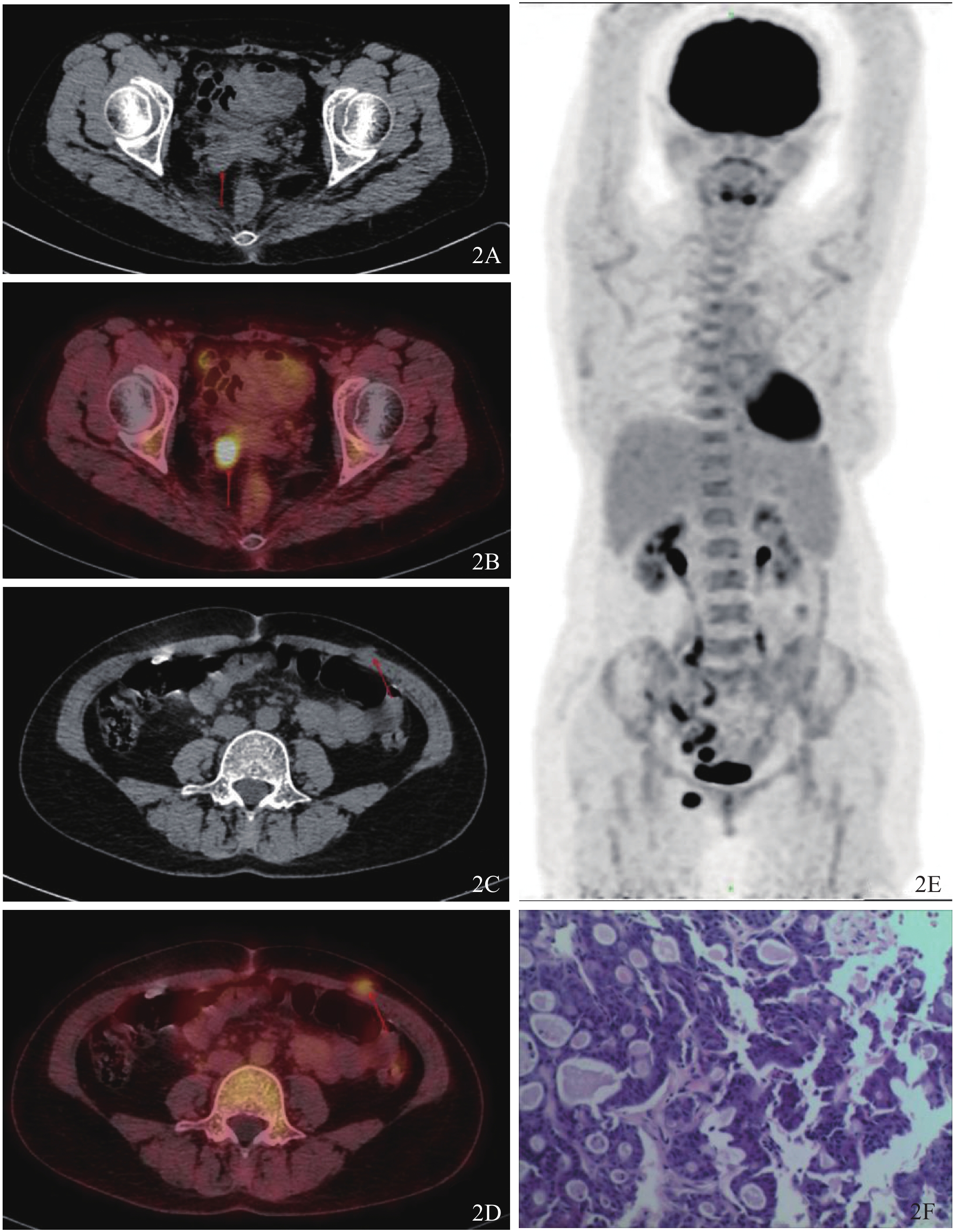
ObjectiveTo investigate the diagnosis value of 18F-FDG PET/CT in patients with carcinoma of unknown primary (CUP). MethodsA total of 121 patients(79 males,42 females, age range 30-86 years) with a diagnosis of CUP who underwent whole body 18F-FDG PET/CT imaging were included in this retrospective study from March 2018 to August 2019. The final diagnoses were confirmed either histopathologically or by clinical follow-up. ResultsThe 18F-FDG-PET/CT successfully detected the primary tumor in 59 out of 121 (49%) patients. The most common primary tumor as detected by 18F-FDG PET/CT was lung cancer (n=31). In a patient, two primary tumors (colon and prostate) were detected on PET/CT imaging. Lung biopsy revealed prostate cancer in this patient and the colon cancer was accepted as a synchronous second primary tumor. 18F-FDG PET/CT findings were false-positive in 11 patients. 18F-FDG PET/CT could not detect any primary lesion in 51 patients, whose conventional work-up detected a primary tumor in 11 and thus considered as false-negative. The sensitivity, specificity rate and accuracy of 18F-FDG PET/CT in detection of primary tumor were identified as 84%, 78% and 82%, respectively. Conclusion18F-FDG PET/CT of total body is an effective method for detecting the primary tumor in patients with CUP. In addition to detect the primary tumor, it can help determine disease extent and contribute to patient management.
2020, 43(1): 144-148.
doi: 10.12122/j.issn.1674-4500.2020.01.30
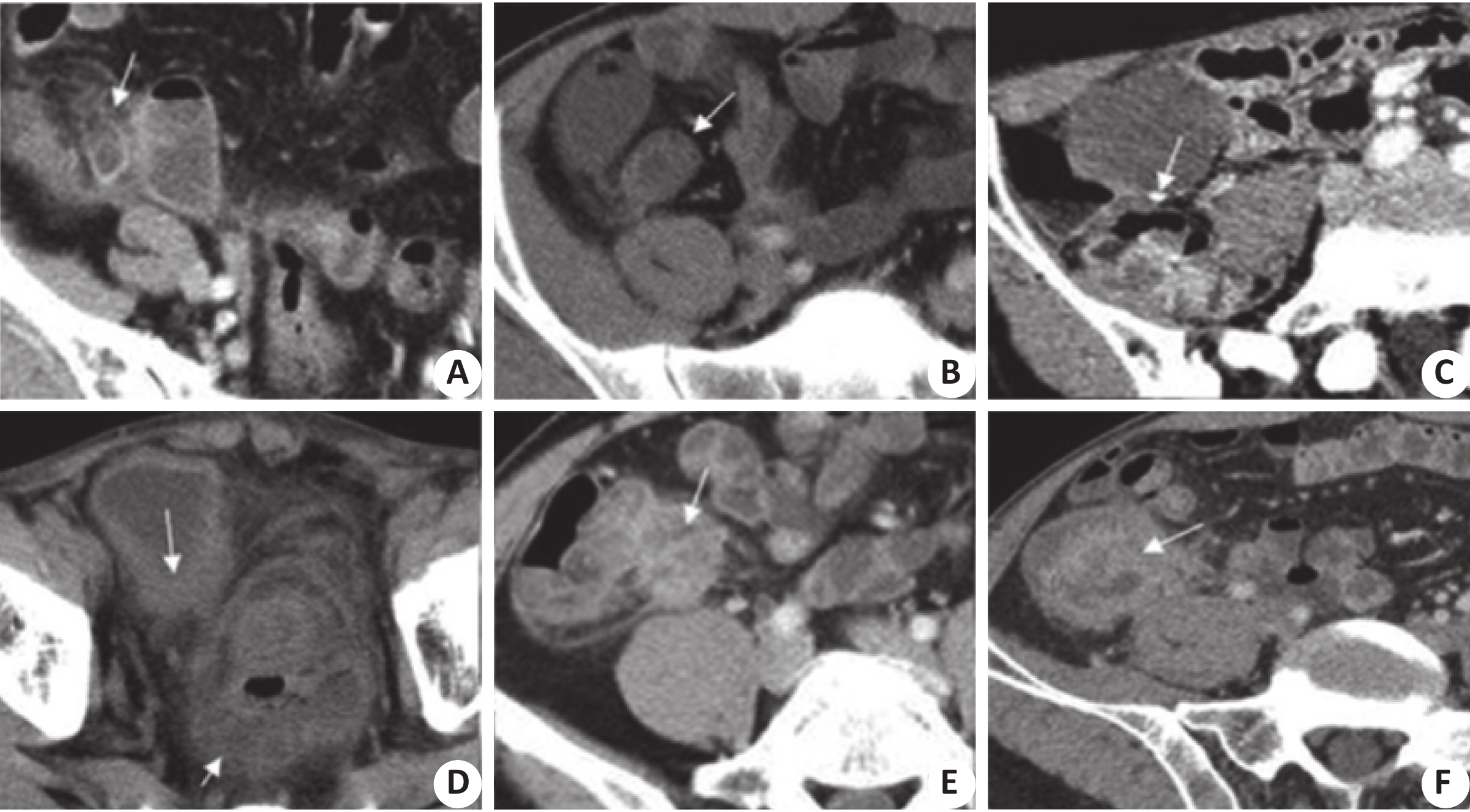
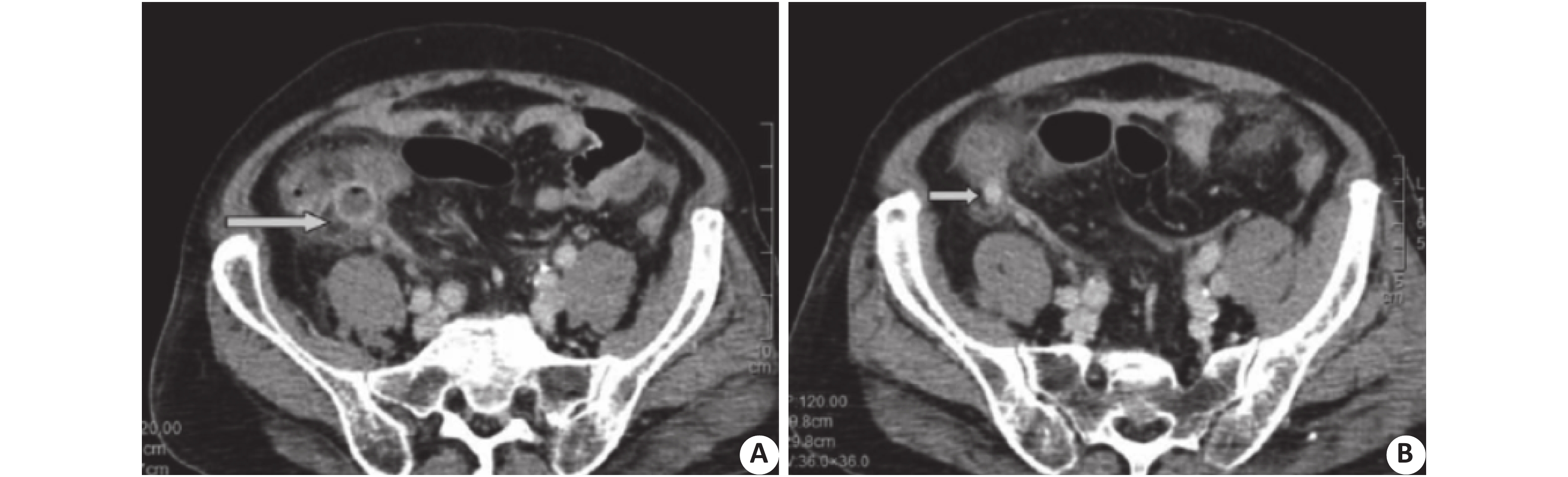
Abstract:
ObjectiveTo explore the CT differential diagnosis of mucinous cystadenoma and carcinoid of appendix. MethodsWe choosed 50 cases of patients with appendiceal mucous cystadenoma and 20 cases of patients with carcinoid. There were 32 males and 38 females with an average age of 57.15±5.28 years old. We analyzed image of patients appendix appendiceal wall thickness, diameter, calcification, infringement, appendix around abnormal wall thickening, wall reinforcement, incidence of bezoar and detection rate and accuracy by CT detection imaging system mucous cystadenoma and carcinoid appendix presenting symptoms. The CT detection heterogeneity analysis and related influence factors were analyzed. ResultsThere were significant differences between the CT images of the patients with appendiceal mucinous adenoma and carcinoid in the thickness, diameter, calcification, peripheral invasion, wall thickening, abnormal wall strengthening, fecality incidence, detection rate and accuracy (P<0.05). The patients in the carcinoid group had more significant symptoms than those in the cystadenoma group. The sensitivity of the carcinoid group, specific degrees, masculine, feminine and diagnostic odds ratio was the largest. The course, age and disease range and CT evaluation carcinoid appendix mucous cystadenoma, and diagnostic value of the highest correlation, and the capsule gland tumors had product and evaluation of CT carcinoid appendix mucous cystadenoma and diagnostic value of correlation were low. ConclusionCT has a high value in the differential diagnosis of appendiceal mucinous cystadenoma and carcinoid.
2020, 43(1): 149-152.
doi: 10.12122/j.issn.1674-4500.2020.01.31
Abstract:
ObjectiveTo study the level change of CA-125 in preeclampsia and the occurrence of reversible leukoencephalopathy syndrome. Methods40 pregnant women with mild preeclampsia, 40 pregnant women with severe preeclampsia and 40 healthy pregnant women admitted to our hospital from January 2018 to December 2019 were selected as subjects, then compare the levels of CA-125 in peripheral blood of pregnant women in each group and evaluate the value of CA-125 in predicting the onset of preeclampsia. The characteristics of brain MRI in all patients were analyzed and the relationship between CA-125 level and the incidence of reversible leukoencephalopathy syndrome in preeclampsia pregnant women was compared. ResultsThe level of CA-125 in peripheral blood of pregnant women in the three groups was higher in the severe preeclampsia group (69.25±25.70 U/mL) than in the mild preeclampsia group (44.40±20.69 U/mL), and lowest in the healthy group (22.58±9.32 U/mL). There was a significant difference between the three groups (F=55.684, P<0.001). Use peripheral blood CA-125 to predict the onset of preeclampsia, and the AUC was 0.873 (95% CI: 0.812−0.934, P<0.05); A total of 7 out of 80 pregnant women developed reversible leukoencephalopathy syndrome, including 6 women with severe preeclampsia and 1 woman with mild preeclampsia. The incidence of reversible leukoencephalopathy syndrome in pregnant women was significantly different between the high-level group of CA-125 and the low-level group of CA-125 (χ2=9.774, P<0.05). ConclusionThe level of CA-125 is significantly higher in pregnant women with preeclampsia, and the more severe the condition, the higher the level. CA-125 can be used as a predictor of the onset of preeclampsia, and the high level of CA-125 increases the incidence of reversible leukoencephalopathy syndrome.
2020, 43(1): 153-156.
doi: 10.12122/j.issn.1674-4500.2020.01.32
Abstract:
ObjectiveTo explore the effect of psychological intervention on preoperative anxiety in patients with percutaneous transforaminal endoscopic discectomy (PTED). MethodsThe clinical data of patients with preoperative anxiety score and PTED under local anesthesia between January 2016 and December 2018 were analyzed retrospectively. Patients who met the inclusion criteria were divided into control group and intervention group according to whether they received preoperative psychological intervention. Different time points of APAIS score, VAS score, systolic blood pressure (SBP), amount of bleeding and operation time were compared between the two groups. The influence of psychological intervention on preoperative anxiety and operation related parameters was analyzed. ResultsOne hundred and twenty-eight patients were included, 69 in the control group and 59 in the intervention group. The APAIS score of the intervention group at waiting room (11.83 ± 1.49) was significantly lower than that at admission (14.12 ± 1.83, P=0.000). There was no significant difference in APAIS score of the control group between at waiting room(13.77 ± 1.59) and the admission (14.22 ± 2.03,P = 0.150). The APAIS score of the intervention group at waiting room (11.83 ± 1.49) was lower than that of the control group (13.77 ± 1.59), the difference was significant (P=0.000). The SBP at waiting room, intraoperative SBP, amount of bleeding and operation time of the control group were higher than those of the intervention group (P<0.005). There was no significant difference in postoperative VAS score, extra analgesic between the two groups (P>0.05). ConclusionPsychological intervention can relieve preoperative anxiety, reduce the amount of bleeding, and shorten the operation time.
2020, 43(1): 157-161.
doi: 10.12122/j.issn.1674-4500.2020.01.33
Abstract:
ObjectiveTo investigate the clinical value of serum human epididymis protein 4 (HE4), serum carbohydrate antigen 125(CA125), and β-Human chorionic gonadotropin(β-HCG)levels in combination to predict the efficacy of laparoscopic single-site surgery in the treatment of benign ovarian tumor. MethodsFrom January 2017 to March 2019, 120 cases of benign ovarian tumors were diagnosed after treatment in the department of obstetrics and gynecology of our hospital. The patients were divided into two groups according to different surgical methods. The observation group chosed single-hole laparoscopic surgery for 72 patients, with the age from 21 to 63 years old (average 32.98±9.34) and the tumor diameter from 3.69~9.78 cm (average 5.46±3.65 cm), while the control group chosed traditional multi-site laparoscopic surgery for 48 patients, with the age from 19 to 68 years old (average 33.02±9.35) and the tumor diameter from 3.82 to 9.93 cm (average 5.61±3.68 cm). Serum levels of HE4, CA125 and β-HCG in the two groups were detected, and the effects of the two groups was observed for comparative analysis. ResultsThe operation of the two groups was smooth, without intraoperative or postoperative complications. The difference in intraoperative bleeding and exhaust time between the observation group and the control group was not significant (P>0.05). The operation time of the observation group was longer than that of the control group (P<0.05). The length of time of the observation group was shorter than that of the control group (P<0.05), and the patient satisfaction score was higher than that of the control group (P<0.05). The difference in serum HE4, CA125 and β-HCG between the two groups before surgery was not significant (P>0.05). The postoperative serum HE4, CA125 and β-HCG between the two groups were significantly lower than those before surgery (P<0.05). The levels of HE4, CA125 and β-HCG in the observation group at 12 hours after the operation were significantly lower than those at 1 day and 3 days after the operation (P<0.05). The difference between 1 day and 3 days after the operation was not significant(P>0.05). The differences in the control group at 12 hours, 1 day and 3 days after surgery were significant(P<0.05). ROC analysis showed that the AUC of all indexes were: HE4 (0.901), CA125 (0.820) and β-HCG(0.736), AUC of all indexes was 0.957. ConclusionThe combined detection of serum HE4, CA125 and β-HCG levels can effectively evaluate the clinical efficacy of laparoscopic single-site surgery in the treatment of benign ovarian tumors. It can achieve the expected efficacy and improve patient satisfaction.
2020, 43(1): 162-166.
doi: 10.12122/j.issn.1674-4500.2020.01.34
Abstract:
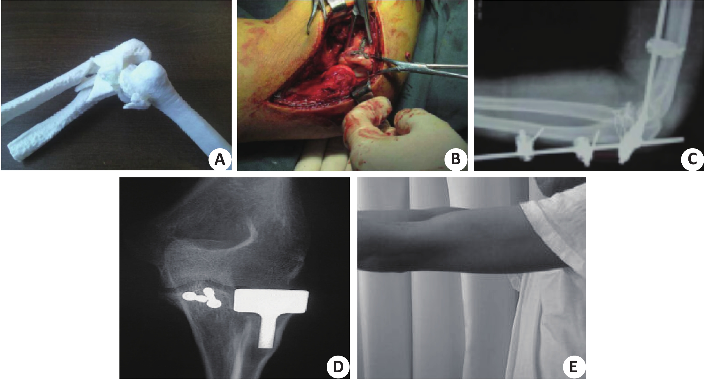
ObjectiveTo evaluate the application effect of three dimensional (3D) printing in the treatment of terrible triad of the elbow. MethodsForty patients with terrible triad of the elbow who were admitted to the hospital between April 2017 and January 2019 were selected as subjects. They were randomly divided into the observation group and the control group, 20 cases in each group. For the control group, preoperative design was completed through X-ray, CT plain scan and 3D reconstruction. The finite element software mimics and 3D printing were applied for the observation group to visualize fracture reduction and internal fixation, by which preoperative design was completed. All the medial and lateral ligament complexes of both groups were repaired with suture rivets. The surgical effect, elbow function (Broberg-Morrev elbow function scores) and pain degree [pain visual analogue scale (VAS) scores] before and after surgery, and incidence of postoperative complications were compared between the two groups. ResultsThe operation time, amount of intraoperative blood loss, frequency of intraoperative fluoroscopy and fracture healing time of the observation group were shorter or less than those of the control group (P<0.05). Broberg-Morrev scores of the observation group at 3 months, 6 months and 9 months after surgery were higher than those of the control group (P<0.05). VAS scores of observation group at 1 month, 3 months and 6 months after surgery were lower than those of the control group (P<0.05). There was no significant difference in incidence rate of complications between the two groups within 9 months after surgery (P>0.05). ConclusionThe application of 3D printing in the treatment of terrible triad of the elbow can improve the surgical effect, promote postoperative recovery of elbow function, and alleviate pain, without significantly increasing the risk of complications.
2020, 43(1): 167-170.
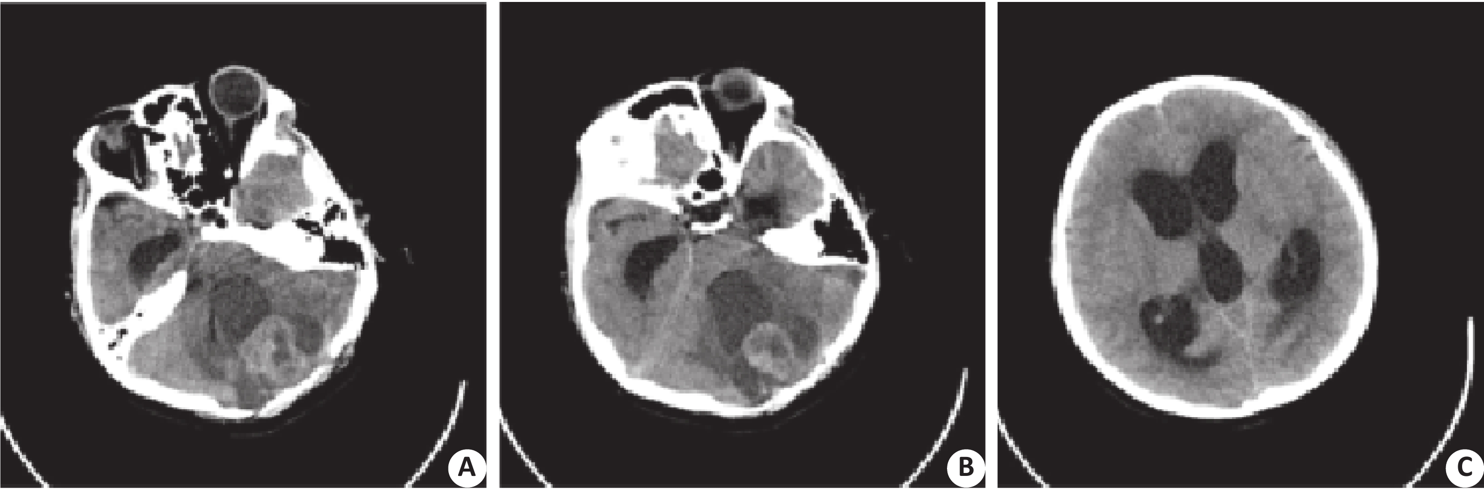
doi: 10.12122/j.issn.1674-4500.2020.01.35
Abstract:
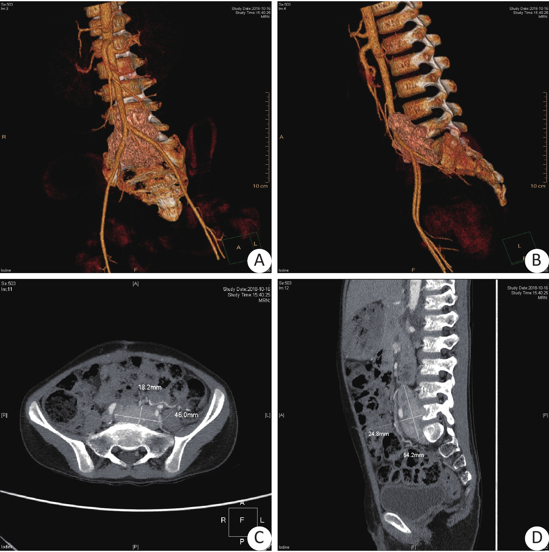
ObjectiveTo analyze the diagnosis and treatment of a child of giant retroperitoneal paraganglioma with metastase, and summarize diagnosis and treatment experience of paraganglioma in children. MethodsWe retrospectivly analyzed the case history, diagnosis, treatment and prognosis of a case of paraganglioma with multiple metastases in urology department of children's hospital affiliated to chongqing medical university. ResultsThe main manifestations of this case were high blood pressure, occasional dizziness and headache, and occasional shortness of breath.Large primary lesions were treated with surgery, and pulmonary metastases and vertebral infiltration were treated with chemotherapy. ConclusionFor malignant paraganglioma with metastasis, especially for patients with atypical clinical symptoms and unclear diagnosis, the nature of the tumor and the relationship between the tumor and surrounding tissues and blood vessels should be clarified if surgical treatment is performed.The key to ensure the success of the operation is to reduce the touch and to ensure the integrity of the tumor capsule.Chemotherapy has a good short-term effect, but the long-term effect needed follow-up.
2020, 43(1): 171-173.
doi: 10.12122/j.issn.1674-4500.2020.01.36
Abstract:
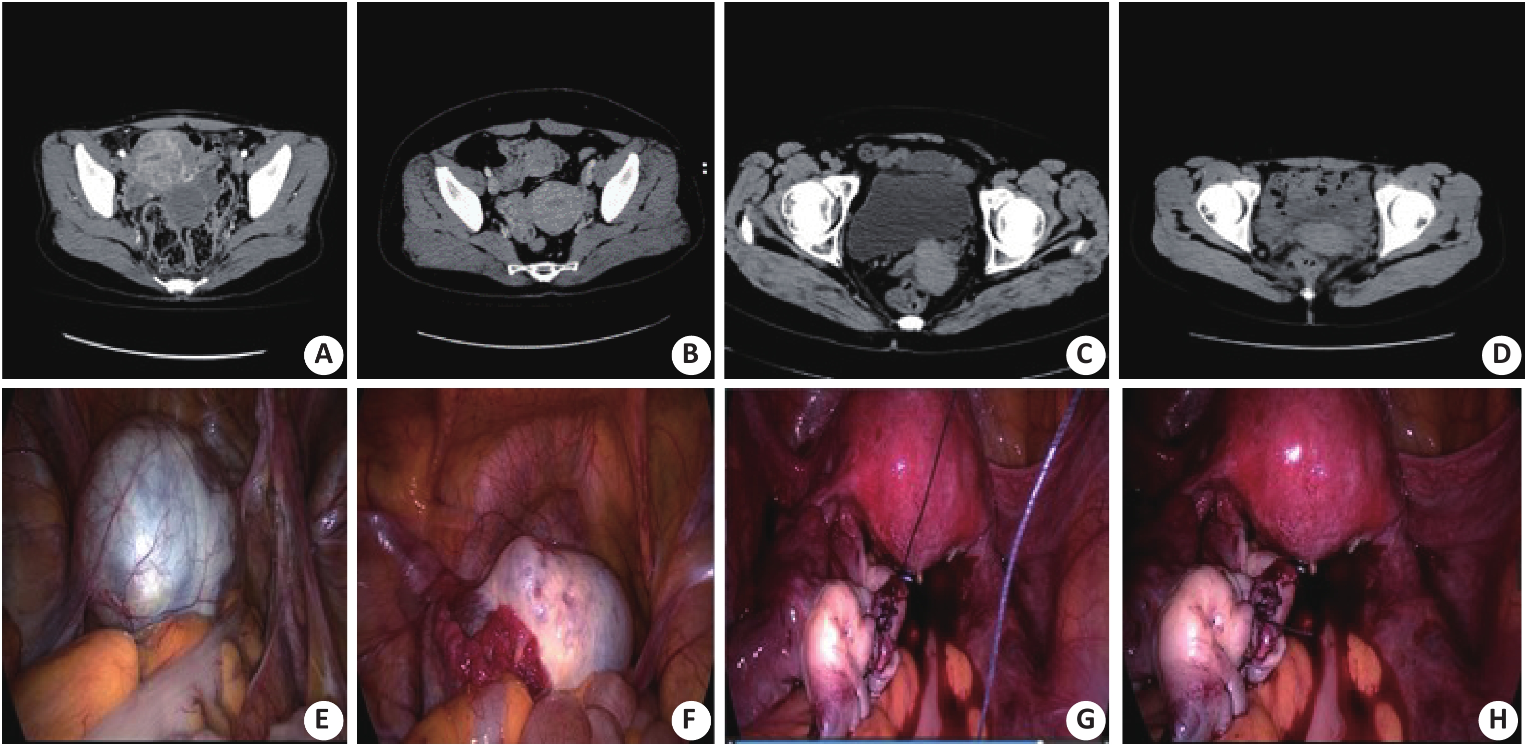
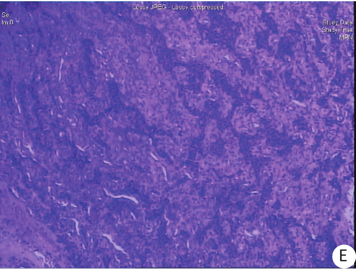
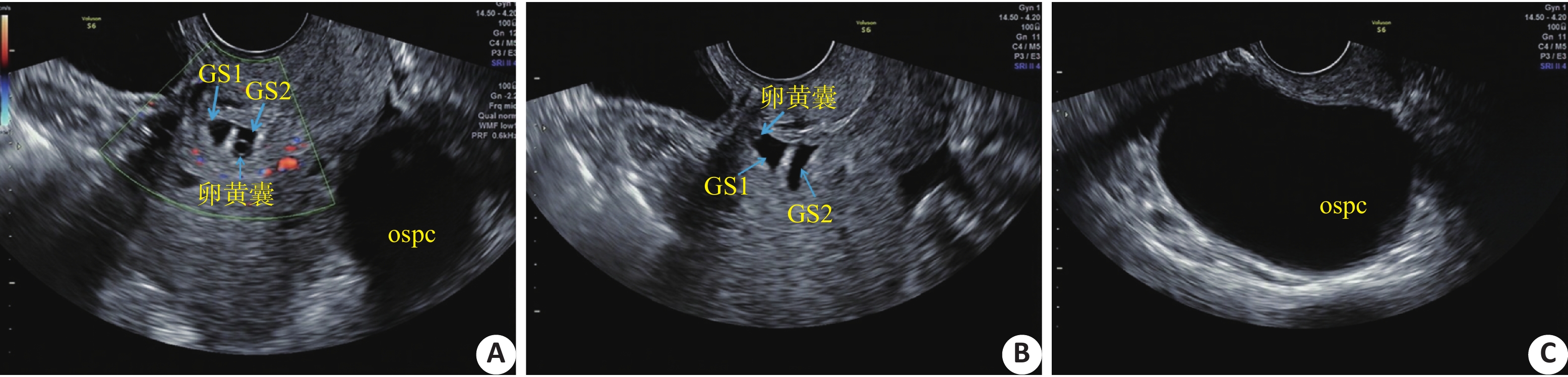
Cesarean scar ectopic pregnancy is defined as the fetus implantes on the previous uterine scar. As a life-threatening disease,cesarean scar ectopic pregnancy is easy to complicated with uterine rupture and bleeding. There were few studied on cesarean scar ectopic dichorionic twins pregnancy worldwide. Here we reported one extremely case with cesarean scar ectopic dichorionic twins pregnancy. We discussed clinical diagnosis and differential diagnosis of cesarean scar ectopic pregnancy with ovarian serous papillary cystadenoma by reviewing the literatures. Early detection of cesarean scar ectopic pregnancy is essential for providing important diagnostic information for correct diagnosis and treatments. In the early stage of pregnancy, conventional transvaginal color Doppler ultrasound scan is helpful to detect cesarean scar ectopic pregnancy and reduce the risk of uterine rupture and bleeding.
Cesarean scar ectopic pregnancy is defined as the fetus implantes on the previous uterine scar. As a life-threatening disease,cesarean scar ectopic pregnancy is easy to complicated with uterine rupture and bleeding. There were few studied on cesarean scar ectopic dichorionic twins pregnancy worldwide. Here we reported one extremely case with cesarean scar ectopic dichorionic twins pregnancy. We discussed clinical diagnosis and differential diagnosis of cesarean scar ectopic pregnancy with ovarian serous papillary cystadenoma by reviewing the literatures. Early detection of cesarean scar ectopic pregnancy is essential for providing important diagnostic information for correct diagnosis and treatments. In the early stage of pregnancy, conventional transvaginal color Doppler ultrasound scan is helpful to detect cesarean scar ectopic pregnancy and reduce the risk of uterine rupture and bleeding.
2020, 43(1): 174-178.
doi: 10.12122/j.issn.1674-4500.2020.01.37
Abstract:
Coronaviruses are mainly divided into 4 types: α-coronavirus, β-coronavirus, γ-coronavirus and δ-coronavirus. Coronaviruses which are pathogenic to humans are mainly concentrated in the genus β-coronavirus. Since the 21st century, there have 3 main outbreaks in humans: SARS-CoV, MERS-CoV and SARS-CoV-2. They are closely related to bat coronavirus in species evolution, which can cause human-to-human transmission, pneumonia, acute respiratory distress syndrome, and even shock and organ failure. They have a higher mortality rate when they develop into severe illness. The outbreak of coronaviruses would cause immeasurable harm to human health and cause great losses to the global society and economy. It is a major problem in public health in the world at present. The new coronaviruses is highly pathogenic and infectious.The treatment is mainly prevention-oriented, with no effective medicines. This article reviews the research progress of the outbreak of coronaviruses since the 21st century, and provides new ideas for better clinical prevention and treatment of new coronaviruses.
Coronaviruses are mainly divided into 4 types: α-coronavirus, β-coronavirus, γ-coronavirus and δ-coronavirus. Coronaviruses which are pathogenic to humans are mainly concentrated in the genus β-coronavirus. Since the 21st century, there have 3 main outbreaks in humans: SARS-CoV, MERS-CoV and SARS-CoV-2. They are closely related to bat coronavirus in species evolution, which can cause human-to-human transmission, pneumonia, acute respiratory distress syndrome, and even shock and organ failure. They have a higher mortality rate when they develop into severe illness. The outbreak of coronaviruses would cause immeasurable harm to human health and cause great losses to the global society and economy. It is a major problem in public health in the world at present. The new coronaviruses is highly pathogenic and infectious.The treatment is mainly prevention-oriented, with no effective medicines. This article reviews the research progress of the outbreak of coronaviruses since the 21st century, and provides new ideas for better clinical prevention and treatment of new coronaviruses.
2020, 43(1): 179-183.
doi: 10.12122/j.issn.1674-4500.2020.01.38
Abstract:
ObjectiveTo analyze the characteristics and distribution of bacterial infections in patients with AMI and the value of using predictive interventions. MethodsA total of 160 patients with AMI who were admitted to our hospital from December 2016 to January 2019 were included. The patients were divided into the infected group (n=90, 53 males, 37 females, 56.39±8.12 years old) and the non-infected group (n=70, 41 males, 29 females, 57.01±9.47 years old) according to whether the patients had bacterial infection. The patients in the infected group were divided into observation group (n=45, 28 males, 17 females, 57.01±9.03 years old) and control group (n=45, 25 males, 20 females, 55.77±10.83 years old) according to the random envelope method. Clinical data such as age, gender, and medical history of the patients were recorded, and the infected strains of the infected group were investigated. The control group was treated with routine nursing intervention, and the observation group was treated with predictive intervention. ResultsDiabetes, chronic obstructive disease, cardiac function, percutaneous coronary intervention, prophylactic antibiotics, hospitalization days, and age-related infection rates were significantly different(P<0.05). The diabetes, chronic Obstructive disease, cardiac function, percutaneous coronary intervention, prophylactic antibiotics, length of hospital stay and age were independent factors influencing bacterial infection, and the difference was significant (P<0.05). There were 61 cases of negative bacteria, accounting for 67.78%, which was the highest proportion. The normal blood pressure, normal blood, normal blood glucose rate in the observation group were higher than the control group. The incidence of angina pectoris and myocardial infarction were significantly lower than the control group (P < 0.05). The scores of physical function, role function, emotional function, social function and cognitive function of the observation group were significantly higher than those of the control group (P < 0.05). ConclusionThe bacterial infections in patients with AMI are mainly Gram-negative bacteria. The factors such as diabetes, chronic obstructive diseases, cardiac function, prophylactic antibiotics, hospitalization days and age are the independent factors that cause patients with nosocomial infection. Interventional care mode intervention can help improve the therapeutic efficacy of patients with AMI complicated with bacterial infection.