


Diagnostic value of whole body magnetic resonance diffusion weighted imaging combined with multi-slice spiral CT in multiple myeloma
-
摘要:
目的 探究全身磁共振弥散加权成像(WB-DWI)联合多层螺旋CT对多发性骨髓瘤的诊断价值。 方法 选取2019年6月~2023年1月于我院收治的80例多发性骨髓瘤患者为研究对象, 并按照检测方式将其分为WB-DWI组(n=25)、多层螺旋CT组(n=25)、联合组(n=30)。分析WB-DWI、多层螺旋CT单一及联合对患者累及部位的检出率, 分析两种检查方式单一及联合对多发性骨髓瘤的检出情况, 通过ROC曲线分析WB-DWI、多层螺旋CT联合诊断多发性骨髓瘤的临床价值。 结果 检测结果主要与Durie-Salmon分期、国际骨髓瘤分期相关(P < 0.05)。与WB-DWI、多层螺旋CT单一检测对比, 联合检测患者累及部位的检出率较高(P < 0.05)。在多发性骨髓瘤诊断中WB-DWI、多层螺旋CT联合检测高于单一检测(P < 0.05)。与WB-DWI、多层螺旋CT单项诊断对比, 两项联合诊断多发性骨髓瘤的敏感度、特异性、准确性均较高(P < 0.05)。 结论 相较于WB-DWI、多层螺旋CT单项检测, 联合检测可有效提升患者病理检出情况, 提高对多发性骨髓瘤的诊断价值。 -
关键词:
- 多发性骨髓瘤 /
- 全身磁共振弥散加权成像 /
- 多层螺旋CT /
- 国际肿瘤分期
Abstract:Objective To explore the diagnostic value of whole body magnetic resonance diffusion weighted imaging (WB-DWI) combined with multi-slice spiral CT in multiple myeloma. Methods A total of 80 patients with multiple myeloma admitted to our hospital from June 2019 to January 2023 were selected as study subjects and divided into WB-DWI group (n=25), multi-slice spiral CT group (n=25) and combined group (n=30). The detection rates of WB-DWI and multi-slice spiral CT alone and combined in the involved sites of patients were analyzed, and the detection of multiple myeloma by the two methods alone and combined was analyzed. The clinical value of WB-DWI and multi-slice spiral CT combined in the diagnosis of multiple myeloma was analyzed by ROC curve. Results The results were mainly correlated with Durie-Salmon stage and international myeloma stage (P < 0.05). Compared with the single detection of WB-DWI and multi-slice spiral CT, the detection rate of the involved sites in the combined detection was higher (P < 0.05). The combined detection of WB-DWI and multi-slice spiral CT was higher than that of single detection in the diagnosis of multiple myeloma (P < 0.05). Compared with the single diagnosis of WB-DWI and multi-slice spiral CT, the sensitivity, specificity and accuracy of the combined diagnosis of multiple myeloma were higher (P < 0.05). Conclusion Compared with WB-DWI and multi-slice spiral CT single tests, the combined test can effectively improve the pathological detection of patients and improve the diagnostic value of multiple myeloma. -

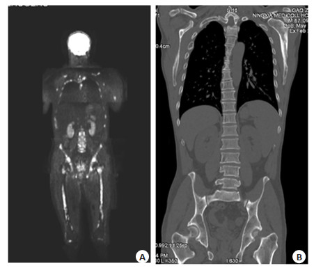
图 1 患者WB-DWI(A)及CT(B)图像
Figure 1. WB-DWI (A) and CT (B) image of patient. A: Multiple spotted high-signal foci in the collarbones, scapula, ribs, thoracic vertebra, pelvis and bilateral femur; B: Multiple patchy low-density bone destruction of bilateral shoulder blades, part of the ribs on the right side, thoracic vertebra, lumbar vertebra and pelvis.

图 2 WB-DWI、多层螺旋CT联合诊断多发性骨髓瘤的ROC曲线
Figure 2. ROC curve of WB-DWI combined with multi-slice spiral CT in diagnosis of multiple mye-loma.
表 1 3组一般资料
Table 1. General data among the three groups
Index WB-DWI group (n=25) Multislice spiral CT group (n=25) Combined group (n=30) χ2/t P Age (years, Mean±SD) 56.17±5.25 56.05±5.16 56.11±5.21 0.082 0.917 Disease course (month, Mean±SD) 4.23±1.06 4.18±1.03 4.29±1.11 0.378 0.621 Gender [n(%)] 0.092 0.761 Male 16(64.00) 15(63.75) 18(60.00) Female 9(36.00) 10(36.25) 12(40.00) Durie-Salmon stage [n(%)] 2.337 0.126 I 11(44.00) 9(36.00) 17(56.67) Ⅱ 9(36.00) 10(40.00) 8(26.67) Ⅲ 5(20.00) 6(24.00) 5(16.67) International myeloma stage staging [n(%)] 2.183 0.140 I 12(48.00) 10(40.00) 18(60.00) Ⅱ 7(28.00) 9(36.00) 7(23.33) Ⅲ 6(24.00) 6(24.00) 5(16.67) Disease type [n(%)] 1.087 0.297 Normal type 10(40.00) 9(36.00) 15(50.00) Focal type 5(20.00) 6(24.00) 4(13.33) Dispersive type 3(12.00) 3(12.00) 5(16.67) Mixed type 7(28.00) 7(28.00) 6(20.00)  下载: 导出CSV
表 2 WB-DWI、多层螺旋CT单一与联合检测对患者累及部位的检出率对比
Table 2. Comparison of the detection rates of multi-slice spiral CT alone and WB-DWI combined with multi-slice spiral CT for the involved sites [n(%)]
Detection method Pelvis (n=18) Spine (n=24) Ribs (n=11) Skull (n=14) Sternum (n=6) Humerus (n=7) Multislice spiral CT 10(55.56) 13(59.09) 5(45.45) 6(42.86) 1(16.67) 2(28.57) WB-DWI 13(72.22) 17(70.83) 8(72.73) 9(64.29) 3(50.00) 4(57.14) Combined detection 16(88.89) 22(91.67) 10(90.91) 12(85.71) 5(83.33) 6(85.71) χ2 4.985 6.695 5.238 5.600 5.333 4.667 P 0.026 0.010 0.022 0.018 0.021 0.031
下载: 导出CSV
表 3 WB-DWI、多层螺旋CT单一及联合对多发性骨髓瘤的检出情况对比
Table 3. Comparison of single and combined detection of multiple myeloma by WB-DWI and multi-slice spiral CT [n(%)]
Pathological findings WB-DWI Multislice spiral CT Combined detection Feminine character Masculine Feminine character Masculine Feminine character Masculine Feminine character 7(61.22) 12(37.84) 6(63.64) 11(32.26) 10(67.19) 13(30.00) Masculine 3(38.78) 3(62.16) 4(36.36) 4(67.74) 3(22.81) 4(70.00) Total 10(40.74) 15(59.26) 10(40.74) 15(59.26) 13(40.74) 17(59.26)
下载: 导出CSV
表 4 ROC曲线分析WB-DWI、多层螺旋CT单项及联合对多发性骨髓瘤的诊断价值
Table 4. ROC curve analysis of the diagnostic value of WB-DWI, multi-slice spiral CT and their c-ombination in multiple myeloma
Detection method AUC(95% CI) P Sensitivity (%) Specificity (%) Accuracy (%) WB-DWI 0.767(0.569-0.964) 0.027 73.33(11/15) 80.00(8/10) 76.00(19/25) Multislice spiral CT 0.783(0.584-0.983) 0.018 86.67(13/15) 70.00(7/10) 80.00(20/25) Combination of two terms 0.817(0.626-0.999) 0.008 94.12(16/17) 69.23(9/13) 83.33(25/30)
下载: 导出CSV
-
[1] Wang SZ, Zhao RH, Tan KY, et al. Multiple myeloma with cardiac involvement accompanied by partial superior vena cava obstruction: a case report and literature review[J]. Echocardiography, 2021, 38(9): 1652-6. doi: 10.1111/echo.15169 [2] Vrábel D, Pour L, Ševčíková S. The impact of NF-κB signaling on pathogenesis and current treatment strategies in multiple myeloma[J]. Blood Rev, 2019, 34: 56-66. doi: 10.1016/j.blre.2018.11.003 [3] Wallington-Beddoe CT, Mynott RL. Prognostic and predictive biomarker developments in multiple myeloma[J]. J Hematol Oncol, 2021, 14(1): 1-15. doi: 10.1186/s13045-020-01025-7 [4] Diaz-delCastillo M, Chantry AD, Lawson MA, et al. Multiple myeloma—a painful disease of the bone marrow[J]. Semin Cell Dev Biol, 2021, 112: 49-58. doi: 10.1016/j.semcdb.2020.10.006 [5] 郑梦龙, 翁春娇, 李文娟, 等. 3.0 T MRI全身扩散加权成像在多发性骨髓瘤诱导治疗疗效评估中的应用[J]. 磁共振成像, 2019, 10(3): 201-5. [6] 郭卫, 牛晓辉, 肖建如, 等. 多发性骨髓瘤骨病外科治疗循证医学指南[J]. 中华骨与关节外科杂志, 2018, 11(4): 252-9, 275. [7] Allegra A, Di Gioacchino M, Tonacci A, et al. Multiple myeloma cell-derived exosomes: implications on tumorigenesis, diagnosis, prognosis and therapeutic strategies[J]. Cells, 2021, 10(11): 2865. doi: 10.3390/cells10112865 [8] 武晓倩, 李晨, 冯玉虎. 血清β2-微球蛋白联合C反应蛋白与白蛋白比值对首诊断多发性骨髓瘤预后的预测价值[J]. 临床和实验医学杂志, 2022, 21(17): 1833-6. doi: 10.3969/j.issn.1671-4695.2022.17.011 [9] 章双林, 陈陽, 刘冲, 等. 99Tcm-MDP全身骨显像与全身低剂量CT诊断多发性骨髓瘤的对比研究[J]. 现代肿瘤医学, 2019, 27(9): 1609-13. [10] 吴晓颖, 施菊妹, 陶怡, 等. IGF-I、β2-MG和SF在多发性骨髓瘤患者中的诊断价值及其与临床分期的关系[J]. 中国实验血液学杂志, 2018, 26(3): 802-6. [11] Guerrero C, Puig N, Cedena MT, et al. A machine learning model based on tumor and immune biomarkers to predict undetectable MRD and survival outcomes in multiple myeloma[J]. Clin Cancer Res, 2022, 28(12): 2598-609. doi: 10.1158/1078-0432.CCR-21-3430 [12] 郑庆中, 苏洁敏, 李小玲, 等. 18F-FDG PET/CT显像对多发性骨髓瘤与骨转移瘤的鉴别诊断价值[J]. 中国实验血液学杂志, 2020, 28(4): 1267-71. [13] 邓成文, 张晓莹, 吕中伟, 等. 18F-FDG PET/CT显像对多发性骨髓瘤与原因不明溶骨性转移瘤的鉴别诊断价值[J]. 中华核医学与分子影像杂志, 2022, 42(5): 269-73. [14] 王根杰, 田颖. MRI、CT及X线在诊断多发性骨髓瘤中的应用比较[J]. 中国CT和MRI杂志, 2019, 17(3): 127-9. [15] 周存凉, 丁勇生, 沈月红. PET/CT和MRI在多发性骨髓瘤诊断中的应用价值及影像表现分析[J]. 中国CT和MRI杂志, 2019, 17(12): 143-5, 152. [16] 胡青竹, 田颖, 王根杰, 等. X线和MRI在多发性骨髓瘤诊断中的应用价值及影像表现分析[J]. 中国CT和MRI杂志, 2018, 16(1): 141-3. [17] 林志畑, 蔡宋浩, 黄锦桂, 等. 初诊多发性骨髓瘤患者 18F-FDG PET/CT影像学表现与高危细胞遗传学异常的相关性及预后评估[J]. 国际放射医学核医学杂志, 2022, 46(4): 203-9. [18] 洪乐旻, 王信峰, 郭丹, 等. 18F-FDG PET/CT显像特征与多发性骨髓瘤患者临床因素的相关性分析[J]. 临床血液学杂志, 2020, 33(9): 609-13. [19] 陈冲, 王雪, 王亚非. IgD型多发性骨髓瘤的研究进展[J]. 分子影像学杂志, 2019, 42(4): 510-3. doi: 10.12122/j.issn.1674-4500.2019.04.21 [20] 王明, 刘莹, 王亚丽, 等. 全身磁共振扩散加权成像对多发性骨髓瘤的诊断价值[J]. 河北医科大学学报, 2022, 43(2): 198-202. [21] 王亚丽, 刘莹, 王明, 等. 全身扩散加权成像对多发性骨髓瘤化疗后疗效评估[J]. 实用放射学杂志, 2022, 38(9): 1508-12. [22] 李鹏, 王国旗. WB-DWI在初诊多发性骨髓瘤诊断及疗效评估中的应用价值[J]. 中国CT和MRI杂志, 2021, 19(3): 148-50, 170. [23] 李淑娥, 葛洪峰, 孙晓星, 等. 不同国际分期体系分期的多发性骨髓瘤患者WB-DWI表现及预后[J]. 分子影像学杂志, 2021, 44(3): 492-5. doi: 10.12122/j.issn.1674-4500.2021.03.15 [24] 张立, 刘璋, 吴恩柱, 等. 磁共振全身弥散加权成像结合磁共振常规扫描在恶性肿瘤个体化介入治疗中的应用[J]. 中国数字医学, 2018, 13(2): 98-100. -


 点击查看大图
计量
- 文章访问数: 92
- HTML全文浏览量: 35
- PDF下载量: 8
- 被引次数: 0