


18F-PSMA-1007 PET/CT examination can noninvasively diagnose and stage the vast majority of prostate cancer
-
摘要:
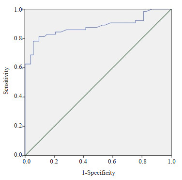
目的 探讨基于18F-PSMA-1007的PET/CT显像技术在前列腺癌(PCa)无创精准诊断中的应用价值。 方法 选择2020年11月~2022年4月梅州市人民医院收治的117例疑似PCa患者,在其穿刺活检前行18F-PSMA-1007 PET/CT检查,并通过勾画感兴趣区域的方法测量病灶和肝的标准摄取最大值,并以肝为背景计算肿瘤背景比(TBR),结合穿刺后的病理结果(PCa 64例,良性53例),比较良恶性疾病TBR组间差异,绘制ROC曲线评价其诊断效能,从而得到最佳截断值。 结果 PCa患者的TBR水平高于良性,两组间差异有统计学意义(P < 0.001)。以TBR诊断PCa绘制ROC曲线,测得ROC曲线下面积为0.881(P < 0.001),截断值取0.955时,敏感度和特异性分别为78.1%和94.3%。TBR低于截断值的14例PCa患者中,7例已出现淋巴结和/或骨转移,可间接诊断为PCa。 结论 18F-PSMA-1007 PET/CT TBR对鉴别前列腺的良恶性具有较高的应用价值,以TBR=0.955作为截断值可获得很好的诊断效能,即使TBR低于截断值,转移灶的发现可作为PCa的补充诊断,进一步提高诊断准确率。行18F-PSMA-1007 PET/CT检查可无创诊断绝大多数的PCa并确定分期。 -
关键词:
- 18F-PSMA-1007 /
- PET/CT /
- 前列腺癌 /
- 无创性诊断
Abstract:Objective To investigate the application value of PET/CT imaging technology based on 18F-PSMA-1007 in the non-invasive and accurate diagnosis of prostate cancer (PCa). Methods A total of 117 patients with suspected PCa admitted to Meizhou People 's Hospital from November 2020 to April 2022 were examined by 18F-PSMA-1007 PET/CT before biopsy. The maximum standardized uptake value of the lesion and liver was measured by delineating the region of interest, and the tumor background ratio (TBR) was calculated with the liver as the background. Combined with the pathological results after biopsy (64 cases of PCa and 53 cases of benign), the TBR differences between benign and malignant diseases were compared, and the ROC curve was drawn to evaluate their diagnostic efficacy, so as to obtain the best cutoff value. Results The TBR level of PCa patients was higher than that of benign patients, and there was significant difference between the two groups (P < 0.001). The ROC curve was plotted by TBR to diagnose PCa, and the area under the ROC curve was 0.881 (P < 0.001), and the sensitivity and specificity were 78.1% and 94.3%, respectively, when the cutoff value was taken as 0.955. Moreover, among the 14 PCa patients with TBR below the cutoff value, 7 had developed lymph nodes and/or bone metastases, which could be indirectly diagnosed as PCa. Conclusion 18F-PSMA-1007 PET/CT TBR has a high application value in distinguishing benign and malignant prostate lesions. Using TBR=0.955 as the cut-off value can achieve good diagnostic efficiency. Even if the TBR is lower than the cutoff value, the detection of metastases can be used as a supplementary diagnosis of PCa and further improve the diagnostic accuracy. 18F-PSMA-1007 PET/CT examination can noninvasively diagnose and stage the vast majority of PCa. -
Key words:
- 18F-PSMA-1007 /
- PET/CT /
- prostate cancer /
- noninvasive diagnosis
-

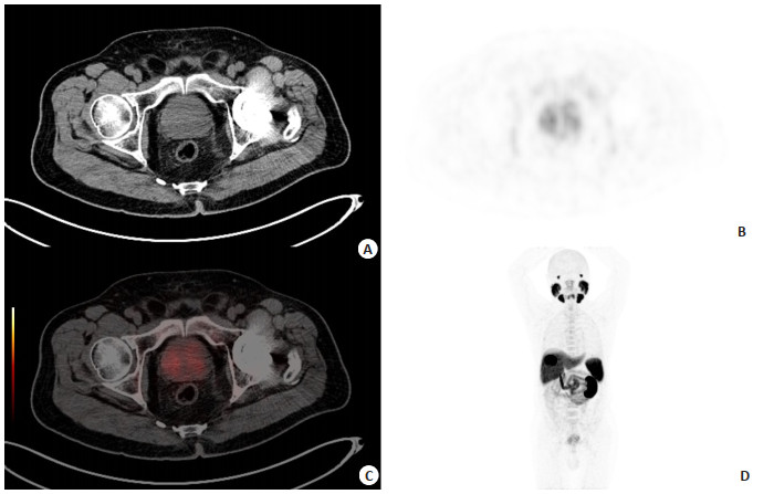
图 1 PCa患者图像
Figure 1. Images of prostate cancer patients. A: CT images; B: 18F-PSMA-1007 PET images; C: 18F-PSMA-1007 PET/CT images shows prostate cancer; D: Distribution of 18F-PSMA-1007 in the body.

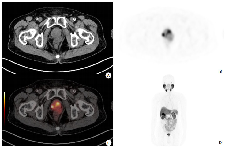
图 2 前列腺良性疾病患者图像
Figure 2. Images of patients with benign prostatic disease. A: CT images; B: 18F-PSMA-1007 PET images; C: 18F-PSMA-1007 PET/CT images shows non prostate cancer; D: Distribution of 18F-PSMA-1007 in the body.

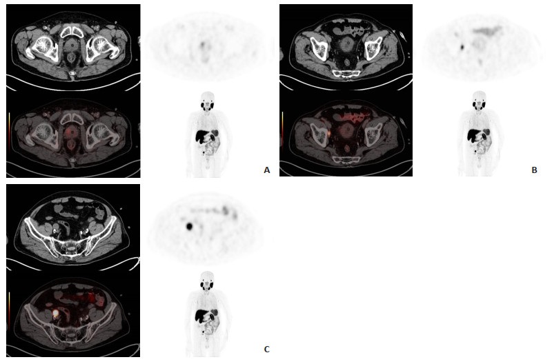
图 4 TBR低于截断值的PCa患者
Figure 4. Prostate cancer patients with TBR below cutoff value. A: Radioactive uptake in the prostate is generally normal, TBR=0.48; B-C: Two radioactive abnormal high uptake lesions were seen next to the right iliac artery, which are considered as lymph node metastases of prostate cancer. It indirectly helps diagnose prostate cancer.
表 1 PCa组与良性组各参数比较
Table 1. Comparison of parameters between PCa group and benign group
Index PCa Benign t/Z P Age(year, Mean±SD) 72.66±7.812 67.13±7.429 3.893 < 0.001 tPSA[ng/mL, M(P25,P75)] 41.87(12.515, 95.512) 12.158±6.841 -5.328 < 0.001 fPSA[ng/mL, M(P25,P75)] 6.896(1.725, 14.128) 1.942(0.843, 3.806) -3.806 < 0.001 TBR[M(P25,P75)] 2.1(0.992, 3.485) 0.626±0.259 -7.072 < 0.001 tPSA: Total prostate specific antigen; fPSA: Free prostate specific antigen; TBR: Tumor background ratio. PCa: Prostate cancer.  下载: 导出CSV
表 2 两种显像对淋巴结转移灶的检出能力比较
Table 2. Comparison of the detection ability of two types of imaging for lymph node metastasis [n(%)]
MRI 18F-PSMA-1007 PET/CT Total + - + 20(31.3) 0(0) 20(31.3) - 8(12.5) 36(56.2) 44(68.7) Total 28(43.8) 36(56.2) 64(100.0)
下载: 导出CSV
表 3 两种显像对骨转移灶的检出能力比较
Table 3. Comparison of the detection ability of two types of imaging for bone metastasis[n(%)]
MRI 18F-PSMA-1007 PET/CT Total + - + 15(23.4) 0(0) 15(23.4) - 9(14.1) 40(62.5) 49(76.6) Total 24(37.5) 40(62.5) 64(100.0)
下载: 导出CSV
-
[1] Sung H, Ferlay J, Siegel RL, et al. Global cancer statistics 2020: GLOBOCAN estimates of incidence and mortality worldwide for 36 cancers in 185 countries[J]. CA A Cancer J Clinicians, 2021, 71(3): 209-49. doi: 10.3322/caac.21660 [2] Chen WQ, Zheng RS, Baade PD, et al. Cancer statistics in China, 2015[J]. CA A Cancer J Clinicians, 2016, 66(2): 115-32. doi: 10.3322/caac.21338 [3] 中国抗癌协会泌尿男生殖系统肿瘤专业委员会前列腺癌学组. 前列腺癌筛查中国专家共识(2021年版)[J]. 中国癌症杂志, 2021, 31(5): 435-40. [4] 孙健, 赵学媛, 于阳. 前列腺癌早期影像学诊断进展[J]. 癌症进展, 2020, 18(6): 560-2, 587. [5] Loeb S, Carter HB, Berndt SI, et al. Complications after prostate biopsy: data from SEER-medicare[J]. J Urol, 2011, 186(5): 1830-4. doi: 10.1016/j.juro.2011.06.057 [6] Loeb S, van den Heuvel S, Zhu XY, et al. Infectious complications and hospital admissions after prostate biopsy in a European randomized trial[J]. Eur Urol, 2012, 61(6): 1110-4. doi: 10.1016/j.eururo.2011.12.058 [7] Nam RK, Saskin R, Lee YN, et al. Increasing hospital admission rates for urological complications after transrectal ultrasound guided prostate biopsy[J]. J Urol, 2010, 183(3): 963-9. doi: 10.1016/j.juro.2009.11.043 [8] 李红星, 高宛生, 杨彦峰, 等. 经直肠超声引导下前列腺穿刺活检术后全身炎症反应综合征的危险因素[J]. 现代泌尿外科杂志, 2016, 21(5): 353-6. doi: 10.3969/j.issn.1009-8291.2016.05.008 [9] Thompson JE, Moses D, Shnier R, et al. Multiparametric magnetic resonance imaging guided diagnostic biopsy detects significant prostate cancer and could reduce unnecessary biopsies and over detection: a prospective study[J]. J Urol, 2014, 192(1): 67-74. doi: 10.1016/j.juro.2014.01.014 [10] 陈锐, 谢立平, 周利群, 等. 中国前列腺癌联盟成员医院前列腺穿刺活检现状的调查报告[J]. 中华泌尿外科杂志, 2015, 36(5): 342-5. [11] Combes AD, Palma CA, Calopedos R, et al. PSMA PET-CT in the diagnosis and staging of prostate cancer[J]. Diagnostics, 2022, 12(11): 2594. doi: 10.3390/diagnostics12112594 [12] Sprute K, Kramer V, Koerber SA, et al. Diagnostic accuracy of 18F-PSMA-1007 PET/CT imaging for lymph node staging of prostate carcinoma in primary and biochemical recurrence[J]. J Nucl Med, 2021, 62(2): 208-13. doi: 10.2967/jnumed.120.246363 [13] Sanli Y, Sanli O, Has Simsek D, et al. 68Ga-PSMA PET/CT and PET/MRI in high-risk prostate cancer patients[J]. Nucl Med Commun, 2018, 39(10): 871-80. doi: 10.1097/MNM.0000000000000888 [14] 周文瑶, 张俊. 前列腺癌PSMA PET显像剂研究进展[J]. 中华核医学与分子影像杂志, 2020, 40(12): 755-60. [15] Awenat S, Piccardo A, Carvoeiras P, et al. Diagnostic role of 18F-PSMA-1007 PET/CT in prostate cancer staging: a systematic review[J]. Diagnostics, 2021, 11(3): 552. doi: 10.3390/diagnostics11030552 [16] Kesch C, Vinsensia M, Radtke JP, et al. Intraindividual comparison of 18F-PSMA-1007 PET/CT, multiparametric MRI, and radical prostatectomy specimens in patients with primary prostate cancer: a retrospective, proof-of-concept study[J]. J Nucl Med, 2017, 58(11): 1805-10. doi: 10.2967/jnumed.116.189233 [17] Mittlmeier LM, Brendel M, Beyer L, et al. Feasibility of different tumor delineation approaches for 18F-PSMA-1007 PET/CT imaging in patients with metastatic prostate cancer[J]. Frontiers in Oncology, 2021, 11: 663631. doi: 10.3389/fonc.2021.663631 [18] Zhou X, Li YC, Jiang X, et al. Intra-individual comparison of 18F-PSMA-1007 and 18F-FDG PET/CT in the evaluation of patients with prostate cancer[J]. Front Oncol, 2021, 10: 585213. doi: 10.3389/fonc.2020.585213 [19] 马婧, 宋争放, 王霄. 前列腺癌美国、欧洲、日本指南与中国诊疗指南对比研究[J]. 四川医学, 2022, 43(5): 511-4. [20] Kesch C, Schütz V, Dieffenbacher S, et al. Multiparametric MRI fusion-guided biopsy for the diagnosis of prostate cancer[J]. Curr Opin Urol, 2018, 28(2): 172-7. doi: 10.1097/MOU.0000000000000461 [21] 张志强, 杨琳琳, 谢栋栋, 等. 前列腺穿刺活检患者疾病不确定感和焦虑抑郁症状的前瞻性研究[J]. 中国男科学杂志, 2019, 33(2): 41-6. [22] Gutiérrez-Cardo AL, Pérez Duarte A, García-Argüello SF, et al. Assessment of 68Ga-PSMA-11 PET positivity predictive factors inprostate cancer[J]. Rev Española De Med Nucl E Imagen Mol Engl Ed, 2019, 38(1): 22-8. [23] Giesel FL, Cardinale J, Schäfer M, et al. 18F-Labelled PSMA-1007 shows similarity in structure, biodistribution and tumour uptake to the theragnostic compound PSMA-617[J]. Eur J Nucl Med Mol Imaging, 2016, 43(10): 1929-30. doi: 10.1007/s00259-016-3447-9 [24] 王卓楠, 吴开杰, 郑安琪, 等. 18F-PSMA PET/CT在前列腺癌诊断与预后预测中的价值初探[J]. 现代泌尿外科杂志, 2021, 26(4): 305-8. [25] 楼云龙, 陈丹丹, 陈南辉. 18F-PSMA-1007 PET/CT在PCa术前诊断中的价值[J]. 现代泌尿生殖肿瘤杂志, 2022, 14(5): 313-5. [26] Schwarzenboeck SM, Rauscher I, Bluemel C, et al. PSMA ligands for PET imaging of prostate cancer[J]. J Nucl Med, 2017, 58(10): 1545-52. doi: 10.2967/jnumed.117.191031 [27] Hiroaki I, Kouji I, Yoshifumi K, et al. Incidences of visceral metastases from prostate cancer increase after progression of castration-resistant status[J]. J Clin Oncol, 2018, 36(6_suppl). 291. doi: 10.1200/JCO.2018.36.6_suppl.291 [28] Bronsert P, Reichel K, Ruf J. Loss of PSMA expression in non-neuroendocrine dedifferentiated acinar prostate cancer[J]. Clin Nucl Med, 2018, 43(7): 526-8. doi: 10.1097/RLU.0000000000002100 -

 点击查看大图
计量
- 文章访问数: 135
- HTML全文浏览量: 106
- PDF下载量: 24
- 被引次数: 0