


Correlation between molecular subtypes and imaging features of breast cancer in women aged 18-30 years
-
摘要:
目的 探讨18~30岁女性乳腺癌的分子亚型与影像学特征的相关性。 方法 回顾性分析195例经术后病理确诊的原发年轻乳腺癌的影像及病理资料,用免疫组织化学检测ER、PR、HER2及Ki-67,HER2(++)者再行荧光原位杂交检测,分为Luminal A型(n=55)、Luminal B型(n=79)、HER2过表达型(n=41)和三阴性乳腺癌(n=20)。比较各亚型的临床病理特征、X线以及超声特点。 结果 195例年轻乳腺癌的分子分型比例依次为Luminal B型>Luminal A型>HER2过表达型>三阴性,Luminal A型乳腺癌患者更多为临床T1期和组织学Ⅰ级(P < 0.05),Luminal B型更多为临床T2期和组织学Ⅱ级(P < 0.05),三阴性乳腺癌更多为临床T3期和组织学Ⅲ级(P < 0.05)。X线结果显示:Luminal A型以肿块伴钙化,细小多形性,边缘毛刺为多(P < 0.05)。Luminal B型以肿块,细小多形性和线样,边缘模糊为多(P < 0.05),HER2过表达型以钙化,线样或线样分枝状,边缘模糊为多(P < 0.05),三阴性乳腺癌以肿块,细小多形性,边缘模糊为多(P < 0.05)。超声结果显示:三阴性乳腺癌内部回声以均匀为多,其它三种类型以不均匀为多(P < 0.05)。 结论 18~30岁青年女性乳腺癌的免疫组织化学分子亚型与部分影像学特征密切相关。术前可根据X线及超声综合表现预测乳腺癌分子分型。 Abstract:Objective To investigate the correlation between molecular subtypes and imaging features of breast cancer in women aged 18-30 years. Methods The images and pathological data of 195 patients with primary breast cancer diagnosed by postoperative pathology were retrospectively analyzed. According to the expression levels of ER, PR HER2 and Ki-67 they were divided into Luminal A group (n=55), Luminal B group (n=79), HER2 overexpression group (n=41) and triple negative group (n=20). The clinical and pathological data of each subtype were analyzed retrospectively. Results The molecular typing proportion of 195 cases of young breast cancer was Luminal B type >Luminal A>HER2 overexpression >triple negative type. More patients with Luminal A breast cancer were in clinical T1 stage and histological grade Ⅰ(P < 0.05). More patients with Luminal B stage were in clinical T2 stage and biopsy grade (P < 0.05). More patients with TNBC were in stage T3 stage and histological grade (P < 0.05). TNBC had more clinical T3 and histological grade Ⅲ (P < 0.05). X-ray results showed that Luminal type A was characterized by Luminal mass with calcification, parvopleomorphism, and marginal burrs (P < 0.05). Luminal B tumor, parvopleomorphism & line-like, with blurred margins were more common (P < 0.05). HER2 overexpression tumor, with calcification, line-like or line-like branching, with blurred margins were more common (P < 0.05). Triple negative tumor, parvopleomorphism, with blurred margins were more common (P < 0.05). Ultrasonographic results showed that most of the three negative internal echoes were uniform, while the other three types were inhomogeneous (P < 0.05). Conclusion The immunohistochemical subtypes of breast cancer in young women under 18 to 30 years old are closely related to some imaging features. Therefore, preoperative molecular typing could be preliminarily determined according to the comprehensive findings of X-ray and ultrasound. -
Key words:
- youth /
- breast cancer /
- molecular typing /
- X-ray /
- ultrasonic
-

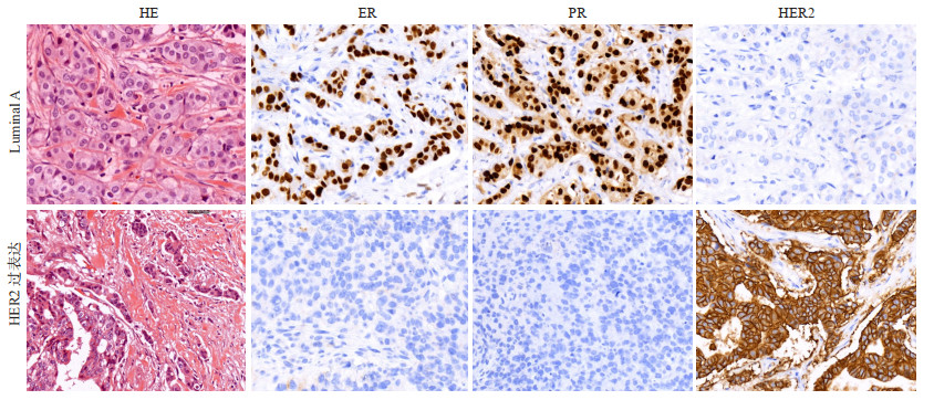
图 1 LuminalA及HER2过表达型乳腺癌组织学表现及IHC表达
Figure 1. Histological characteristics and IHC expression of LuminalA and HER2 overexpression breast cancer

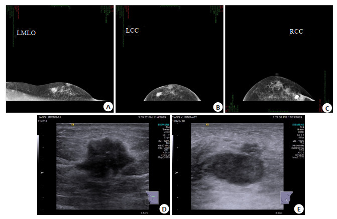
图 2 不同分子亚型患者的X线及超声特点
A: LuminalA型伴钙化(X线); B: Luminal B型伴有钙化(X线); C: 不伴有钙化(X线); D: 非三阴性乳腺癌为不均匀回声(超声); E: 三阴性乳腺癌超声为均匀回声.
Figure 2. X-ray and ultrasound characteristics of breast cancer patients with different molecular subtypes
表 1 不同分子亚型患者病理特征
Table 1. Clinicopathological characteristics of molecular subtypes of breast cancer [n(%)]
特征 Luminal A(n=55) Luminal B(n=79) HER2过表达(n=41) 三阴性(n=20) t/χ2 P 年龄(岁,Mean±SD) 26.26±2.93 27.41±2.17 26.84±2.11 27.5±2.18 0.262 0.124 患侧 0.241 0.152 左侧 27 (49.09) 38 (48.10) 22 (53.66) 10 (50.00) 右侧 28 (50.91) 41 (51.90) 19 (46.34) 10 (50.00) 家族史 0.274 0.135 是 20 (36.36) 35 (44.30) 15 (36.59) 8 (40.00) 否 35 (63.64) 44 (55.70) 26 (63.41) 12 (60.00) 肿瘤直径(cm) 0.299 0.117 <5 25 (45.45) 37 (46.83) 18 (43.90) 9 (45.00) ≥5 30 (54.55) 42 (53.17) 23 (56.10) 11 (55.00) TNM分期 6.131 < 0.001 T1 34 (61.82) 25 (31.65) 12 (29.27) 3 (15.00) T2 15 (27.27) 48 (60.76) 15 (36.59) 7 (35.00) T3 6 (9.09) 6 (7.59) 14(34.15) 10 (50.00) 组织学分级 6.862 < 0.001 Ⅰ 33 (60.00) 21 (38.18) 13 (31.71) 2 (10.00) Ⅱ 14 (22.45) 52 (65.82) 15 (36.59) 5 (25.00) Ⅲ 8 (14.55) 6 (7.59) 13 (31.71) 13 (65.00) 组织学类型 4.356 0.640 浸润性导管癌 46 (83.64) 69 (87.34) 33 (80.49) 9 (45.00) 导管原位癌 4 (7.26) 3 (3.80) 3 (7.32) 7 (35.00) 其它 5 (9.10) 7 (8.86) 5 (12.19) 4 (20.00) 淋巴结转移 0.261 0.137 是 26 (47.27) 38 (48.10) 19 (46.33) 9 (45.00) 否 29 (52.73) 41 (51.90) 22 (53.67) 11 (55.00)  下载: 导出CSV
表 2 不同分子亚型患者的X线特点
Table 2. X-ray characteristics of breast cancer patients with different molecular subtypes
特征 Luminal A(n=55) Luminal B(n=79) HER2过表达(n=41) 三阴性(n=20) χ2 P 病变类型 20.338 0.012 单纯肿块 14 (25.45) 36 (45.57) 8(19.51) 9 (45.00) 肿块伴钙化 25 (45.45) 24 (30.38) 11 (26.83) 5 (25.00) 单纯钙化 10(18.18) 15 (18.98) 19 (46.34) 4 (20.00) 其它 6(10.91) 4 (7.27) 3 (7.32) 2 (10.00) 肿块形态 0.421 0.127 圆/椭圆 21 (38.18) 30 (37.79) 18 (43.09) 8 (40.00) 不规则 34 (61.0) 49 (62.21) 23 (56.91) 12(60.000) 钙化形态 6.651 < 0.001 细小多形性 45 (81.82) 24 (30.38) 5 (12.20) 15 (75.00) 线样或线样分枝状 6(10.91) 22 (27.85) 21 (51.22) 4 (20.00) 细小多形性 & 线样 4 (7.27) 33 (41.77) 15 (36.59) 1 (5.00) 边缘 62.982 < 0.001 清晰 11 (20.00) 20 (25.32) 1 (2.44) 0 (0.00) 微分叶 10(18.18) 21 (26.58) 2 (4.88) 1 (2.44) 模糊 9 (16.36) 32 (40.51) 28 (68.29) 10 (50.00) 毛刺 25 (45.45) 6 (7.59) 10(18.18) 9 (45.00) 分布 0.503 0.094 成簇 1 (2.44) 18 (22.78) 10 (24.39) 5 (25.00) 段样 54 (97.56) 61 (77.22) 31 (75.61) 15 (75.00) 超声 内部回声 6.296 < 0.001 均匀 19 (34.55) 26 (32.91) 15 (36.59) 14 (70.00) 不均匀 36 (65.45) 53 (67.09) 26(63.41) 6 (30.00) 后方回声 0.459 0.105 增强或无改变 23 (41.82) 35 (44.30) 18 (43.90) 8 (40.00) 衰减 32 (58.18) 44 (55.70) 23 (66.01) 12 (60.00) 血流 0.532 0.087 丰富 28 (50.91) 39 (49.79) 21 (51.22) 10 (50.00) 不丰富 27 (49.09) 40 (50.21) 20 (48.78) 10 (50.00)
下载: 导出CSV
-
[1] Limbach KE, Leon E, Pommier RF, et al. Comparison of breast cancer incidence, clinicopathologic features, and risk factor prevalence in women aged 20-29 at diagnosis to those aged 30-39 [J]. Breast J, 2020, 26(5): 1069-70. doi: 10.1111/tbj.13783 [2] An YY, Kim SH, Kang BJ, et al. Breast cancer in very young women (<30 years): Correlation of imaging features with clinicopathological features and immunohistochemical subtypes[J]. Eur J Radiol, 2015, 84(10): 1894-902. doi: 10.1016/j.ejrad.2015.07.002 [3] Siegel RL, Miller KD, Jemal A. Cancer statistics, 2020[J]. CA Cancer J Clin, 2020, 70(1): 7-30. doi: 10.3322/caac.21590 [4] 曾辉, 陈卫国, 徐泽园, 等. 不同分子分型原发乳腺癌临床病理特征及X线、超声表现: 140例23~35岁患者[J]. 分子影像学杂志, 2020, 43(3): 387-93. doi: 10.12122/j.issn.1674-4500.2020.03.05 [5] Murphy BL, Day CN, Hoskin TL, et al. Adolescents and young adults with breast cancer have more aggressive disease and treatment than patients in their forties[J]. Ann Surg Oncol, 2019, 26(12): 3920-30. doi: 10.1245/s10434-019-07653-9 [6] Durhan G, Azizova A, Önder Ö, et al. Imaging findings and clinicopathological correlation of breast cancer in women under 40 years old[J]. Eur J Breast Health, 2019, 15(3): 147-52. doi: 10.5152/ejbh.2019.4606 [7] 田林, 李玉娜, 胡啸, 等. 6种影像学技术在致密型乳腺诊断中的应用[J]. 分子影像学杂志, 2020, 43(1): 7-11. doi: 10.12122/j.issn.1674-4500.2020.01.02 [8] Cai S, Yao M, Cai D, et al. Association between digital breast tomosynthesis and molecular subtypes of breast cancer[J]. Oncol Lett, 2019, 17(3): 2669-76. http://www.ncbi.nlm.nih.gov/pubmed/30867729 [9] 吕琪, 吴军刚, 贾春梅, 等. 年轻女性乳腺浸润性导管癌及其不同分子亚型的超声声像图特征分析[J]. 中国全科医学, 2020, 23(3): 358- 62. https://www.cnki.com.cn/Article/CJFDTOTAL-QKYX202003024.htm [10] Sabiani L, Houvenaeghel G, Heinemann M, et al. Breast cancer in young women: Pathologic features and molecular phenotype[J]. Breast, 2016, 29: 109-16. doi: 10.1016/j.breast.2016.07.007 [11] 梁旭, 徐忠孜, 廖雪芮, 等. 3种乳腺癌分子亚型与数字乳腺断层合成显像X线特征的关系: 基于BI-RADS[J]. 分子影像学杂志, 2021, 44 (4): 567-73. doi: 10.12122/j.issn.1674-4500.2021.04.01 [12] 黄峻琳, 林青, 崔春晓, 等. 青年女性乳腺癌的影像学特征与免疫组织化学分子亚型相关性分析[J]. 中国癌症杂志, 2020, 30(10): 812-20. https://www.cnki.com.cn/Article/CJFDTOTAL-ZGAZ202010013.htm [13] 许凤锐, 江泽飞. 人表皮生长因子受体2阳性乳腺癌诊疗新进展及解析[J]. 中华医学杂志, 2020, 7(30): 2324-7. doi: 10.3760/cma.j.cn112137-20200226-00455 [14] Mercado CL. BI-RADS update[J]. Radiol Clin North Am, 2014, 52 (3): 481-7. doi: 10.1016/j.rcl.2014.02.008 [15] Coates AS, Winer EP, Goldhirsch A, et al. Tailoring therapiesimproving the management of early breast cancer: St gallen international expert consensus on the primary therapy of early breast cancer 2015[J]. Ann Oncol, 2015, 26(8): 1533-46. doi: 10.1093/annonc/mdv221 [16] 杨圆, 王守满, 陈飞宇. 湖南省单中心518例年轻乳腺癌临床病理学特征分析[J]. 中国普通外科杂志, 2020, 29(11): 1303-10. doi: 10.7659/j.issn.1005-6947.2020.11.003 [17] Han W, Kang SY, Society TKBC. Relationship between age at diagnosis and outcome of premenopausal breast cancer: age less than 35 years is a reasonable cut-off for defining young age-onset breast cancer[J]. Breast Cancer Res Treat, 2009, 119(1): 193-200. [18] Keegan TH, Press DJ, Tao L, et al. Impact of breast cancer subtypes on 3-year survival among adolescent and young adult women[J]. Breast Cancer Res, 2013, 15(5): R95. doi: 10.1186/bcr3556 [19] 张琦, 宋富桂, 吕哲昊, 等. 影像组学在预测乳腺癌分子分型中的研究进展[J]. 放射学实践, 2020, 35(11): 1476-8. https://www.cnki.com.cn/Article/CJFDTOTAL-FSXS202011031.htm [20] Darwish AD, Helal AM, Aly El-Din NH, et al. Breast cancer in women aging 35 years old and younger: The Egyptian National Cancer Institute (NCI) experience[J]. Breast, 2017, 31: 1-8. doi: 10.1016/j.breast.2016.09.018 [21] Collins LC, Marotti JD, Gelber S, et al. Pathologic features and molecular phenotype by patient age in a large cohort of young women with breast cancer[J]. Breast Cancer Res Treat, 2012, 131 (3): 1061-6. doi: 10.1007/s10549-011-1872-9 [22] Wu M, Ma J. Association between imaging characteristics and different molecular subtypes of breast cancer[J]. Acad Radiol, 2017, 24(4): 426-34. doi: 10.1016/j.acra.2016.11.012 [23] 贾志莺, 张银华, 冷晓玲, 等. 三阴性及非三阴性乳腺癌超声、临床病理特征的回顾性分析[J]. 中国临床医学影像杂志, 2017, 28(1): 23-6. https://www.cnki.com.cn/Article/CJFDTOTAL-LYYX201701007.htm -

 点击查看大图
计量
- 文章访问数: 200
- HTML全文浏览量: 198
- PDF下载量: 4
- 被引次数: 0