

 Find Duplicates
 Check Document
 Submission(new)
 Experts Office
 Editorial Office
2022 Vol. 45, No. 1
column
Display Method:
2022, 45(1): 1-7.
doi: 10.12122/j.issn.1674-4500.2022.01.01
Abstract:
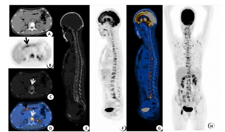
Objective To explore the clinical characteristics of patients with bone metastases from gastric cancer and the clinical application of 18F-FDG PET/CT in bone metastasis from gastric cancer. Methods From January 2010 to December 2020, 21 gastric cancer patients with bone metastases from gastric cancer who had undergone 18F-FDG PET/CT before treatment at the Cancer Prevention and Treatment Centre of Sun Yat-sen University and whose primary foci were pathologically confirmed, including 9 males and 12 females with a median age of 57.0 (28.0, 81.0) years old. The clinical data and the characteristics of the 18F-FDG PET/CT qualitative and semi-quantitative (maximum standardized uptake value SUVmax) index were retrospectively analyzed. According to the tissue differentiation of the primary foci, the patients were divided into: 13 cases (65.0%) in the hypofractionated group and 7 cases (35.0%) in the intermediate-hypofractionated group. According to the Lauren typing, the patients were divided into: 4 cases (20.0%) in the intestinal group, 9 cases (45.0%) in the mixed group and 7 cases (35.0%) in the diffuse group. According to Soloway classification, the number of bone metastases was divided into 3 groups (class Ⅰ≤5, class Ⅱ 6-20, class Ⅲ > 20), including 6 cases of class (group) Ⅰ, 4 cases of class (group) Ⅱ, and 11 cases of class (group) Ⅲ. Bone metastasis types were divided into 3 groups according to the manifestations on PET/CT (osteolytic, mixed, and osteogenic). There were 6 cases of pure osteolytic metastasis, 11 cases of mixed(osteolytic/osteogenic), and 4 cases of osteogenic metastasis. Results In this group of patients with gastric cancer, bone metastases occurred in the spine (19/21), scapula, ribs, collarbone, sternum (18/21), and pelvis (17/21), of which 3 cases were accompanied by bone marrow infiltration. The serum alkaline phosphatase and lactate dehydrogenase were elevated in 11 cases (57.9%) and 7 cases (36.8%) during the same period. There were differences in lactate dehydrogenase levels between different types of bone metastasis (χ2=6.823, P=0.047), with elevated lactate dehydrogenase being more common in mixed metastases. There were statistically significant differences in the SUVmax of metastatic bone lesions in gender, Lauren's classification, and Soloway classification (Z=-1.990, H=6.326, H=6.070, all P < 0.05). The median SUVmax 11.6 (7.3, 32.1) in the female group was higher than that in the male group [7.2 (3.7, 17.1)]; The diffuse-type group [12.2 (5.3, 32.1)] and the mixed-type group [10.8 (7.2, 17.2)] were higher than the intestinal-type group [6.7 (3.7, 7.3)]. The higher the Soloway classification, i.e. the higher the number of bone metastases, the higher the SUVmax of metastatic bone lesions. There was a difference in the Soloway grading by gender (χ2=6.832, P=0.033), with female patients generally having a higher Soloway grade than males. Primary and bone metastases SUVmax size was age dependent, with the lower age group (< median age 57.0 years) having higher bone metastases SUVmax and lower primary SUVmax. (χ2=5.838, 10.831, P < 0.05). Conclusion 18F-FDG PET/CT can provide a comprehensive assessment of bone metastases from gastric cancer. Bone metastasis from gastric cancer have specific characteristics, such as often occurring in the mid-axis bone, with osteolytic and multifocal metastases commonly and are often associated with elevated lactate dehydrogenase and alkaline phosphatase. Elevated lactate dehydrogenase is more common in patients with mixed bone metastases.
2022, 45(1): 8-12.
doi: 10.12122/j.issn.1674-4500.2022.01.02
Abstract:
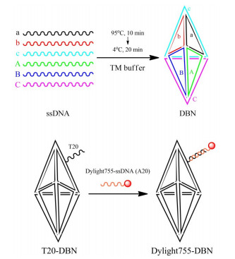
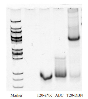
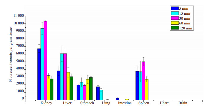
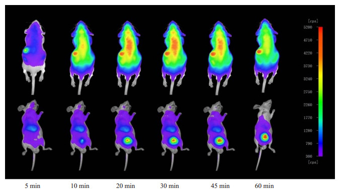
Objective To construct a kind of fluorescent molecular probes based on DNA Bipyramid Nanostructure (DBN), and to explore the biological properties of the probes at the level of living animals. Methods DBN with side arm chains, which was prepared through the one-step annealing method, was mixed with single-stranded oligonucleotide A20 coupled with DyLight 755 (DyLight 755-A20) to prepare fluorescent molecular probe DyLight 755-DBN. DyLight 755-DBN was injected into the mice through the tail vein, then the mice were executed at different time points, and fluorescence counts were taken from organs of interest to explore the in-vivo distribution of the molecular probes. DyLight 755-DBN was injected into the mouse through the tail vein, and in-vivo imaging studies of small animals were performed at different time points. Results The DBN with side arm chains was successfully prepared and characterized by polyacrylamide gel electrophoresis. The fluorescent molecular probe DyLight 755-DBN was successfully prepared by the equimolar hybridization of DBN with side arm chains and DyLight 755-A20. In vivo distribution experiments showed that after DyLight 755-DBN was entering the body, the fluorescent signal was mainly concentrated in the kidney, liver, spleen and stomach. There was a certain fluorescent signal in the lungs of experimental mouse during 5-15 min, but the fluorescence signal was almost absent after 15 min. In vivo imaging results showed that the fluorescent signal of DyLight 755-DBN was mainly concentrated in the liver and stomach, and a strong fluorescent signal in the bladder. Conclusion The fluorescent molecular probe DyLight 755-DBN is a kind of molecular imaging probe with excellent properties.
2022, 45(1): 13-17.
doi: 10.12122/j.issn.1674-4500.2022.01.03
Abstract:
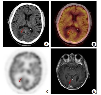
Objective To analyze and discuss the diagnostic value and differences of 18F-FDG PET/CT+head enhanced CT and 18F-FDG PET/CT + head enhanced MRI for brain metastasis of lung cancer. Methods Clinical data of 327 patients with lung cancer were randomly selected to compare the imaging data of 18F-FDG PET/CT, head-enhanced CT and head-enhanced MRI to analyze the effects of 18F-FDG PET/CT combined with head enhanced CT and 18F-FDG PET/CT combined with head-enhanced MRI on the staging of lung cancer. Comparison of the value of 18F-FDG PET/CT, head-enhanced CT and head-enhanced MRI in the detection of brain metastases from lung cancer; Comparison between 18F-FDG PET/CT with head-enhanced MRI, and the difference in the presentation of cystic lesions and oedema between the group with and without missed brain metastases from lung cancer. Results The combined examination methods of 18F-FDG PET/CT+head enhanced CT and 18F-FDG PET/CT + head enhanced MRI had a statistically significant difference in the detection of lung cancer brain metastasis (χ2=305.58, P < 0.01). The detection rates of brain metastases from lung cancer by PET/CT, head enhanced CT and head enhanced MRI were 7.34%, 12.23% and 19.88%, respectively. The difference in the detection of brain metastases from lung cancer among the three methods was significant(χ2=22.867, P < 0.01). There was no significant difference in cystic degeneration and edema between the missed group and the unmissed group (χ2=0.657, P > 0.05;χ2=0.023, P > 0.05). The rate of cystic degeneration was 31.70% (13/41) in the missed diagnosis group and 41.67% (10/24) in the unmissed diagnosis group, and the rates of edema was 56.09% (23/41) in the missed diagnosis group and 54.17% (13/24) in the unmissed diagnosis group. Conclusion Both 18F-FDG PET/CT + head enhanced CT and 18F-FDG PET/CT + head enhanced MRI can improve the detection rate of lung cancer brain metastasis, which is important for accurately determining the stage of lung cancer, grasping the details of brain metastasis, and reducing the missed diagnosis rate of brain metastasis. The highest value has been found in the combination of 18F-FDG PET/CT+head MRI.
2022, 45(1): 18-22.
doi: 10.12122/j.issn.1674-4500.2022.01.04
Abstract:
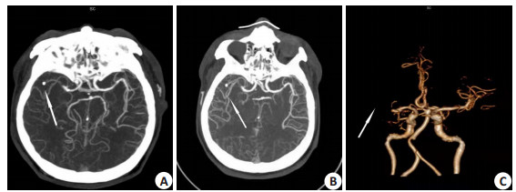
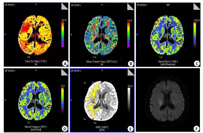
Objective To explore the correlation between the distribution of cerebral infarction caused by severe stenosis or occlusion of the middle cerebral artery and the establishment of collateral circulation using dual-source CT cerebral perfusion imaging (CTP), and further explore the state of microcirculation in the area of ischemia established by different collateral vessels. Methods 80 patients with unilateral middle cerebral artery stenosis > 70% and symptomatic sudden stroke confirmed by CT angiography were collected. On the diffusion-weighted image, the types of cerebral infarction were divided into perforating artery infarction, large area infarction, cortical blood supply area infarction, watershed infarction and multiple cerebral infarction and no infarcts. After CTP imaging, all patients were divided into two groups with good collateral circulation and poor circulation, and differences in perfusion parameters and related risk factors between the two groups were analyzed. Penumbra calculation software was used to analyze the ischemic penumbra area and core infarct area, and to compare whether the diffusion-weighted image high signal area was different from the core infarct area and whether distribution of cerebral infarction correlates with collateral circulation. Results The proportion of males and the proportion of hypertension patients were significantly higher in the group with poor collateral circulation than that in the group with good collateral circulation (P < 0.05). CTP results of all patients were positive, 66 cases were positive for core infarct area in 68 cases in diffusion weighted image. Two methods were consistent in the diagnosis of acute cerebral infarction. The type of cerebral infarction in the group with good collateral circulation being predominantly penetrating artery infarction, and the group with poor collateral circulation being predominantly watershed infarction. For the comparison of the perfusion parameters of the ischemic penumbra area between the two groups, the difference in cerebral blood flow value was not significant (P > 0.05), while the increase in cerebral blood volume, and the decrease in mean transit time and time to peak in the group with good collateral circulation were statistically significant (P < 0.05). Conclusion The distribution type of cerebral infarction caused by severe stenosis or occlusion of the middle cerebral artery is related to the establishment of collateral circulation. CTP imaging can detect the ischemic areas earlier, accurately assess the degree of stenosis of the responsible blood vessel and local hemodynamic information. The shadow calculation mode allows faster assessment of the ischemic core infarct area and the ischemic penumbra area.
2022, 45(1): 23-28.
doi: 10.12122/j.issn.1674-4500.2022.01.05
Abstract:
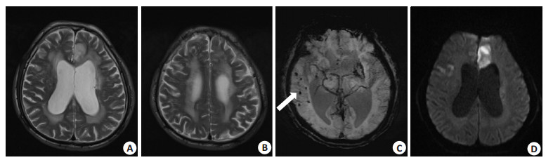
Objective To investigate the relationship between cerebral small vessel disease (CSVD) load and 3-month poor prognosis in mild acute cerebral infarction (ACI) treated with recombinant tissue plasminogen activator. Methods 161 patients with mild ACI who received intravenous thrombolysis with recombinant tissue plasminogen activator from 2016 to 2019 were retrospectively analyzed. Clinical variables studied included age, gender, vascular risk factors, National Institute of Health Stroke Score (NIHSS) as well as initial hematologic and MR parameters. Mild acute cerebral infarction was defined as a baseline NIHSS score ≤7 and a 3-month poor prognosis was defined as a modified Rankin score ≥3. CSVD load included white matter hyperintensities (WMHs), lacunes, cerebral microbleeds (CMBs) and enlarged perivascular space. Total CSVD load was evaluated based on the clinical MR images, and severity WMHs was assessed according to the Fazekas criteria. Results Of the 161 patients, 117 (72.7%) were male, and the number of patients with a poor 3-month prognosis was 29 (18.0%). Univariate analysis showed that baseline NIHSS, atrial fibrillation, symptomatic intracranial artery stenosis (SIAS), WMHs and CMBs were correlated with 3-month poor prognosis (P < 0.05). WMHs and CMBs were entered into two logistic regression equation models, respectively. In Model 1, baseline NIHSS (OR=1.601, 95% CI: 1.203-2.130, P=0.001), symptomatic intracranial artery stenosis (OR=2.658, 95% CI: 1.013-6.978, P=0.047) and WMHs (OR=1.449, 95% CI: 1.033-2.031, P=0.032) were significantly associated with 3-month poor prognosis. In Model 2, baseline NIHSS (OR=1.650, 95% CI: 1.232- 2.210, P=0.001), symptomatic intracranial artery stenosis (OR=3.732, 95% CI: 1.435-9.702, P=0.007) and CMBs (OR=1.242, 95% CI: 1.062-1.452, P=0.007) were significantly correlated with poor prognosis at 3 months after intravenous thrombolysis for mild ACI. Conclusion WMHs and CMBs are predictors of poor prognosis at 3 months after intravenous thrombolysis in mild ACI.
Scale for predicting early hematoma expansion in intracerebral hemorrhage based on CT-specific signs
2022, 45(1): 29-34.
doi: 10.12122/j.issn.1674-4500.2022.01.06
Abstract:
Objective To explore multiple risk factors which may trigger early hematoma expansion and to establish a predictive scale of early hematoma expansion for intracerebral hemorrhage. Methods The clinical data of 107 patients with spontaneous cerebral hemorrhage were analyzed retrospectively. The relationship between multiple risk factors and early haematoma enlargement was analysed by univariate analysis and multivariate logistic regression. Results Univariate analysis and multiple Logistic regression analysis showed that six indicators were statistically significant (P < 0.05): use of anticoagulant or antiplatelet drugs, baseline intracerebral hemorrhage volume, time to initial CT, acute blood pressure, CT special signs, recurrent intracerebral hemorrhage were independent risk factors for early hematoma expansion (P < 0.05). According to the above data, a scoring table was established, in which scores ≥16 points at extremely high- risk group, 11-15 at high-risk group, 6-10 at medium risk group and ≤5 at low-risk group. Conclusion The prediction scale can accurately predict the enlargement of haematoma in the early stage of intracerebral hemorrhage, which is independent of other clinical and imaging predictors, and can be used as a reliable reference basis for correct clinical treatment plan.
2022, 45(1): 35-39.
doi: 10.12122/j.issn.1674-4500.2022.01.07
Abstract:
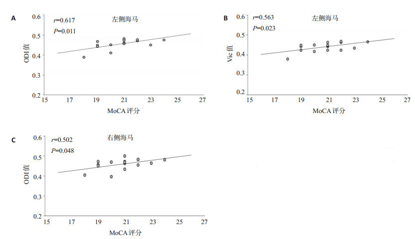
Objective To explore the changes of hippocampal microstructure in patients with Parkinson's disease cognitive impairment (PD-CI) by using neurite orientation dispersion and density imaging (NODDI), and explore its relationship with cognitive function. Methods Thirty-six patients with Parkinson's disease in our hospital were selected as Parkinson's disease group, and 20 healthy volunteers were selected as control group. The patients with PD were given Montreal Cognitive Score (MoCA) before examination. Among them, 16 patients with MoCA score < 26 were divided into PD-CI group (PD-CI), and 20 patients with MoCA score ≥26 were divided into normal Parkinson's cognition group. According to the results of NODDI scanning, the related parameters, such as orientation dispersion index (ODI), intracellular volume fraction (Vic) and isotropic water molecule volume fraction (Viso) were obtained, and the differences in ODI, Vic and Viso values in the region of interest were analyzed. The indicators with statistically significant differences were each subjected to Pearson correlation analysis with MoCA scale scores to investigate the correlation between each diffusion index and MoCA scale scores. Results The values of ODI and Vic in hippocampus of PD patients were significantly lower than those in healthy control group (P < 0.05), and the values of ODI and Vic in PD-CI group were lower than those in cognitive normal of Parkinson's disease group, with a statistically significant difference (P < 0.05), but there was no significant difference between Viso and healthy control group. The left hippocampal ODI value correlated most strongly with the MoCA scale score (r=0.617, P < 0.05). Conclusion NODDI technique can reflect the changes of hippocampal microstructure of PD patients, and to a certain extent, reflecting the clinical cognitive status of PD patients, thus providing important reference value in its auxiliary diagnosis.
2022, 45(1): 40-43.
doi: 10.12122/j.issn.1674-4500.2022.01.08
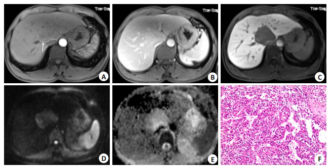
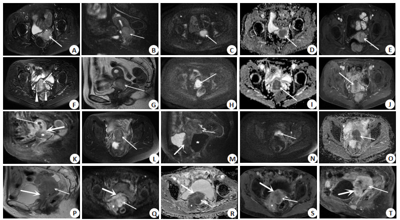
Abstract:
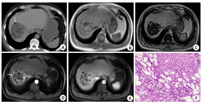
Objective To investigate the CT and MRI features of hepatic perivascular epithelioid cell differentiation tumors (PEComa) in different types. Methods The imaging data of 12 patients with liver PEComa confirmed by surgical pathology from July 2014 to July 2018 in our hospital were retrospectively analyzed to analyze the location, density and signal characteristics, and form of enhancement of the lesions on CT and MRI images. Results The fat-containing lesions showed no significant enhancement of the fatty component with the naked eye, while the soft-tissue component was significantly enhanced. Dynamic enhancement scans showed significant enhancement in the arterial phase and intra-tumoural vessels, and moderate to mild enhancement in the portal and delayed phases, with a circularly enhanced pseudo-envelope. In fat-deficient type, which was mainly characterized by a substantial soft tissue mass, the fat components are invisible to the naked eye. Dynamic enhancement scan showed marked homogeneous enhancement in the arterial phase of the lesion. with coarse distorted malformed vessels and aneurysmal nodular enhancement visible in the portal and equilibrium phases, delayed enhancement in the portal and equilibrium phases. Due to lack of normal hepatocytes in the lesions, the hepatobiliary specific phase showed low signal without contrast uptake. The fat-containing type tumors need to be differentiated from angiomyolipoma, but the lack of specificity in imaging means that the non- fatty type is almost always misdiagnosed as hepatocellular carcinoma. Conclusion There is unique immunohistochemical features in hepatic PEComa. If it is staged according to the presence or absence of a fat component, the imageology manifestations have certain characteristics. However, the lesions have no specificity in ADC or hepatobiliary specific phase, and cannot be used as a separate diagnostic basis. The probability of misdiagnosis before clinical procedures is relatively high in fat-deficient type, so it is important for radiologists to be more aware of this condition.
2022, 45(1): 44-48.
doi: 10.12122/j.issn.1674-4500.2022.01.09
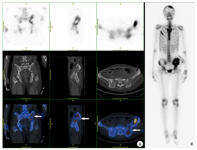
Abstract:
Objective To explore the diagnostic value and imaging characteristics of whole-body bone imaging and SPECT/CT in bone destruction caused by Talaromyces marneffei. Methods We retrospectively analyzed 47 patients with Talaromyces marneffei who underwent whole-body bone imaging and SPECT/CT tomographic fusion imaging in the Department of Nuclear Medicine in our hospital from June 2016 to June 2021, including 25 males and 22 females, aged from 17 to 72 years old, with a median age of 51 years. We compared the diagnostic efficacy of whole-body bone imaging and SPECT/CT in bone destruction, and discussed the imaging characteristics of bone destruction. Results There was a significant difference in the coincidence rate of whole-body bone imaging and SPECT/CT tomographic fusion imaging in the diagnosis of bone destruction caused by Talaromyces marneffei (P < 0.05). The sensitivity of whole-body bone imaging in the diagnosis of bone destruction was 84.4%, the specificity was 100%, and the diagnostic coincidence rate was 85.1%. The diagnostic sensitivity of SPECT/CT for TM bone destruction was 97.8%, the specificity was 100%, and the diagnostic compliance rate was 98.9%. Bone destruction of Talaromyces marneffei mainly affects the axial bones and is associated with the involvement of several locations throughout the body, with the bones of the extremities and the skull being the relatively specific sites of invasion. Conclusion Whole-body bone imaging can observe the whole-body focus at one time, and the abnormal activity of bone metabolism can be detected in the early stage of the lesion. The coincidence rate of SPECT/CT diagnosis is high. The combination of SPECT/CT has a high diagnostic yield and is of good use in the case of bone destruction caused by Talaromyces marneffei.
2022, 45(1): 49-54.
doi: 10.12122/j.issn.1674-4500.2022.01.10
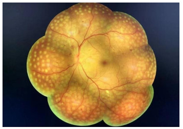
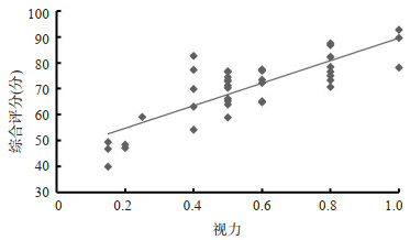
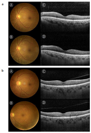
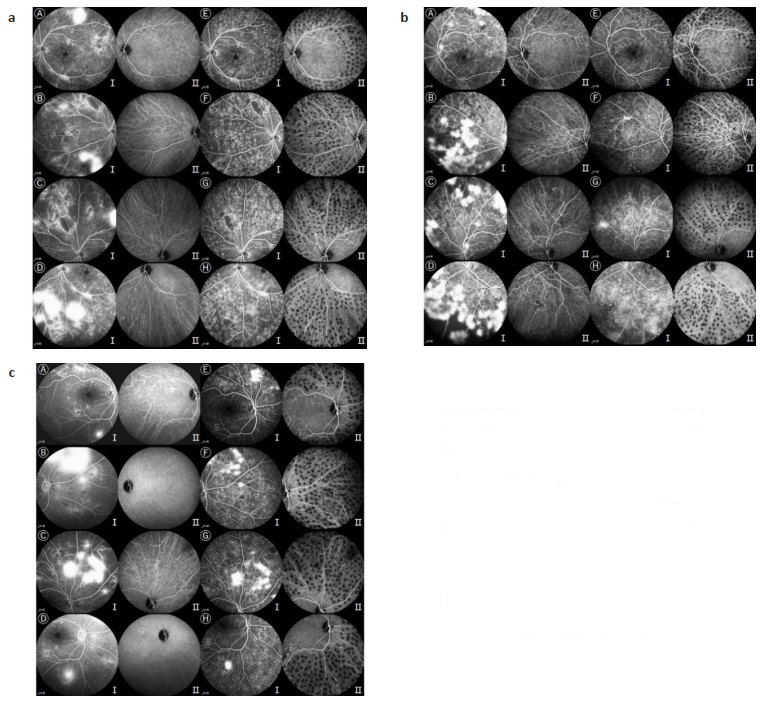
Abstract:
Objective To evaluate the safety, effectiveness and the effect on vision-related quality of life of panretinal photocoagulation (PRP), which is used as a conventional method for the treatment of severe non-proliferative diabetic retinopathy (NPDR) and proliferative diabetic retinopathy (PDR). Methods Forty-one patients who were diagnosed with severe NPDR or PDR from January to December 2020 in the ophthalmology department of the Second Affiliated Hospital of Xi'an Jiaotong University were retrospectively collected, and patients with PDR requiring surgical treatment were excluded. Based on the fluorescein fundus angiography, the patients were divided into severe NPDR group (n=20) and PDR group (n=21). Fundus photography, fluorescein fundus angiography, and optical coherence tomography were performed before PRP treatment and 1 month after PRP. Chinese version of the Visual Function-related Quality of Life Scale questionnaire was used to evaluate their quality of life before and 6 months after PRP treatment. Results Forty-one patients completed PRP treatment and follow-up, 13 of whom were treated with PRP combined with vitreous cavity injection of anti-vascular endothelial growth factor drugs. No serious complications occurred in 41 patients after PRP treatment, and neovascularization was significantly reduced, with no statistically significant differences in preoperative and postoperative best corrected visual acuity (P > 0.05). In the severe NPDR group, the scores of role limitations (65.63±18.97 vs 68.75±20.48, P=0.021) and social function (73.75±15.12 vs 76.25±15.12, P=0.042) was increased after PRP treatment. In the PDR group, the scores of peripheral visual acuity (72.62±19.21 vs 67.86±19.60, P=0.042) was decreased after PRP treatment. There was no statistically significant difference in any other subscale scores between the two groups before and after treatment (P > 0.05). Conclusion PRP is a safe and effective method in the treatment of severe NPDR and PDR. To a certain extent, PRP could improve the quality of life of patients in relation to their vision.
2022, 45(1): 55-60.
doi: 10.12122/j.issn.1674-4500.2022.01.11
Abstract:
Objective To analyze the diagnostic value of MRI combined with Federation International of Gynecology and Obstetrics (FIGO) staging system in different stages of cervical cancer. Methods A total of 85 patients with cervical cancer who attended our hospital from January 2014 to December 2020 were selected, and the patients' clinical data and imaging data, etc. were retrospectively analyzed and compared with the pathological findings. The FIGO staging system and MRI combined with FIGO staging system were used for diagnosis. The sensitivity, specificity and consistencies tests were performed to compare, the accuracy rates and consistencies of MRI combined with clinical FIGO staging and clinical FIGO staging alone in diagnosing different stages of cervical cancer. Results The accuracy of FIGO staging was 92.94% for stage Ⅰa, 90.59% for stage Ⅰb, 85.88% for stage Ⅱa, 82.35% for stage Ⅱb, 97.65% for stage Ⅲb and 96.47% for stage Ⅳ. The overall accuracy rate was 72.94%. The consistency of FIGO staging was good in the diagnosis of cervical cancer at stages Ⅰa and Ⅰb (Kappa > 0.60), while the consistency was poor in the diagnosis of stages Ⅱa, Ⅱb and Ⅲb (Kappa < 0.60). The accuracy rate of MRI combined with clinical FIGO staging tests was 92.94% for stage Ⅰa, 90.59% for stage Ⅰb, 94.12% for stage Ⅱa, 96.47 % for stage Ⅱb, 100.00% for stage Ⅲb, 100.00% for stage Ⅳ and 88.24% for overall. The consistency of MRI combined with clinical FIGO staging in the diagnosis of cervical cancer at stages Ⅰa, Ⅰb, Ⅱa, Ⅱb, Ⅲb and Ⅳ was good (Kappa > 0.60). The accuracy rates of MRI combined with clinical FIGO staging were significantly higher than those of clinical FIGO staging alone in the diagnosis of stages Ⅱa, Ⅱb and overall (P < 0.05). Conclusion MRI combined with clinical FIGO staging can effectively staging cervical cancer, and its accuracy is higher than that of clinical FIGO staging alone.
2022, 45(1): 61-65.
doi: 10.12122/j.issn.1674-4500.2022.01.12
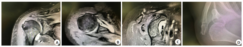
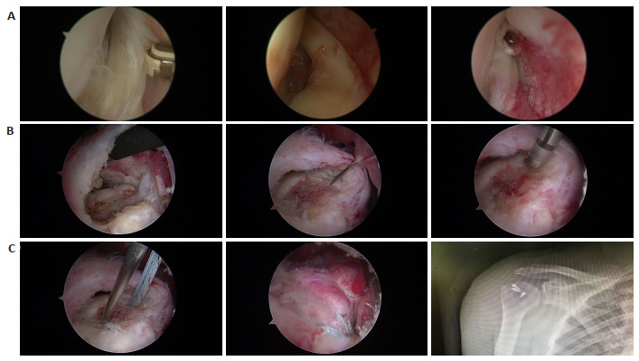
Abstract:
Objective To study the clinical efficacy of arthroscopic biceps long head tendon transection combined with suture bridge double-row repair in the treatment of huge rotator cuff tears. Methods Fifty patients with huge rotator cuff tear admitted to our hospital from June 2019 to March 2021 were retrospectively analyzed as research samples, and they were divided into two groups based on the differences of operation methods; 25 patients in the control group were treated with double-row suture bridge repair under arthroscopy, and 25 patients in the experimental group were treated with double row suture bridge repair under arthroscopy. The differences in constant Murley shoulder function score, short-term efficacy and retear rates between the two groups were compared. Results The pain score, activity of daily living (ADL) score, range of motion (ROM) score, muscle strength score and Constant-Murley total scores of the two groups after treatment were higher than those before treatment, the difference was statistically significant (P < 0.05); the differences in Constant-Murley scores of each dimension and total scores between the two groups before treatment were not statistically significant (P > 0.05), but the experimental group after treatment Constant-Murley scores and total scores of each dimension were higher in the experimental group than in the control group (P < 0.05). The total excellent and good rate of the experimental group (96.00%) was higher than that of the control group (64.00%), the difference was statistically significant (P < 0.05); the difference in the retear rate between the two groups was not statistically significant (P > 0.05). Conclusion In the field of surgical treatment of patients with huge rotator cuff tear, the treatment of biceps brachii long head tendon cutting combined with suture bridge double-row repair under arthroscopy can further improve the patients' pain symptoms, joint mobility and muscle strength, and enhance the quality of life in the short term, with definite overall efficacy and low risk of re-tear.
2022, 45(1): 66-70.
doi: 10.12122/j.issn.1674-4500.2022.01.13
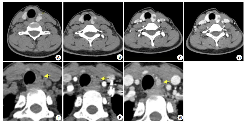
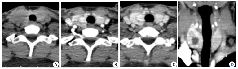
Abstract:
Objective To observe the application value of dual-phase CT enhancement combined with three-dimensional reconstruction in the qualitative diagnosis of thyroid tumors. Methods The clinical data of 98 patients with thyroid tumors admitted to the hospital from January 2017 and May 2021 were retrospectively analyzed. All patients underwent dual-phase CT enhancement combined with three-dimensional reconstruction, and surgical pathological examination was used as the gold standard. The diagnostic value of dual-phase CT enhancement and dual-phase CT enhancement combined with threedimensional reconstruction for thyroid tumors was explored, and the diagnostic value of dual-phase CT enhancement combined with three-dimensional reconstruction for pathological features of thyroid tumors was analyzed. Results The diagnostic sensitivity of dual-phase CT enhancement for thyroid tumors was 84.91%, the specificity was 82.22% and the accuracy rate was 83.67%, the diagnostic sensitivity of duplex CT enhancement combined with 3D reconstruction technique for thyroid tumors was 92.45%, the specificity was 80.00% and the accuracy was 86.73%. The diagnostic accuracy rates of dualphase CT enhancement combined with three-dimensional reconstruction for unilateral or bilateral lymph node enlargement, coarse calcification, microcalcification and the number of lesions were 94.23%, 72.73%, 89.74% and 87.12% respectively. Dualphase CT enhancement combined with three-dimensional reconstruction showed that the unilateral or bilateral lymph node enlargement in patients with thyroid cancer accounted for 71.43% of the total detection, and the unilateral or bilateral lymph node enlargement in thyroid adenoma accounted for 28.57% of the total detection, and calcification accounted for 52.86% of the 70 lesions in patients with thyroid cancer and 31.11% of the 45 lesions in patients with thyroid adenoma. Conclusion Dualphase CT enhancement combined with three-dimensional reconstruction are of high value in the qualitative diagnosis of thyroid tumors, and cervical lymph node enlargement and calcification are of value as an aid to diagnosis.
2022, 45(1): 71-75.
doi: 10.12122/j.issn.1674-4500.2022.01.14
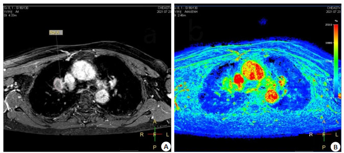
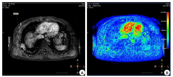
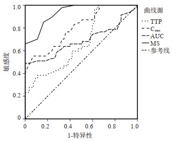
Abstract:
Objective To investigate the clinical dynamic contrast enhancement magnetic resonance imaging (DCE-MRI) for the semi-quantitative differential diagnosis of benign and malignant pulmonary occupying lesions. Methods Clinical data of 102 patients with pulmonary occupying lesions in our hospital from March 2020 to March 2021 were retrospectively analyzed. Based on the pathological findings, patients with pulmonary cancer were considered as the malignant group (n=47) and patients with benign pulmonary occupying lesions were considered as the benign group (n=55). All patients received DCE and MRI scans to obtain semi-quantitative parameters including time to peak, maximum concentration, AUC of time-concentration curve, maximum slope were obtained. The differences in semi-quantitative parameters between benign and malignant groups were compared, and ROC curve was used to evaluate the discriminatory value of the each of these parameters. Results In the malignant group, the lesions were mainly cystic solidity, heterogeneous enhancement, irregular morphology and unclear borders, while those in benign group, the lesions were mainly cystic, homogeneous enhancement, regular morphology and clear or poorly defined borders. The lesion features of two groups showed no significant difference in conventional MRI (P > 0.05). the levels of the parameters of time to peak in malignant group were lower than those in benign group (P < 0.05), and the levels of the parameters of maximum concentration, AUC and maximum slope were higher than those in benign group (P < 0.05); Based on pathological results, ROC curve of semi-quantitative parameters showed that maximum slope curve, and the maximum area under the curve of maximum linear slope was 0.937. The diagnostic efficacy was higher than the rest of the parameters, with a maximum linear slope accuracy of 84.4%, a maximum concentration specificity of 92.7%, and a peak attainment time sensitivity of 97.9%. Conclusion Semi-quantitative parameters of DCE-MRI are effective in the differential diagnosis of benign and malignant pulmonary occupying lesions.
2022, 45(1): 76-80.
doi: 10.12122/j.issn.1674-4500.2022.01.15
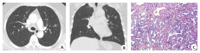
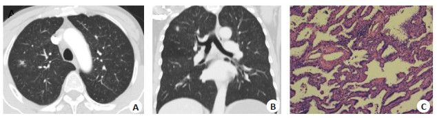
Abstract:
Objective To analyze the prediction model of benign and malignant pulmonary sub-centimeter nodules based on enhanced dual-phase CT imaging. Methods Ninty-eight patients with pulmonary sub-centimeter nodules treated in our hospital from January 2019 to March 2021 were selected as the research objects, and divided into benign group (n=64) and malignant group (n=34) according to the pathological diagnosis. All the subjects underwent enhanced two-phase CT imaging, and a logistic regression model was used to analyze the predictive model of benign and malignant nodules by enhanced dual phase CT imaging, and ROC curve was drawn to analyze the application value of the predictive model of benign and malignant pulmonary sub-centimeter nodules by enhanced dual phase CT imaging. Results There were significant differences in the incidence of burr, clear nodule boundary, upper lobe, lobar sign, vacuole sign, pleural depression sign, vascular bundle sign and ground glass density between benign lesions and malignant lesions (P < 0.05). The prediction model for predicting the benignity and malignancy of pulmonary sub-centimeter nodules by enhanced duplex CT imaging was Log(P) =1.211×burr + 2.843×lobulation+1.981×groundglass+0.793×unclearboundary+1.326; The area under curve of enhanced dual phase CT imaging in predicting the benign and malignant of pulmonary sub-centimeter nodules was 0.930 (P < 0.05). Conclusion the model for predicting the benignity and malignancy of pulmonary sub-centimeter nodules based on enhanced dual phase CT imaging has high clinical value and high predictive value.
2022, 45(1): 81-85.
doi: 10.12122/j.issn.1674-4500.2022.01.16
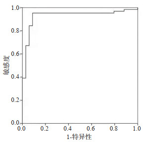
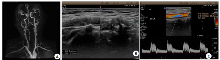
Abstract:
Objective To explore the relationship between high-resolution MRI combined with carotid ultrasound and the detection of carotid stenosis in patients with acute cerebral infarction and the risk factors affecting the prognosis of patients. Methods In this prospective study, 67 patients diagnosed with cerebral infarction from January 2018 to December 2020 were selected as the research objects, including 35 patients with carotid stenosis and 32 patients with non-carotid stenosis. The differences of carotid ultrasound and 3.0 T MRI diagnosis between the two groups were compared, and the risk factors affecting the prognosis of patients were analyzed. Results There was no significant difference in gender, age and BMI between patients with carotid stenosis and non-carotid stenosis group (P > 0.05). The proportion of patients with diabetes mellitus carotid stenosis group was not significant (χ2=7.102, P=0.007), hypertension (χ2=5.902, P=0.015) and hyperlipidemia (χ2=4.532, P= 0.033) was significantly higher in the carotid stenosis group than that in the non-carotid stenosis group. The maximum systolic velocity (PSV) (t=4.992, P < 0.001)and end-diastolic velocity (EDV) (t=15.210, P < 0.001) in the carotid stenosis group were significantly lower than that in the non-carotid stenosis group, and the intima-media thickness (IMT) (t=16.835, P < 0.001) in the patients with carotid stenosis was significantly higher than those in the non-carotid stenosis group. The difference in caroid PSV (F=4.968, P < 0.001), IMT (F=16.468, P < 0.001), EDV (F=14.791, P < 0.001) between patients with different degrees of carotid stenosis were statistically significant. After comparison between the two groups, PSV and EDV in the three groups were ranked from high to low were mild, moderate and severe, and IMT was in the order of severe, moderate and mild groups. There was strong diagnostic agreement between carotid ultrasound and MRI, with different prognoses for age (t=4.532, P < 0.001), BMI (t=2.324, P=0.023), diabetes (χ2=10.602, P=0.001), hypertension (χ2=7.502, P=0.006), hyperlipidemia (χ2=7.202, P= 0.007), PSV (t=20.013, P < 0.001), IMT (t=4.708, P < 0.001), EDV (t=22.018, P < 0.001). Through multivariate analysis, age, BMI, hyperlipidemia, diabetes mellitus, hypertension, PSV, IMT and EDV were all the risk factors for poor prognosis. Conclusion Carotid ultrasound combines with 3.0T MRI examination in patients with cerebral infarction is significance for the severity of the disease, while older age, high weight, hypertension, diabetes and hyperlipidemia are all important risk factors for disease progression.
2022, 45(1): 90-93.
doi: 10.12122/j.issn.1674-4500.2022.01.18
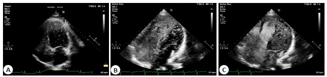
Abstract:
Objective To analyze the value of P-selection combined with right echocardiography (cTTE) diagnosing patients with migraine combined with unclosed foramen ovale. Methods A total of 64 migraine patients admitted to our hospital from April 2019 to April 2021 were selected and divided into the occurrence group (n=47) and the non-occurrence group (n=17) according to the complication of patent foramina ovale. The serum P-selection levels of all patients was compared, and right echocardiography was performed to compare the diagnostic accuracy of different examination methods. Results The levels of P selectin were significant higher in patients with patent foramen ovale than in those without patent foramen ovale, with a statistically significant difference (P < 0.05). Using intraoperative closure as the gold standard, the sensitivity of P-selectin for the diagnosis of migraine combined with oval foramen nonocclusion was 68.09%, specificity was 82.35%, positive predictive value was 91.43%, negative predictive value was 48.28%, and diagnostic compliance rate was 71.88% and concordance 0.412. Using intraoperative closure as the gold standard, the sensitivity of P-selectin combined with cTTE for the diagnosis of migraine combined with patent foramen ovale was 82.98%, specificity was 88.24%, positive predictive value was 95.12%, negative predictive value was 65.22%, diagnostic compliance was 84.38%, with a consistency of 0.640. Conclusion P-selection combined with right aspiration imaging can improve the diagnostic accuracy in patients with migraine combined with unclosed foramen ovale.
2022, 45(1): 94-100.
doi: 10.12122/j.issn.1674-4500.2022.01.19
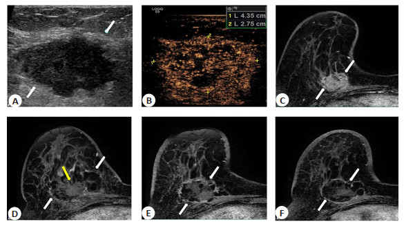
Abstract:
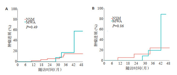
Objective To compare the feasibility and effectiveness of ultrasound-guided microwave ablation (MWA) and nipple sparing mastectomy (NSM) for early breast cancer (T0/1/2 N0/1 M0). Methods Retrospective analysis of 83 patients with early breast cancer who underwent MWA or NSM from January 2014 to January 2020 in our hospital. including 30 patients in the MWA group and the other 53 patients in the NSM group. Propensity score matching was performed at 1∶1 to balance the baseline characteristics. The primary evaluation metric was tumor progression in the intention-to-treat population, and secondary evaluation metrics included overall survival, cosmetic satisfaction and complications. Results Before propensity score matching The mean tumor size was 2.3 cm (0.3-5.0 cm). After propensity score matching, each group had 19 patients and the baseline characteristic were balanced. Median follow-up was 34.1 months (IQR 23.1-41.1 months). All the tumors achieved technique effectiveness. For the management of metastatic lymph nodes, 4 axillary lymph nodes were ablated/resected in both groups (4/19, 21.0%). There were fewer adjuvant systemic postoperative treatments in the MWA group than in the NSM group (36.8% vs 73.7%, P=0.02). A total of 6 cases of tumour progression occurred, including 3 in the MWA and 3 in the NSM. One local tumor progression occurred in the MWA group at 42 mouths, one ipsilateral breast recurrence occurred at 28 months and 1 case of brain metastasis at 32.8 months after MWA. 1 case of ipsilateral breast metastasis at 10.2 months and 1 case of bone metastasis at 31.2 months and 1 case of bone metastasis at 34 months postoperatively in the NSM group. Two groups had no significant difference in tumor progression (P=0.68). No participants in both groups developed cancer related death and major complications (P>0.05). MWA achieves similar outcomes to NSM and better cosmetic satisfaction during median follow-up in patients with early-stage breast cancer, and that who do not want or are intolerant of surgery.However, MWA had a shorter operation time (32 min vs 133 min, P < 0.001), a shorter postoperative hospital stay (3 d vs 5 d, P < 0.001) and better beauty satisfaction (P < 0.001). Conclusion Preliminary study showed that in selected early breast cancer patients, MWA has achieved similar outcomes to early breast cancer treatment and better aesthetic satisfaction as NSM during an intermediate follow-up. MWA offers a minimally invasive, safe and tolerable local treatment option for patients who reluctant or intolerant to breastconserving surgery.
2022, 45(1): 101-105.
doi: 10.12122/j.issn.1674-4500.2022.01.20
Abstract:
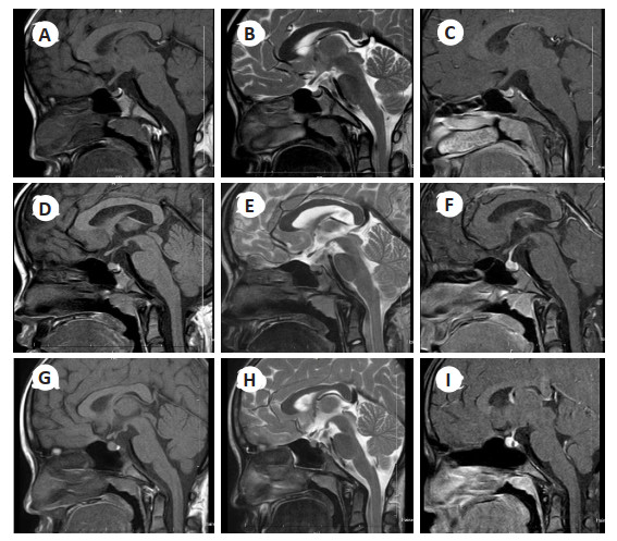
Objective To investigate the imaging features of pituitary MRI and its correlation with bone age in children with central precocity (CPP). Methods A total of 96 children with central precocity admitted to our hospital from November 2019 to November 2021 were selected as the research group, and another 90 children with healthy physical examination during the same period were selected as the control group. The MRI imaging manifestations and bone age X- ray manifestations were compared between the two groups, and the correlation between MRI imaging features and bone age X-ray in children with CPP was analyzed. Results In MRI parameters, the length of coronal high diameter and anterior and posterior diameter in the study group was longer than that in the control group (P < 0.05), and the width diameter of coronal was shorter than that in the control group (P < 0.05). There was no significant difference in sagittal height between the two groups (P>0.05). The proportion of children in the research group with a "flat shape" superior pituitary margin was lower than that in the control group (P < 0.05), and the proportion of children with a "raised shape" superior pituitary margin was higher than that in the control group (P < 0.05); There was no significant difference in the proportion of "depressed shape" in the superior pituitary margin between the two groups (P>0.05). X-ray results of bone age in the study group showed that the proportion of "normal bone age" was significantly lower than that in the control group (P < 0.05), and the proportion of "advanced bone age" was higher than that in the control group (P < 0.05). There was no significant difference in the proportion of "delayed bone age" between the two groups (P>0.05). In the MRI parameters, the coronal height and anterior-posterior diameter of the study group were longer than In the study group, the coronal height diameter, coronal width diameter, sagittal height diameter and anterior and posterior diameter of the pituitary were negatively correlated with the advance of bone age (r=-0.216, -0.345, -0.539, -0.478, P < 0.05), and positively correlated with the delayed bone age (r=0.516, 0.609, 0.784, 0.542, P < 0.05); The correlation between pituitary parameters and normal bone age was weak, with coronal height diameter and sagittal height diameter correlating with it (r=0.490, 0.241, P < 0.05), and no significant correlation with coronal width diameter and anterior and posterior diameter (P>0.05). Conclusion Excluding organic causes, the pituitary morphology and height of children with CPP were found to be significantly abnormal, mostly in the shape of "bulge", and positively correlated with both early and delayed bone age. The combination of pituitary imaging features and bone age can provide guidance for the clinical diagnosis and treatment of CPP.
2022, 45(1): 106-109.
doi: 10.12122/j.issn.1674-4500.2022.01.21
Abstract:
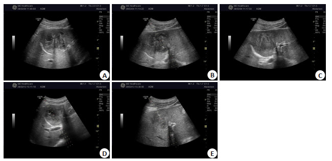
Objective To investigate the effects of ultrasound-guided percutaneous catheter drainage on oxidative stress index and imaging characteristics of patients with bacterial liver abscess. Methods Seventy patients with bacterial liver abscess in our hospital from October 2018 to July 2021 were selected. They were divided into control group (traditional open abscess incision and drainage) and observation group (ultrasound-guided percutaneous catheter drainage) using random number table method (n=35/group). The control group underwent traditional open abscess incision and drainage, while the observation group underwent ultrasound-guided percutaneous puncture and drainage. The preoperative and postoperative imaging manifestations of the observation group were compared as well as the clinical treatment results. The oxidative stress indicators (malondialdehyde, superoxide dismutase, serum cortisol) levels and the incidence of complications were compared between the two groups before and after treatment. Results The total effective rate of observation group was higher than control group (97.14% vs 77.14%, P < 0.05). Imaging results showed that there were mixed echo areas and liquid dark areas in the liver area before surgery, while the intraoperative and ultrasound guided puncture needles entered the mixed echo area of liver abscess during surgery, and liquid dark areas disappeared immediately after surgery. The operation, hospitalization and recovery time of indexes in the observation group were shorter than those in the control group (P < 0.05). Before treatment, there were no statistically significant differences in superoxide dismutase, malondialdehyde and cortisol levels between the two groups (P> 0.05). After treatment, superoxide dismutase level in both groups increased, and the observation group was higher than the control group (P < 0.05), malondialdehyde and cortisol levels decreased in both groups, and were lower in the observation group than in the control group (P < 0.05). The overall incidence of postoperative complications such as biliary fistula, pneumothorax, wound infection, hemorrhage from intrahepatic pus cavity and diffuse peritonitis in the observation group was significantly lower than that in the control group (20.00% vs 2.86%, P < 0.05). Conclusion Ultrasound-guided percutaneous catheter drainage is effective in the treatment of bacterial liver abscess, and can effectively improve the levels of serum oxidative stress index with a low complication rate.
2022, 45(1): 110-113.
doi: 10.12122/j.issn.1674-4500.2022.01.22
Abstract:
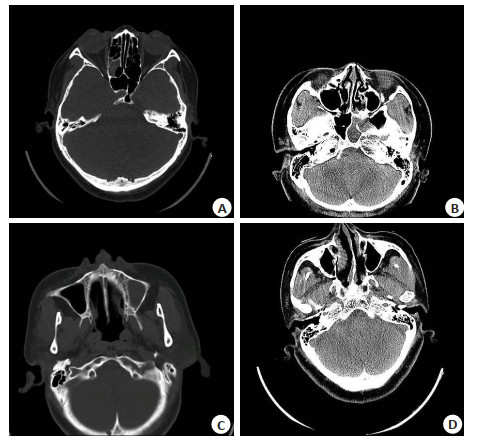
Objective To analyze the preoperative CT performance and surgical treatment effect of patients with rhinogenic headache caused by nasal septum deviation. Methods A total of 132 patients with rhinogenic headache caused by nasal septum deviation admitted to our hospital from January 2018 to December 2020 were selected as the research objects. Combined with CT imaging examination, the location, degree distribution, CT manifestations and measurement results of inferior turbinate of nasal septum deviation were analyzed to compare the headache degree of patients and analyze the surgical treatment effect of patients with different headache grades. Results In this study, CT imaging results showed that in 132 patients with deviated nasal septum, 24 cases were located anterior, 62 cases were located centrally, 40 cases in the middle and lower part, 2 cases in the middle and upper part, 1 case in the lower part and 3 cases in the lower part, 10 cases were mildly deviated, 114 cases were moderately deviated and 8 cases were severely deviated. CT results showed that 62 cases showed "C" shape (2 cases with acuminate apex), 59 cases showed reverse "C" shape (2 cases of apex sharpening), and 6 cases showed "S" shape, 1 case showed reverse "S" shape, 1 case showed " < " shape, 2 cases showed ">" shape, 1 case showed mixed shape. According to the classification of headache grade by VAS score, among 132 patients, 11 patients had grade I headache, 33 patients had grade II headache, 80 patients had grade III headache and 8 patients had grade IV headache. The angle and area of inferior turbinate on the opposite side of nasal septum deviation were larger than those on the same side, and the difference between them was statistically significant (P < 0.05). The follow-up results after the treatment showed that the total cure rate was 68.94%, the inefficiency rate was 7.58%, among which the cure rate of grade III and below headache was more than 66.00%, and the total effective rate was 92.42%. Conclusion Preoperative CT examination of patients with rhinogenic headache caused by nasal septum deviation can clearly and accurately reflect the real situation of nasal deviation, which is conducive to effective operation and improve the clinical treatment effect.
2022, 45(1): 114-118.
doi: 10.12122/j.issn.1674-4500.2022.01.23
Abstract:
Objective To analyze the relationship between CT values of renal calculus and the treatment effect of percutaneous nephroscopic holmium laser lithotripsy in patients. Methods Ninety patients with renal calculus treated by percutaneous lithotripsy with holmium laser in our hospital from October 2016 to August 2021 were selected for retrospective analysis. All patients underwent preoperative spiral CT scan to determine the size of calculi and calculate the average CT value, and were divided into two groups according to the mean CT value: Group A (n=43) with CT value < 1000 Hu, group B (n=47) with CT value ≥1000 Hu. Patients in both groups were treated with percutaneous nephroscopic holmium laser lithotripsy. Operation time, single stone removal rate, postoperative hospital stay and complications were compared between the two groups. Pearson correlation was used to analyze the correlation between CT value and operation time, Spearman correlation was used to analyze the correlation between CT value and single stone removal rate, and Logistic multifactor analysis was used to analyze the independent risk factors affecting stone residual after percutaneous holmium laser lithotripsy. Results The operation time in group A was shorter than that in group B, the single stone removal rate was higher than that of group B, and the postoperative complications were lower than that in group B, with statistically significant differences (P < 0.05). There was no significant difference in postoperative hospital stay between the two groups (P>0.05). Correlation analysis showed that CT value was positively proportional to operation time and inversely proportional to stone removal rate (P < 0.05). Further Logistic regression analysis showed that CT value and stone surface area were independent risk factors for residual stones after percutaneous with holmium laser lithotripsy. Conclusion CT value of kidney stones is significantly correlated with stone removal efficiency and stone removal rate in patients undergoing percutaneous nephroscopic holmium laser lithotripsy, and CT value and stone surface area are independent risk factors affecting stone residual after percutaneous nephroscopic holmium laser lithotripsy.
2022, 45(1): 119-123.
doi: 10.12122/j.issn.1674-4500.2022.01.24
Abstract:
Objective To explore the application value of magnetic resonance in diagnosis of idiopathic central precocity in girls and its correlation with sex hormones. Methods Fifty-eight girls with idiopathic central precocious puberty admitted to our hospital from September 2019 to October 2021 were selected as the precocious puberty group, and 58 girls with simple breast development and 58 healthy girls who were examined in our hospital during the same period were selected as the breast development group and the healthy control group respectively. All girls underwent pituitary MRI. The clinical signs, serum levels of sex hormone and pituitary status of the three groups were compared, and the correlation between pituitary status, physical signs and serum levels of sex hormone of girls with idiopathic central precocious puberty was analyzed by Pearson correlation analysis. Results The height, weight, bone age advance index and pituitary height of the precocious puberty group were higher than those of the breast development group and the healthy control group (P < 0.05). The level of E2 in the precocious puberty group was higher than that in the breast development group and the healthy control group, and that in the breast development group was higher than that in the healthy control group (P < 0.05). The level of serum luteinizing hormone and luteinizing hormone/follicle stimulating hormone in the precocious puberty group were higher than that in the breast development group (P < 0.05). The proportion of patients with pituitary morphology of grade Ⅳ-Ⅴ in the precocious puberty group (84.48%) was higher than that in the breast development group and the healthy control group (15.52%, 20.69%, P < 0.05). Pearson correlation analysis showed that height, body mass, advanced bone age index, serum levels of E2, luteinizing hormone, luteinizing hormone/follicle stimulating hormone were positively correlated with pituitary height and morphology in girls with idiopathic central precocious puberty (r=0.694, 0.884, 0.838, 0.937, 0.895, 0.799 and 0.784, 0.895, 0.835, 0.947, 0.884, 0.905, P < 0.05). Conclusion Compared to girls with simple breast development and healthy girls, girls with idiopathic central precocious puberty have higher height, weight, advanced bone age index, serum sex hormone level, pituitary height and pituitary morphological classification, and girls with idiopathic central precocity had a significant positive correlation between pituitary height and pituitary morphological grading and height, weight, premature bone age index and serum sex hormone levels.
2022, 45(1): 128-133.
doi: 10.12122/j.issn.1674-4500.2022.01.26
Abstract:
Coronary thrombosis caused by rupture or erosion of coronary atherosclerotic plaques results in myocardial infarction events. Understanding how plaques change from stable to life-threatening, high-risk vulnerable plaques and seeking effective intervention is a pressing need. Currently, intra-cavitary imaging technology used in clinical practice allows visualization of detailed morphological features of plaques, however, it is not reliable to predict which type of stable plaques will transform into high-risk vulnerable plaques or rupture secondary to myocardial infarction. This article collates advances in single-modality imaging of high-risk plaques, intravascular molecular imaging to the progress of multimodality intravascular imaging, and summarizes studies in recent years to assess high-risk and vulnerable plaques. The possibility and potential of multimodal imaging for plaque identification in clinical transformation are reviewed.
Coronary thrombosis caused by rupture or erosion of coronary atherosclerotic plaques results in myocardial infarction events. Understanding how plaques change from stable to life-threatening, high-risk vulnerable plaques and seeking effective intervention is a pressing need. Currently, intra-cavitary imaging technology used in clinical practice allows visualization of detailed morphological features of plaques, however, it is not reliable to predict which type of stable plaques will transform into high-risk vulnerable plaques or rupture secondary to myocardial infarction. This article collates advances in single-modality imaging of high-risk plaques, intravascular molecular imaging to the progress of multimodality intravascular imaging, and summarizes studies in recent years to assess high-risk and vulnerable plaques. The possibility and potential of multimodal imaging for plaque identification in clinical transformation are reviewed.
2022, 45(1): 134-139.
doi: 10.12122/j.issn.1674-4500.2022.01.27
Abstract:
Plaque erosion is one of the important pathogenesis of acute coronary syndrome. For patients with acute coronary syndrome with plaque erosion, antithrombotic therapy rather than stent implantation is a safer and more effective treatment option, so the accurate clinical identification of plaque erosion in clinic is quite critical. The development of technology has greatly advanced the diagnosis of plaque erosion. This article reviews and summarizes the diagnostic methods from the human autopsy studies, optical coherence tomography, intravascular ultrasound, near-infrared spectroscopy, coronary computed tomography angiography, and biomarkers.
Plaque erosion is one of the important pathogenesis of acute coronary syndrome. For patients with acute coronary syndrome with plaque erosion, antithrombotic therapy rather than stent implantation is a safer and more effective treatment option, so the accurate clinical identification of plaque erosion in clinic is quite critical. The development of technology has greatly advanced the diagnosis of plaque erosion. This article reviews and summarizes the diagnostic methods from the human autopsy studies, optical coherence tomography, intravascular ultrasound, near-infrared spectroscopy, coronary computed tomography angiography, and biomarkers.
2022, 45(1): 140-145.
doi: 10.12122/j.issn.1674-4500.2022.01.28
Abstract:
Patients with mild cognitive impairment (MCI) are considered as the high-risk group of Alzheimer's disease. Intervention treatment in MCI stage is beneficial to delay the progress of disease and even reverse the cognitive impairment, so it is of great clinical significance to study MCI. MRI technology includes multiple sequential imaging, which can detect structural and functional brain abnormalities in MCI from different angles, facilitating early diagnosis, predicting disease progression and revealing cognitive mechanisms. It is also useful for early diagnosis, prediction of disease progression and revealing pathological mechanisms to facilitate the prevention and treatment of MCI and Alzheimer's disease. In this paper, we mainly discuss current status of research on structural magnetic resonance imaging, functional magnetic resonance imaging, diffusion tensor imaging, arterial spin labeling and proton magnetic resonance spectroscopy in the diagnosis, classification and prediction of MCI in recent years, hoping to provide reference for future clinical diagnosis and scientific research.
Patients with mild cognitive impairment (MCI) are considered as the high-risk group of Alzheimer's disease. Intervention treatment in MCI stage is beneficial to delay the progress of disease and even reverse the cognitive impairment, so it is of great clinical significance to study MCI. MRI technology includes multiple sequential imaging, which can detect structural and functional brain abnormalities in MCI from different angles, facilitating early diagnosis, predicting disease progression and revealing cognitive mechanisms. It is also useful for early diagnosis, prediction of disease progression and revealing pathological mechanisms to facilitate the prevention and treatment of MCI and Alzheimer's disease. In this paper, we mainly discuss current status of research on structural magnetic resonance imaging, functional magnetic resonance imaging, diffusion tensor imaging, arterial spin labeling and proton magnetic resonance spectroscopy in the diagnosis, classification and prediction of MCI in recent years, hoping to provide reference for future clinical diagnosis and scientific research.
2022, 45(1): 146-150.
doi: 10.12122/j.issn.1674-4500.2022.01.29
Abstract:
As a product of social technological progress, laser acupuncture is a treatment that applies low-level laser therapy to the meridian system, which broadens the application and development of traditional acupuncture and moxibution, and the research of its brain effect has risen up in the past 20 years. The author consulted relevant literature and discussed the research progress and future prospects of brain effects on laser acupuncture at the level of three techniques, namely functional magnetic resonance imaging, electroencephalogram and multidirectional transcranial doppler-sonography that are being used currently. It is shown that the brain effects produced by laser acupuncture when stimulating different acupuncture points can reflect the specificity of the acupuncture points and correlate with their main treatment, while there is a correlation between the brain effects produced by acupuncture points with similar effects. At present, the research on the brain effects of laser acupuncture provide a new perspective for exploring the efficacy in the treatment of certain diseases represented by pain as it is still unexplored. Although there could be many challenges, it has a promising future.
As a product of social technological progress, laser acupuncture is a treatment that applies low-level laser therapy to the meridian system, which broadens the application and development of traditional acupuncture and moxibution, and the research of its brain effect has risen up in the past 20 years. The author consulted relevant literature and discussed the research progress and future prospects of brain effects on laser acupuncture at the level of three techniques, namely functional magnetic resonance imaging, electroencephalogram and multidirectional transcranial doppler-sonography that are being used currently. It is shown that the brain effects produced by laser acupuncture when stimulating different acupuncture points can reflect the specificity of the acupuncture points and correlate with their main treatment, while there is a correlation between the brain effects produced by acupuncture points with similar effects. At present, the research on the brain effects of laser acupuncture provide a new perspective for exploring the efficacy in the treatment of certain diseases represented by pain as it is still unexplored. Although there could be many challenges, it has a promising future.
2022, 45(1): 151-156.
doi: 10.12122/j.issn.1674-4500.2022.01.30
Abstract:
Deep learning is currently one of the most rapidly developing branches of artificial intelligence. Deep learning can automatically extract good feature expressions in large sample data, effectively improve the performance of various machine learning tasks, which is widely used in image signal processing, computer vision, and natural language processing. With the development of digital imaging, deep learning has become one of the essential techniques for the clinical application of medical image analysis with the advantages of automatically extracting features and efficiently processing high-dimensional medical image data. At present, this technology has reached the level of radiologists in analyzing certain medical images, such as detecting and recognizing lung nodules and classifying knee joint degeneration, which will provide a new chance for the development of computer science in medical applications. Due to the wide variety of diseases in the orthopedics field, clear image data features, and complex and rich contents in the orthopedic field, related learning tasks and application scenarios put forward a new challenge for deep learning. This article reviews the application research progress of deep learning in orthopedics from five clinical image processing and analysis tasks including bone and joint critical parameter measurement, lesion detection, disease grading, image segmentation, and image registration and outlook the development for the reference of orthopedics-related researchers.
Deep learning is currently one of the most rapidly developing branches of artificial intelligence. Deep learning can automatically extract good feature expressions in large sample data, effectively improve the performance of various machine learning tasks, which is widely used in image signal processing, computer vision, and natural language processing. With the development of digital imaging, deep learning has become one of the essential techniques for the clinical application of medical image analysis with the advantages of automatically extracting features and efficiently processing high-dimensional medical image data. At present, this technology has reached the level of radiologists in analyzing certain medical images, such as detecting and recognizing lung nodules and classifying knee joint degeneration, which will provide a new chance for the development of computer science in medical applications. Due to the wide variety of diseases in the orthopedics field, clear image data features, and complex and rich contents in the orthopedic field, related learning tasks and application scenarios put forward a new challenge for deep learning. This article reviews the application research progress of deep learning in orthopedics from five clinical image processing and analysis tasks including bone and joint critical parameter measurement, lesion detection, disease grading, image segmentation, and image registration and outlook the development for the reference of orthopedics-related researchers.