


Diagnostic value of combined application of multiple ultrasound probes and CT angiography in subclavian steal syndrome
-
摘要:
目的 分析多种超声探头联合应用与CT血管造影对锁骨下动脉盗血综合征(SSS)的诊断价值。 方法 将我院2015年11月~2020年11月收治的102例SSS患者作为观察组,同期选择100例健康体检者作为对照组;采用IE Elite彩色多普勒超声诊断仪对患者行超声检查,采用高频线阵探头、相控阵探头及凸阵探头对患者进行检查;对受试者进行CT血管造影检查;采用Logistic回归模型分析多种超声探头联合CT血管造影对SSS进行诊断的联合应用模型,以数字减影血管造影确诊的分组作为标准,通过ROC曲线分析各指标单独及联合应用预测SSS的价值。 结果 采用多种超声探头联合应用与CT血管造影对SSS进行诊断的模型为Log(P)=0.732×超声探查+0.811×CT血管造影+0.473;采用超声探查和CT血管造影分别检测出阳性患者数92例和92例,采用联合应用检测阳性患者数为101例;采用多种超声探头联合应用与CT血管造影的联合应用诊断SSS的敏感度、特异性和曲线下面积均明显高于各指标单独应用(P < 0.05)。 结论 多种超声探头联合应用与CT血管造影具有较高的评估和诊断SSS价值。 -
关键词:
- 多种超声探头 /
- CT血管造影 /
- 锁骨下动脉盗血综合征 /
- 诊断
Abstract:Objective To analyze the diagnostic value of ultrasound probe combined with CT angiography in subclavian steal syndrome. Methods A total of 102 patients with subclavian steal syndrome in our hospital from November 2015 to November 2020 were selected as the observation group, and 100 healthy people were selected as the control group. Toshiba color Doppler ultrasound diagnostic instrument was used to examine the patients. High frequency linear array probe, phased array probe and convex array probe were used to examine the patients. The subjects were examined by GE Revolution 64 slice spiral CT angiography. Logistic regression model was used to analyze the combined application model of multiple ultrasound probes combined with CT angiography in the diagnosis of subclavian steal syndrome. The ROC curve was drawn to analyze the value of each index in predicting subclavian steal syndrome. Results The diagnostic model of subclavian steal syndrome using multiple ultrasound probes combined with CT angiography was log(P)=0.732×ultrasound exploration+0.811×CT angiography+ 0.473. The number of positive patients detected by ultrasound exploration and CT angiography was 92 and 92, respectively, and the number of positive patients detected by combined application was 101. The sensitivity, specificity and area under curve of combined application of multiple ultrasound probes and CT angiography in the diagnosis of subclavian steal syndrome were significantly higher than those of single application of each index (P < 0.05). Conclusion The combined application of multiple ultrasound probes and CT angiography has high value in the evaluation and diagnosis of subclavian steal syndrome. -
Key words:
- various ultrasonic probes /
- CT angiography /
- subclavian steal syndrome /
- diagnosis
-

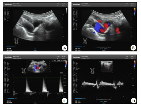
图 1 典型案例:男性,63岁,既往高血糖病史,行颈部彩超检查,发现患者有部分型锁骨下动脉盗血
A: 为患者右锁骨下动脉起始处二维图,示右锁骨下动脉斑块形成并狭窄; B: 为右锁骨下动脉起始处彩色多普勒图像; C: 为右锁骨下动脉起始狭窄处频谱多普勒图像; D: 为右侧椎动脉频谱多普勒图像
Figure 1. Typical case diagram: male patient, 63 years old, with a history of hyperglycemia, underwent neck color Doppler ultrasonography and found that the patient had partial subclavian artery steal.
表 1 联合应用模型分析结果
Table 1. Analysis results of combined application model
方法 b SE χ2 P OR 95% CI 下限 上限 超声探查 0.732 0.264 7.688 0.006 2.079 1.239 3.488 CT血管造影 0.811 0.259 9.805 0.002 2.25 1.354 3.738 常数项 0.473 0.102 21.504 < 0.001 1.605 1.314 1.96  下载: 导出CSV
表 2 不同方法鉴别锁骨下动脉盗血综合征结果
Table 2. Comprehensive results of identifying subclavian artery steal by different methods (n)
方法 鉴别结果 分组 合计 阳性 阴性 超声探查 阳性 74 18 92 阴性 28 82 110 CT血管造影 阳性 83 10 93 阴性 19 90 109 联合应用 阳性 97 4 101 阴性 5 96 101 合计 102 100
下载: 导出CSV
表 3 各指标单独及联合应用预测SSS价值
Table 3. Predicted SSS value of individual and combined application of each index
方法 敏感度 特异性 AUC SE P 95% CI 下限 上限 超声探查 72.55 82 0.749 0.036 < 0.001 0.679 0.82 CT血管造影 81.37 90 0.85 0.028 < 0.001 0.796 0.904 联合应用 95.1 96 0.933 0.022 < 0.001 0.89 0.976
下载: 导出CSV
-
[1] Coles M, Mareddy C, Arora V. Don't ignore that chest pain: positionally dependent coronary subclavian steal syndrome[J]. J Invasive Cardiol, 2021, 33(2): E145. http://www.ncbi.nlm.nih.gov/pubmed/33531448 [2] van Noort K, Schuurmann RCL, Post Hospers G, et al. A new methodology to determine apposition, dilatation, and position of endografts in the descending thoracic aorta after thoracic endovascular aortic repair[J]. J Endovasc Ther, 2019, 26(5): 679-87. doi: 10.1177/1526602819859891 [3] 刘静涵, 丁国富. 经颅多普勒超声评价锁骨下动脉盗血综合征的临床意义[J]. 影像研究与医学应用, 2020, 4(12): 114-5. doi: 10.3969/j.issn.2096-3807.2020.12.073 [4] Steinberger J, Hachem A, Jayyousi BA, et al. Siphoning coronary flow: a case of coronary subclavian steal syndrome[J]. J Am Coll Cardiol, 2020, 75(11): 2442. doi: 10.1016/S0735-1097(20)33069-2 [5] 苏峰太. 锁骨下动脉盗血综合征的血管内介入治疗[J]. 现代仪器与医疗, 2019, 25(2): 49-52. https://www.cnki.com.cn/Article/CJFDTOTAL-XDYI201902014.htm [6] Bernardo M, Beasley M, Marciano P, et al. An unlikely suspect: coronary-subclavian steal syndrome[J]. J Am Coll Cardiol, 2019, 73 (9): 2539. doi: 10.1016/S0735-1097(19)33145-6 [7] Cai XQ, Tian F, Zhou SS, et al. A rare case of non-ST-segment elevation myocardial infarction triggered by coronary subclavian steal syndrome[J]. J Geriatr Cardiol, 2019, 16(4): 378-80. http://www.cnki.com.cn/Article/CJFDTotal-JOGC201904008.htm [8] 魏书恒, 杨雪, 车延旭, 等. 头颈部CTA和DSA对头颈动脉狭窄诊断比较[J]. 河北医药, 2020, 42(11): 1673-6. https://www.cnki.com.cn/Article/CJFDTOTAL-HBYZ202011017.htm [9] Arntzen K, Alstadhaug KB. Room tilt illusion and subclavian steal - a case report[J]. BMC Neurol, 2020, 20(1): 369. doi: 10.1186/s12883-020-01947-2 [10] Ahuja C, Joshi M, Mohindra S, et al. Vertebrobasilar junction aneurysm associated with subclavian steal: yet another hemodynamic cause for aneurysm development and associated challenges [J]. Neurol India, 2020, 68(3): 708. doi: 10.4103/0028-3886.288981 [11] 李静, 华扬, 周福波, 等. 锁骨下动脉狭窄合并椎动脉狭窄的窃血类型分析[J]. 中华医学超声杂志: 电子版, 2019, 16(10): 768-73. doi: 10.3877/cma.j.issn.1672-6448.2019.10.010 [12] Zhao BJ, Liu JY, Zhao T, et al. Carotid artery Stenosis after radiation therapy in a patient with lung cancer: a case report and literature review[J]. Neuro Endocrinol Lett, 2019, 40(3): 113-8. http://www.ncbi.nlm.nih.gov/pubmed/31816217 [13] Quinto L, Testolina M, Zanon F, et al. Twiddler's syndrome combined with subclavian crush syndrome: a case of ICD lead failure and potential challenging lead extraction[J]. J Invasive Cardiol, 2019, 31(11): E340. http://www.ncbi.nlm.nih.gov/pubmed/31671067 [14] 苏志向, 谷涌泉. 腔内血管技术治疗无名动脉支架术后闭塞1例[J]. 中国血管外科杂志: 电子版, 2019, 11(2): 145-7. https://www.cnki.com.cn/Article/CJFDTOTAL-XGWK201902019.htm [15] Nowakowski P. TCTAP C-198 patient with Arnold-chiari syndrome and cervical, subclavian and VCS occlusion endovascular treatment [J]. JAm Coll Cardiol, 2019, 73(15): S259-60. doi: 10.1016/j.jacc.2019.03.404 [16] 薛素芳, 石海艳, 杜祥颖, 等. 旋转性椎动脉闭塞综合征致反复后循环梗死一例[J]. 中国脑血管病杂志, 2019, 16(8): 423-5. doi: 10.3969/j.issn.1672-5921.2019.08.006 [17] Vigneswaran TV, Bellsham-Revell HR, Chubb H, et al. Early postnatal echocardiography in neonates with a prenatal suspicion of coarctation of the aorta[J]. Pediatr Cardiol, 2020, 41(4): 772-80. doi: 10.1007/s00246-020-02310-5 [18] Wang W, Zhang X, Liu C, et al. A novel internal and external bypass method to safeguard cerebral blood flow during aortic arch endovascular repair requiring triple in situ fenestrations[J]. J Endovasc Ther, 2019, 26(5): 652-7. doi: 10.1177/1526602819865503 [19] de Borst GJ. Innominate artery stenting: the continuing Saga of "who, when, and how"?[J]. J Endovascular Ther, 2019, 26(3): 391-3. http://www.onacademic.com/detail/journal_1000042308567099_f923.html [20] Zacharias N, Goodney PP, Stone DH, et al. PC072. innominate artery revascularization: long-term outcomes and comparison of open and endovascular approach[J]. J Vasc Surg, 2019, 69(6): e224-5. http://www.sciencedirect.com/science/article/pii/S0741521419308705 -

 点击查看大图
计量
- 文章访问数: 239
- HTML全文浏览量: 195
- PDF下载量: 2
- 被引次数: 0