


Relationship between DCE-MRI quantitative parameters and HIF-1α levels in patients with primary hepatocellular carcinoma
-
摘要:
目的 探讨磁共振动态对比增强扫描(DCE-MRI)定量参数与原发性肝癌患者病理特征及缺氧诱导因子-1α(HIF-1α)水平的关系。 方法 选取2019年1月~2020年12月收治的94例原发性肝癌患者作为研究对象,并选取同期收治的40例肝脏良性结节患者作为对照组。所有受试者均进行DCE-MRI检查,评估两组微血管密度(MVD)、血管内至血管外细胞间隙的转运系数(Ktrans)、血管外细胞外间隙转运至血管内的速率常数(Kep)、血管外细胞外间隙体积百分比(Ve)及HIF-1α。分析肝癌患者中不同肿瘤分期、不同分化程度、是否有转移、不同HIF-1α水平者DCE-MRI定量参数的差异。 结果 肝癌患者MVD、Ktrans、Kep、Ve及HIF-1α水平均高于对照组(P < 0.05);肝癌患者Ⅲ/Ⅳ期者MVD、Ktrans、Kep、Ve水平均高于Ⅰ/Ⅱ期者(P < 0.05);肝癌低分化患者MVD、Ktrans、Kep、Ve水平均高于中高分化者(P < 0.05);有淋巴结转移的肝癌患者MVD、Ktrans、Kep、Ve水平均高于无淋巴结转移者(P < 0.05);HIF-1α≥162.96 ng/L的肝癌患者MVD、Ktrans、Kep、Ve水平均高于HIF-1α < 162.96 ng/L者(P < 0.05);肝癌临床分期与MVD、Ve呈正相关关系(P < 0.05),肝癌淋巴结转移与Ktrans、Ve呈正相关关系(P < 0.05)。HIF-1α与MVD、Ktrans、Kep、Ve呈正相关关系(P < 0.05)。 结论 DCE-MRI定量参数与原发性肝癌患者病理特征及HIF-1α均密切相关,可利用DCE-MRI定量参数评估肝癌患者病情进展情况。 -
关键词:
- 磁共振动态对比增强扫描 /
- 血管内至血管外细胞间隙的转运系数 /
- 血管外细胞外间隙转运至血管内的速率常数 /
- 微血管密度 /
- 缺氧诱导因子-1α
Abstract:Objective To investigate the relationship between quantitative parameters of dynamic contrast-enhanced magnetic resonance (DCE-MRI) scanning and pathological features and HIF-1α levels in patients with primary hepatocellular carcinoma (HCC). Methods A total of 94 patients with primary liver cancer admitted from January 2019 to December 2020 were selected as the study subjects, and 40 patients with benign liver nodules admitted during the same period were selected as the control group. All subjects underwent DCE-MRI to assess microvascular density (MVD), transport coefficient from intracellular space to extracellular space (Ktrans), rate constant of extracellular space to intracellular space (Kep), percentage of extracellular space volume (Ve), and HIF-1α in both groups. The quantitative parameters of DCE-MRI were analyzed among patients with different tumor stages, different degrees of differentiation, whether there was metastasis or not, and different levels of HIF-1α. Results The MVD, Ktrans, Kep, Ve and HIF-1α in HCC patients were higher than those in control group (P < 0.05). The MVD, Ktrans, Kep and Ve in stage Ⅲ/Ⅳ were higher than those in stage Ⅰ/Ⅱ (P < 0.05). The MVD, Ktrans, Kep and Ve in patients with low differentiation were higher than those in patients with high differentiation (P < 0.05). The MVD, Ktrans, Kep and Ve in HCC patients with lymph node metastasis were higher than those without lymph node metastasis (P < 0.05). The MVD, Ktrans, Kep and Ve in patients with HIF-1α≥162.96 ng/L were higher than those in patients with HIF-1α < 162.96 ng/L (P < 0.05). The clinical stage of HCC was positively correlated with MVD and Ve (P < 0.05), and lymph node metastasis of HCC was positively correlated with Ktrans and Ve (P < 0.05). The HIF-1α was positively correlated with MVD, Ktrans, Kep and Ve (P < 0.05). Conclusion DCE-MRI quantitative parameters are closely related to the pathological characteristics and HIF-1α of HCC patients, and they can be used to evaluate the disease progression of HCC patients. -

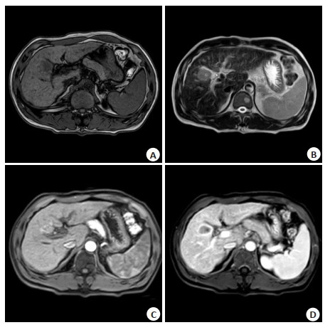
图 1 肝癌患者,男性,58岁,Ⅲ期,低分化
A: T1WI示病灶呈稍低信号,边界欠清; B: T2WI示病灶呈不均匀高信号; C: DCE-MRI动脉期造影示边缘结节状明显强化, 病灶强化程度高于背景肝; D: DCE-MRI延迟期造影示病灶强化减退, 边缘见明显强化包膜影.
Figure 1. A58-year-old male patient with liver cancer, stage Ⅲ, poorly differentiated.
表 1 两组MVD、Ktrans、Kep、Ve及HIF-1α水平
Table 1. Levels of MVD, Ktrans, Kep, Ve and HIF-1α in the two groups (Mean±SD)
组别 MVD Ktrans(min-1) Kep(min-1) Ve HIF-1α(ng/L) 肝癌(n=94) 66.91±9.25 0.47±0.10 1.31±0.50 0.38±0.10 165.84±69.94 对照组(n=40) 50.45±12.60 0.26±0.11 1.08±0.44 0.26±0.06 29.12±8.62 t 8.422 10.794 2.522 7.059 12.297 P < 0.001 < 0.001 0.013 < 0.001 < 0.001 MVD: 微血管密度; Ktrans: 血管内至血管外细胞间隙的转运系数; Kep: 血管外细胞外间隙转运至血管内的速率常数; Ve: 血管外细胞外间隙体积百分比; HIF-1α: 缺氧诱导因子-1α.  下载: 导出CSV
表 2 不同肿瘤分期肝癌患者DCE-MRI定量参数比较
Table 2. Comparison of DCE-MRI quantitative parameters of liver cancer patients with different tumor stages (Mean±SD)
组别 MVD Ktrans(min-1) Kep(min-1) Ve Ⅲ/Ⅳ(n=54) 72.72±6.90 0.52±0.09 1.63±0.36 0.43±0.09 Ⅰ/Ⅱ(n=40) 59.08±5.44 0.41±0.06 0.89±0.28 0.31±0.05 t 10.347 7.118 10.591 8.072 P < 0.001 < 0.001 < 0.001 < 0.001
下载: 导出CSV
表 3 不同分化程度肝癌患者DCE-MRI定量参数比较
Table 3. Comparison of DCE-MRI quantitative parameters of liver cancer patients with different degrees of differentiation (Mean±SD)
组别 MVD Ktrans(min-1) Kep(min-1) Ve 低分化(n=17) 78.88±4.12 0.60±0.06 1.75±0.36 0.45±0.10 中高分化(n=77) 64.27±7.88 0.45±0.08 1.22±0.47 0.36±0.09 t 7.406 9.192 5.176 3.719 P < 0.001 < 0.001 < 0.001 < 0.001
下载: 导出CSV
表 4 有无淋巴结转移肝癌患者DCE-MRI定量参数比较
Table 4. Comparison of DCE-MRI quantitative parameters of liver cancer patients with and without lymph node metastasis (Mean±SD)
组别 MVD Ktrans(min-1) Kep(min-1) Ve 淋巴结转移(n=36) 73.72±5.92 0.56±0.06 1.49±0.49 0.47±0.06 无淋巴结转移(n=58) 62.69±8.41 0.42±0.07 1.21±0.47 0.33±0.07 t 6.881 9.913 2.753 9.645 P < 0.001 < 0.001 0.007 < 0.001
下载: 导出CSV
表 5 不同HIF-1α水平肝癌患者DCE-MRI定量参数比较
Table 5. Comparison of DCE-MRI quantitative parameters of liver cancer patients with different HIF-1α levels (n=47, Mean±SD)
组别 MVD Ktrans(min-1) Kep(min-1) Ve HIF-1α≥162.96ng/L 68.89±8.65 0.49±0.09 1.43±0.53 0.40±0.10 HIF-1α < 162.96ng/L 64.94±9.50 0.45±0.10 1.20±0.44 0.36±0.09 t 2.112 2.152 2.368 2.172 P 0.037 0.034 0.02 0.032
下载: 导出CSV
表 6 肝癌患者DCE-MRI定量参数与临床病理特征及HIF-1α的关系
Table 6. The relationship between DCE-MRI quantitative parameters and clinicopathological features and HIF-1α in HCC patients
DCE-MRI定量参数 r 临床分期 分化程度 淋巴结转移 HIF-1α MVD 0.416* 0.196 0.219 0.302* Ktrans(min-1) 0.216 0.123 0.361* 0.414* Kep(min-1) 0.398 0.089 0.102 0.379* Ve 0.075* 0.163 0.337* 0.398* *P < 0.05.
下载: 导出CSV
-
[1] Orcutt ST, Anaya DA. Liver resection and surgical strategies for management of primary liver cancer[J]. Cancer Control, 2018, 25 (1): 107-32. http://europepmc.org/abstract/MED/29327594 [2] 程书蕙, 陈波, 李晔雄. 原发性肝癌术后辅助治疗的研究进展[J]. 中华放射肿瘤学杂志, 2019, 28(3): 233-7. doi: 10.3760/cma.j.issn.1004-4221.2019.03.016 [3] Dimitroulis D, Damaskos C, Valsami S, et al. From diagnosis to treatment of hepatocellular carcinoma: an epidemic problem for both developed and developing world[J]. World J Gastroenterol, 2017, 23(29): 5282-94. doi: 10.3748/wjg.v23.i29.5282 [4] 张进伟. 螺旋CT和MRI对原发性肝癌的诊断价值分析[J]. 实用癌症杂志, 2020, 35(2): 226-9. doi: 10.3969/j.issn.1001-5930.2020.02.015 [5] 蒿景龙, 于建奇, 赵丹蕾, 等. DCE-MRI功能成像参数对原发性肝细胞癌和肝转移瘤的鉴别诊断价值[J]. 中国现代普通外科进展, 2020, 23(2): 161-3. https://www.cnki.com.cn/Article/CJFDTOTAL-PWJZ202002023.htm [6] 于巍伟, 于卫永, 刘坤, 等. MRI多期动态对比增强灌注参数与原发性肝癌患者微血管密度、病理分级的关联[J]. 中西医结合肝病杂志, 2021, 31(2): 161-4. https://www.cnki.com.cn/Article/CJFDTOTAL-ZXGB202102018.htm [7] Cong WM, Bu H, Chen J, et al. Practice guidelines for the pathological diagnosis of primary liver cancer: 2015 update[J]. World J Gastroenterol, 2016, 22(42): 9279-87. doi: 10.3748/wjg.v22.i42.9279 [8] 高欣, 周丽娟, 徐孝秋, 等. 动态对比增强磁共振成像定量参数直方图分析与鼻咽癌临床分期的相关性初步研究[J]. 实用放射学杂志, 2019, 35(10): 1590-4. doi: 10.3969/j.issn.1002-1671.2019.10.009 [9] 赵晓艳. DCE-MRI定量参数在宫颈癌放化疗效果评价及其与病理特征的相关性分析[J]. 医学影像学杂志, 2020, 30(11): 2069-73. https://www.cnki.com.cn/Article/CJFDTOTAL-XYXZ202011032.htm [10] Chen J, Qian T, Zhang H, et al. Combining dynamic contrast enhanced magnetic resonance imaging and microvessel density to assess the angiogenesis after PEI in a rabbit VX2 liver tumor model [J]. Magn Reson Imaging, 2016, 34(2): 177-82. doi: 10.1016/j.mri.2015.10.013 [11] Chen J, Chen C, Xia C, et al. Quantitative free-breathing dynamic contrast-enhanced MRI in hepatocellular carcinoma using gadoxetic acid: correlations with Ki67 proliferation status, histological grades, and microvascular density[J]. Abdom Radiol: NY, 2018, 43(6): 1393-403. doi: 10.1007/s00261-017-1320-3 [12] Yang F, Zhao J, Liu C, et al. Superb microvascular imaging technique in depicting vascularity in focal liver lesions: more hypervascular supply patterns were depicted in hepatocellular carcinoma[J]. Cancer Imaging, 2019, 19(1): 92. doi: 10.1186/s40644-019-0277-6 [13] 杨锦锋, 曾荣耀. 血管内皮生长因子和缺氧诱导因子-1α的表达与原发性肝癌患者临床病理特征及预后的关系[J]. 中国慢性病预防与控制, 2020, 28(12): 924-7. https://www.cnki.com.cn/Article/CJFDTOTAL-ZMXB202012011.htm [14] 邓锻炼. 磁共振成像动态对比增强(DCE-MRI)定量参数对肝细胞肝癌和肝良性肿瘤鉴别诊断的价值[J]. 临床医药文献电子杂志, 2018, 5(96): 117. doi: 10.3877/j.issn.2095-8242.2018.96.094 [15] 李宏斌, 石珏, 李娜. 原发性肝癌患者PIVKA-Ⅱ、AFU、HIF-1α的表达及其临床意义[J]. 医学临床研究, 2019, 36(2): 360-2. doi: 10.3969/j.issn.1671-7171.2019.02.053 [16] Chen BB, Hsu CY, Yu CW, et al. Early perfusion changes within 1 week of systemic treatment measured by dynamic contrastenhanced MRI may predict survival in patients with advanced hepatocellular carcinoma[J]. Eur Radiol, 2017, 27(7): 3069-79. doi: 10.1007/s00330-016-4670-2 [17] Zhang W, Chen HJ, Wang ZJ, et al. Dynamic contrast enhanced MR imaging for evaluation of angiogenesis of hepatocellular nodules in liver cirrhosis in N-nitrosodiethylamine induced rat model[J]. Eur Radiol, 2017, 27(5): 2086-94. doi: 10.1007/s00330-016-4505-1 [18] Zhang Y, Tie M, Wang K, et al. Tanshinone Ⅱ improves distribution and anti-tumor efficacy of pegylated liposomal doxorubicin via normalizing the structure and function of tumor vasculature in hepa1- 6 hepatoma mice model[J]. J Tradit Chin Med, 2018, 38(6): 815-22. doi: 10.1016/S0254-6272(18)30980-4 [19] Kim SH, Lee HS, Kang BJ, et al. Dynamic contrast-enhanced MRI perfusion parameters as imaging biomarkers of angiogenesis[J]. PLoS One, 2016, 11(12): e0168632. doi: 10.1371/journal.pone.0168632 [20] 张成成, 唐小琦, 张志国, 等. MRI多期动态对比增强灌注参数与肝癌微血管密度的相关性[J]. 肝脏, 2019, 24(4): 428-31. doi: 10.3969/j.issn.1008-1704.2019.04.026 [21] 苏纯洁, 何前进, 毛伟明, 等. 肝癌组织中PD-L1和HIF-1α的相关性研究[J]. 中国现代普通外科进展, 2019, 22(1): 22-5. https://www.cnki.com.cn/Article/CJFDTOTAL-PWJZ201901008.htm -

 点击查看大图
计量
- 文章访问数: 149
- HTML全文浏览量: 80
- PDF下载量: 2
- 被引次数: 0