


Comparation of low-dose computeirzed tomography angiography and digital subtraction angiography under the guidance of BMI in the diagnosis of moderate to severe coronary stenosis
-
摘要:
目的 探讨低剂量计算机断层摄影血管造影术与数字减影血管造影在冠状动脉中重度狭窄的诊断价值比较。 方法 选取88例确诊冠状动脉粥样硬化性心脏病(冠心病)的患者作为研究对象,应用血管分析软件进行图像重组,分析计算机断层摄影血管造影术检出冠状动脉斑块的性质,评估其狭窄情况,将其结果与对应节段数字减影血管造影检出的病变狭窄度进行对比。 结果 以数字减影血管造影作为金标准,计算机断层摄影血管造影术检出冠状动脉病变斑块所致中重度狭窄855节段,其中软斑块396节段、硬斑块459节段。通过两种检验方法对硬斑块导致中度狭窄、硬斑块导致重度狭窄、软斑块导致中度狭窄和软斑块导致重度狭窄进行检验,差异均无统计学意义(P>0.05)。 结论 低剂量计算机断层摄影血管造影术与数字减影血管造影检查对冠心病诊断都具有较高准确率,而低剂量计算机断层摄影血管造影术检查经济、方便、实用,可作为临床筛查冠心病的首选方法。 -
关键词:
- 冠状动脉狭窄 /
- 低剂量计算机断层摄影血管造影术 /
- 数字减影血管造影
Abstract:Objective To compare the diagnostic value of low-dose computeirzed tomography angiography and digital subtraction angiography in severe coronary artery stenosis. Methods Eighty-eight patients with clinically suspected or confirmed coronary atherosclerotic heart disease were selected as the research objects. Vascular analysis software was applied to reconstruct images to analyze the nature of coronary plaques detected by computed tomography angiography and evaluate them for stenosis. The result were compared with the stenosis of the lesion detected by digital subtraction angiography of the corresponding segment. Results Digital subtraction angiography was taken as the gold standard, computed tomography angiography detected moderate to severe stenosis at 855 segments caused by coronary artery diseased plaques, there were 396 segments of soft plaques and 459 segments of hard plaques among them. The test results of hard plaques leading to moderate stenosis, hard plaques leading to severe stenosis, moderate stenosis caused by soft plaque and severe stenosis caused by soft plaque had no significant difference (P>0.05). Conclusion Both low-dose computeirzed tomography angiography and digital subtraction angiography have high accuracy in the diagnosis of coronary heart disease. Low-dose computer tomography angiography is economical, convenient and practical to be used as the first choice for clinical screening of coronary heart disease. -

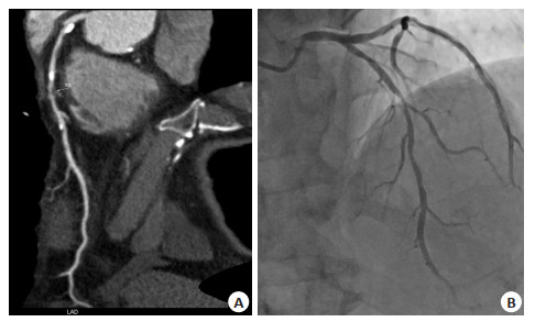
图 1 冠状动脉硬斑块轻度狭窄CTA与相应节段DSA对比
A: CTA显示左冠状动脉回旋支近段硬斑块清晰可见, 管腔呈轻度狭窄; B: 对应DSA节段管腔未见明显狭窄, 且硬斑块未见显示, 而CTA能清晰显示.
Figure 1. Coronary artery hard plaques with mild stenosis CTA and DSA corresponding segments contrast

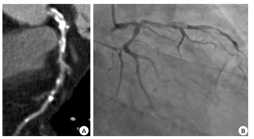
图 2 冠状动脉软斑块CTA与相应节段DSA对比
A: CTA显示左冠状动脉前降支中段软斑块清晰可见,管腔呈重度狭窄; B: 对应DSA节段管腔重度狭窄, 但软斑块未见显示, 而CTA能清晰显示.
Figure 2. Coronary CTA and DSA corresponding segments contrast soft plaques

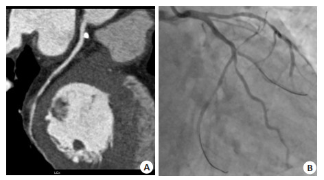
图 3 冠状动脉硬斑块中度狭窄CTA与相应节段DSA对比
A: CTA显示左冠状动脉主干硬斑块清晰可见,管腔呈中度狭窄; B: 左冠状动脉主干硬斑块对应DSA节段管腔重度狭窄,此段狭窄为硬斑块导致, 钙化严重, CTA诊断狭窄度稍受影响, 但DSA检查中硬斑块未见显示,而CTA能清晰显示.
Figure 3. Coronary artery hard plaques with moderate stenosis CTA and DSA corresponding segments contrast
表 1 CTA和DSA检出冠状动脉血管硬斑块致中度狭窄节段的比(节段)
Table 1. CTA and DSAdetection of coronary arteries hard plaques compared to moderate stenosis segment (section)
检查结果 检查方法 阳性 阴性 总数 χ2 P 血管硬斑块致中度狭窄 CTA 113 102 215 0.166 0.683 DSA 121 101 222 血管硬斑块致重度狭窄 CTA 103 141 244 1.683 0.194 DSA 114 123 237 血管软斑块致中度狭窄 CTA 195 43 238 0.024 0.877 DSA 179 38 217 血管软斑块致重度狭窄 CTA 106 52 158 3.276 0.070 DSA 136 43 179 CTA: CT血管造影; DSA: 数字减影血管造影  下载: 导出CSV
-
[1] 王国兴, 肖静, 刘颖, 等. 冠脉CTA对冠心病的诊断价值研究[J]. 影像研究与医学应用, 2020, 4(16): 92-3. https://www.cnki.com.cn/Article/CJFDTOTAL-YXYY202016063.htm [2] Bogaard K, van der Zant FM, Knol RJ, et al. High-pitch prospective ECG-triggered helical coronary computed tomography angiography in clinical practice: image quality and radiation dose[J]. Int J Cardiovasc Imaging, 2015, 31(1): 125-33. doi: 10.1007/s10554-014-0515-8 [3] Lim J, Park EA, Lee W, et al. Image quality and radiation reduction of 320-row area detector CT coronary angiography with optimal tube voltage selection and an automatic exposure control system: comparison with body mass index-adapted protocol[J]. Int J Cardiovasc Imaging, 2015, 31(1): 23-30. http://europepmc.org/abstract/med/25604967 [4] Li CC, Matthews AK, Rywant MM, et al. Racial disparities in eligibility for low-dose computed tomography lung cancer screening among older adults with a history of smoking[J]. Cancer Causes Control, 2019, 30(3): 235-40. doi: 10.1007/s10552-018-1092-2 [5] Huang KL, Wang SY, Lu WC, et al. Effects of low-dose computed tomography on lung cancer screening: a systematic review, metaanalysis, and trial sequential analysis[J]. BMC Pulm Med, 2019, 19 (1): 126. doi: 10.1186/s12890-019-0883-x [6] Dai X, Yu M, Pan J, et al. Image quality and diagnostic accuracy of coronary CT angiography derived from low- dose dynamic CT myocardial perfusion: a feasibility study with comparison to invasive coronary angiography[J]. Eur Radiol, 2019, 29(8): 4349-56. doi: 10.1007/s00330-018-5777-4 [7] Pan YY, Zhou SC, Wang YJ, et al. Application of low tube voltage, low-concentration contrast agent using a 320-row CT in coronary CT angiography: evaluation of image quality, radiation dose and iodine intake[J]. Curr Med Sci, 2020, 40(1): 178-83. doi: 10.1007/s11596-020-2162-8 [8] Plank F, Beyer C, Friedrich G, et al. Sex differences in coronary artery plaque composition detected by coronary computed tomography: quantitative and qualitative analysis[J]. Neth Heart J, 2019, 27(5): 272-80. doi: 10.1007/s12471-019-1234-5 [9] 梁健华, 宋国军, 林永开, 等. BMI指导下改变对比剂流速对冠脉CTA图像质量的影响[J]. 现代医用影像学, 2017, 26(4): 881-6. https://www.cnki.com.cn/Article/CJFDTOTAL-XDYY201704002.htm [10] 杨文兵, 查云飞, 阳朝晖, 等. 冠脉CTA评估冠状动脉斑块性质及其与炎症分子、MMPs/TIMPs的相关性[J]. 海南医学院学报, 2017, 23 (21): 2929-32, 2936. https://www.cnki.com.cn/Article/CJFDTOTAL-HNYY201721009.htm [11] 章辉庆, 刘海燕, 江荣炎, 等. 100 kV管电压双源CT冠状动脉血管成像诊断冠心病的临床价值[J]. 中西医结合心脑血管病杂志, 2020, 18 (16): 2653-5. doi: 10.12102/j.issn.1672-1349.2020.16.022 [12] 王芳, 覃群, 韦迎娜. CT冠状动脉成像在冠心病诊断及预后评估中的应用价值[J]. 中国CT和MRI杂志, 2020, 18(9): 93-5, 99. doi: 10.3969/j.issn.1672-5131.2020.09.029 [13] 张艳婷. 320排CT冠脉成像评估冠状动脉斑块的临床价值分析[J]. 影像研究与医学应用, 2019, 3(7): 87-8. doi: 10.3969/j.issn.2096-3807.2019.07.058 [14] 李洪杰. 双低剂量技术结合CTA对急性冠状动脉综合征患者诊断的意义研究[J]. 中国医药指南, 2020, 18(6): 170-1. https://www.cnki.com.cn/Article/CJFDTOTAL-YYXK202006139.htm [15] 周瑜, 李直苹, 漆钜霞, 等. 胸部CT低剂量扫描与常规扫描对肺癌检查的对照研究[J]. 影像研究与医学应用, 2021, 5(4): 45-6. doi: 10.3969/j.issn.2096-3807.2021.04.022 [16] 葛沿安, 董傲. 双源冠状动脉"双低"CTA检查技术用于冠心病诊断中的临床特异性分析[J]. 影像研究与医学应用, 2021, 5(7): 67-8. doi: 10.3969/j.issn.2096-3807.2021.07.032 [17] 欧阳裕锋, 胡秋根, 刘维松, 等. 冠脉疾病128层螺旋CT血管成像的诊断研究[J]. 现代医用影像学, 2019, 28(10): 2259-60. doi: 10.3969/j.issn.1006-7035.2019.10.042 [18] 吴玉辉, 张惠英. 256层螺旋CTA对冠状动脉斑块及管腔狭窄的诊断价值[J]. 华北理工大学学报: 医学版, 2017, 19(3): 191-4. https://www.cnki.com.cn/Article/CJFDTOTAL-MTYX201703006.htm [19] 王月琦, 宋扬, 孙世元. CT对冠状动脉狭窄及斑块诊断的意义[J]. 中国卫生标准管理, 2020, 11(9): 104-6. doi: 10.3969/j.issn.1674-9316.2020.09.044 [20] 李智明. 探讨西门子64排128层螺旋CT冠脉成像技术与冠状动脉造影在冠心病患者中冠状动脉狭窄病变影像诊断中的应用效果[J]. 世界最新医学信息文摘, 2019, 19(54): 154, 156. https://www.cnki.com.cn/Article/CJFDTOTAL-WMIA201954110.htm [21] 蔡朝君, 潘仙琴, 崔恒武. 64排螺旋CT冠状动脉成像对冠心病诊断准确性的影响因素分析[J]. 中外医疗, 2017, 36(26): 190-2. https://www.cnki.com.cn/Article/CJFDTOTAL-HZZZ201726071.htm [22] Oraby AS, Alarabawy RA, Abd Alla TM, et al. High risk plaque criteria by multislice coronary CT angiography in patients with stable vs. unstable coronary artery disease: analytic cross-sectional study[J]. Egypt J Radiol Nucl Med, 2020, 51(1): 1-11. doi: 10.1186/s43055-019-0116-6 [23] Kaji SZ, Kida S. Overview of image-to-image translation by use of deep neural networks: denoising, super-resolution, modality conversion, and reconstruction in medical imaging[J]. Radiol Phys Technol, 2019, 12(3): 235-48. doi: 10.1007/s12194-019-00520-y [24] Chamberlin J, Kocher MR, Waltz J, et al. Automated detection of lung nodules and coronary artery calcium using artificial intelligence on low-dose CT scans for lung cancer screening: accuracy and prognostic value[J]. BMC Med, 2021, 19(1): 55. doi: 10.1186/s12916-021-01928-3 -

 点击查看大图
计量
- 文章访问数: 286
- HTML全文浏览量: 273
- PDF下载量: 5
- 被引次数: 0