


Evaluation of ultrasound multiple parameters (two-dimensional, color Doppler flow imaging, shear wave elastography, contrast-enhanced imaging) in breast cancer versus pathological control
-
摘要:
目的 分析乳腺癌超声多参数(二维高频超声、彩色多普勒血流显像、剪切波弹性成像、造影)评估与病理对照。 方法 选择2018年4月~2020年9月我院诊治的乳腺占位性病变患者61例(72个病灶),均行二维高频超声、彩色多普勒血流显像、剪切波弹性成像、造影和病理检查,以术后病理检查结果为金标准。计算各种检查方法诊断乳腺癌的敏感度、准确度。 结果 术后病理诊断结果示:61例(72个病灶)中有43例恶性(51个病灶),18例良性(21个病灶);二维高频超声诊断乳腺癌敏感度47.06%,特异性66.67%,准确度52.78%;彩色多普勒血流显像敏感度60.78%,特异性71.43%,准确度63.89%;剪切波弹性成像敏感度82.35%,特异性80.95%,准确度81.94%;超声造影敏感度86.27%,特异性85.71%,准确度86.11%;联合诊断敏感度94.12%,特异性90.48%,准确度93.06%。联合诊断乳腺癌准确率高于单独二维、彩色多普勒血流显像、剪切波弹性成像、造影检查(P < 0.05)。 结论 乳腺癌超声多参数(二维、彩色多普勒血流显像、剪切波弹性成像、造影)联合检查,可明显提高乳腺癌诊断的准确率。 Abstract:Objective To compare the multimodal ultrasonographic parameters (two-dimensional high frequency ultrasound, color Doppler flow imaging (CDFI), shear wave elasticity imaging, contrast-enhanced ultrasound) and clinicopathological appearances of breast cancer. Methods Sixty-one patients (72 lesions) with breast space occupying lesions from April 2018 to September 2020 in our hospital were enrolled. Two-dimensional high frequency ultrasound, color Doppler flow imaging, shear wave elasticity imaging, contrast-enhanced ultrasound and pathological examinations were performed on all patients. Using pathological results as gold standard, the sensitivity and accuracy of each examination in the diagnosis of breast cancer were analyzed. Results Pathological results showed that among 61 patients (72 lesions), 43 were malignant (51 lesions) and 18 were benign (21 lesions). In the diagnosis of breast cancer, the sensitivity, specificity and accuracy were 47.06%, 66.67% and 52.78% through two-dimensional high frequency ultrasound, those of color Doppler flow imaging were 60.78%, 71.43% and 63.89%, and those of shear wave elasticity imaging were 82.35%, 80.95% and 81.94%, and those of contrast-enhanced ultrasound were 86.27%, 85.71% and 86.11%. The sensitivity, specificity and accuracy of combined examinations in the diagnosis of breast cancer were 94.12%, 90.48% and 93.06%, respectively. The accuracy of combined examination in the diagnosis of breast cancer was higher than that of single examination (P < 0.05). Conclusion Combined examinations can significantly improve accuracy of diagnosis of breast cancer, which is highly consist with pathological results. -

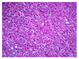
图 1 某女性患者,浸润性导管癌Ⅲ级(HE, ×200)
Figure 1. A female patient, grade III invasive ductal carcinoma (HE, ×200)

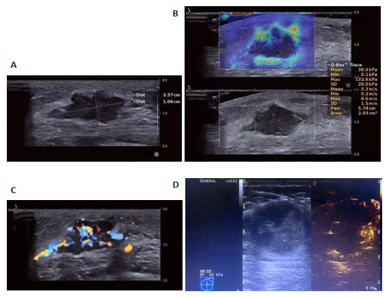
图 2 同病理结果病例的二维高频超声、剪切波弹性成像、CDFI及造影图像
A: 二维高频超声图像显示: 实性低回声包块, 边界清晰, 边缘成角及小分叶改变, 内部回声不均匀, 见微钙化, 后方回声轻度衰减, 纵横比<1; B: 剪切波弹性成像图像显示: 内部彩色部分缺失, 周边以黄色、橘红色为主, 成“硬环征”声像, SWE-Emax 132.6 kPa,SWE-Emean 38.6 kPa,SWE-Emin 0.1 kPa; C: CDFI图像显示:肿块内可见5个及以上点状血管及3个较长血管丰富血流信号,按Adler半定量法分III级; D: 造影图像示:肿块造影后体积明显较二维高频超声测量增大,中部未见增强,边缘增强明显高于周围组织,肿块整体造影增强成不均匀型.
Figure 2. Two-dimensional high frequency ultrasound imaging, shear wave elasticity imaging, CDFI imaging and contrast- enhanced ultrasound imaging of patient with same pathologic result

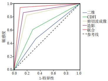
图 3 二维、CDFI、剪切波成像、造影单独超声技术及联合超声多参数诊断的ROC曲线
Figure 3. ROC curves of single and combined examinations of two-dimensional high frequency ultrasound, CDFI, shear wave elasticity imaging, and contrast-enhanced ultrasound.
表 1 二维、CDFI、剪切波成像、造影及联合结果
Table 1. Combined examinations of two-dimensional high frequency ultrasound, CDFI, shear wave elasticity imaging and contrastenhanced ultrasound (n)
项目 诊断 病理 合计 恶性 良性 二维 恶性 24 7 31 良性 27 14 41 CDFI 恶性 31 6 37 良性 20 15 35 剪切波成像 恶性 42 4 46 良性 9 17 26 造影 恶性 44 3 47 良性 7 18 25 联合 恶性 48 2 50 良性 3 19 22 合计 51 21 72  下载: 导出CSV
-
[1] 高军喜, 王雅婷, 杨磊, 等. 乳腺癌超声造影特征及边缘带定量参数与生物学预后因子相关性研究[J]. 中国超声医学杂志, 2019, 35(4): 306-9. https://www.cnki.com.cn/Article/CJFDTOTAL-ZGCY201904007.htm [2] Park HS, Shin HJ, Shin KC, et al. Comparison of peritumoral stromal tissue stiffness obtained by shear wave elastography between benign and malignant breast lesions[J]. Acta Radiol, 2018, 59(10): 1168-75. doi: 10.1177/0284185117753728 [3] 周红梅, 冉海涛, 成涓. 超声新技术结合超声乳腺影像报告和数据系统评估乳腺病变的应用现状[J]. 中国介入影像与治疗学, 2018, 15 (3): 188-91. https://www.cnki.com.cn/Article/CJFDTOTAL-JRYX201803022.htm [4] Correas JM, Halpern EJ, Barr RG, et al. Advanced ultrasound in the diagnosis of prostate cancer[J]. World J Urol, 2021, 39(3): 661-76. doi: 10.1007/s00345-020-03193-0 [5] 陈继赵, 谢春梅, 孙希文. 剪切波弹性成像技术联合超声BI-RADS分类鉴别诊断乳腺良恶性病变的价值[J]. 中国医学计算机成像杂志, 2020, 26(4): 359-63. doi: 10.3969/j.issn.1006-5741.2020.04.012 [6] 中国抗癌协会乳腺癌专业委员会. 中国抗癌协会乳腺癌诊治指南与规范(2015版[) J]. 中国癌症杂志, 2015, 25(9): 692-754. https://www.cnki.com.cn/Article/CJFDTOTAL-ZGAZ201509014.htm [7] Song EJ, Sohn YM, Seo M. Tumor stiffness measured by quantitative and qualitative shear wave elastography of breast cancer[J]. Br J Radiol, 2018, 91(1086): 20170830. http://www.onacademic.com/detail/journal_1000040250104210_022d.html [8] Nofal MN, Yousef AJ. The diagnosis of male breast cancer[J]. Neth J Med, 2019, 77(10): 356-9. http://www.researchgate.net/publication/338215298_The_diagnosis_of_male_breast_cancer [9] Guo R, Lu G, Qin B, et al. Ultrasound imaging technologies for breast cancer detection and management: a review[J]. Ultrasound Med Biol, 2018, 44(1): 37-70. doi: 10.1016/j.ultrasmedbio.2017.09.012 [10] 肖莉玲, 李颖嘉, 马菲, 等. 术前二维灰阶超声、三维容积超声及弹性成像对乳腺癌大小测量的准确性及其影响因素研究[J]. 中华超声影像学杂志, 2021(5): 414-9. doi: 10.3760/cma.j.cn131148-20201029-00840 [11] 闫敏芳, 杜起军, 任路平, 等. 彩色多普勒超声成像联合钼靶X线对早期乳腺癌的诊断意义[J]. 中国妇幼保健, 2018, 33(22): 5273-5. https://www.cnki.com.cn/Article/CJFDTOTAL-ZFYB201822096.htm [12] 杨小玲, 杨文涛. 二维彩超联合超声实时剪切波弹性成像在乳腺疾病中的诊断价值[J]. 河南医学研究, 2020, 29(20): 3791-3. https://www.cnki.com.cn/Article/CJFDTOTAL-HNYX202020060.htm [13] Polat AV, Ozturk M, Polat AK, et al. Efficacy of ultrasound and shear wave elastography for the diagnosis of breast cancer-related lymphedema[J]. J Ultrasound Med, 2020, 39(4): 795-803. doi: 10.1002/jum.15162 [14] 薛永铭, 张静. 乳腺癌诊断中超声弹性成像、常规超声、钼靶X线技术的应用[J]. 中国急救医学, 2016, 36(z2): 144-5. doi: 10.3969/j.issn.1002-1949.2016.z2.098 [15] 梁铭, 欧冰, 吴嘉仪, 等. 剪切波弹性成像和应变弹性成像对乳腺癌诊断价值的研究[J]. 中国超声医学杂志, 2019, 35(4): 310-2. https://www.cnki.com.cn/Article/CJFDTOTAL-ZGCY201904008.htm [16] 陈树新, 胡智慧, 侯颖, 等. 彩色多普勒二维超声联合实时超声剪切波弹性成像用于乳腺癌诊断[J]. 国际生物医学工程杂志, 2018, 41 (4): 305-9. doi: 10.3760/cma.j.issn.1673-4181.2018.04.006 [17] Wu H, Liang W, Jiao Y, et al. A preliminary comparative study of Young's modulus versus shear modulus in the diagnosis of breast cancer[J]. Ultrasound Q, 2019, 35(1): 88-92. doi: 10.1097/RUQ.0000000000000434 [18] Zhou SC, Le J, Zhou J, et al. The role of contrast- enhanced ultrasound in the diagnosis and pathologic response prediction in breast cancer: a meta-analysis and systematic review[J]. Clin Breast Cancer, 2020, 20(4): e490-509. doi: 10.1016/j.clbc.2020.03.002 [19] 徐子杭, 姜珏, 贾琬莹, 等. 超声造影对肉芽肿性小叶性乳腺炎和乳腺癌的鉴别诊断价值[J]. 中国超声医学杂志, 2020, 36(5): 402-4. doi: 10.3969/j.issn.1002-0101.2020.05.006 [20] 刘健, 王宁, 武敬平, 等. 剪切波弹性成像联合常规超声诊断乳腺癌的价值[J]. 医学研究杂志, 2020, 49(11): 122-5, 131. https://www.cnki.com.cn/Article/CJFDTOTAL-YXYZ202011029.htm [21] Jafari SH, Saadatpour Z, Salmaninejad A, et al. Breast cancer diagnosis: Imaging techniques and biochemical markers[J]. J Cell Physiol, 2018, 233(7): 5200-13. doi: 10.1002/jcp.26379 [22] Budny A, Starosławska E, Budny B, et al. Epidemiologia oraz diagnostyka raka piersi[J]. Pol Merkur Lekarski, 2019, 46(275): 195- 204. [23] 王艳, 游岚岚. 乳腺超声自动容积成像联合超声造影微血管成像在乳腺病灶鉴别诊断及肿瘤TNM分期中的应用价值[J]. 中国老年学杂志, 2020, 40(13): 2737-40. doi: 10.3969/j.issn.1005-9202.2020.13.016 -

 点击查看大图
计量
- 文章访问数: 187
- HTML全文浏览量: 149
- PDF下载量: 4
- 被引次数: 0