


Value of diffusion weighted magnetic resonance imaging combined with three- dimensional digital tomography in differential diagnosis of benign and malignant calcified lesions of breast
-
摘要:
目的 评估磁共振弥散加权成像(DWI)结合三维乳腺数字化断层摄影(DBT)钙化分数对于钙化征象在乳腺良恶性病变诊断的应用价值。 方法 回顾性分析我院118例行乳腺DWI检查和DBT检查的患者,DBT检查发现有钙化征象,其中乳腺癌患者51例,病理类型包括:浸润性导管癌、浸润性导管癌、导管原位癌、髓样癌、浸润性小叶癌、黏液腺癌;乳腺良性病变67例,病理类型包括:纤维腺瘤、腺病、导管内乳头状瘤。对所有患者DBT图像的钙化征象进行钙化评分并且测量相应病变区域的表观扩散系数(ADC)值,分析ADC值、钙化分数及二者联合在乳腺含钙化病变的诊断效能。 结果 乳腺癌组ADC值低于乳腺良性病变组,差异有统计学意义(P < 0.05);钙化分数高于良性病变组,差异有统计学意义(P < 0.05)。ADC值对诊断乳腺良恶性病变的曲线下面积(AUC)为0.853,钙化分数对诊断乳腺良恶性病变的AUC值为0.855,采用Delong检验得出两种方法对诊断乳腺含钙化病变的良恶性诊断效能一致,差异无统计学意义(P=0.625)。两种方法(钙化分数+ADC值)的Logistic回归分析显示,两种方法相结合的AUC值为0.903,具有很高的诊断效能。将ROC曲线(钙化分数、ADC值、两种方法结合的预测概率)经两两Delong检验结果显示,两种方法结合的ROC与单一ROC的差异有统计学意义(P=0.041、0.022)。 结论 DWI与DBT钙化分数对于钙化征象在乳腺良恶性病变的鉴别诊断效能无差别,两种方法相结合的诊断效能显著高于单一方法,为乳腺含钙化病灶的诊断与鉴别诊断中提供了更加可靠的依据。 -
关键词:
- 乳腺癌 /
- 弥散加权成像 /
- 三维断层乳腺摄影技术 /
- 表观扩散系数 /
- 钙化
Abstract:Objective To evaluate the value of diffusion- weighted magnetic resonance imaging (DWI) combined with three- dimensional digital mammography (DBT) in the diagnosis of benign and malignant breast lesions. Methods A total of 118 cases of breast DWI and DBT in our hospital were retrospectively analyzed. The signs of calcification were found by DBT. There were 51 cases with breast cancer. The pathological types included invasive ductal carcinoma, invasive ductal carcinoma, ductal carcinoma in situ, medullary carcinoma, invasive lobular carcinoma, mucinous adenocarcinoma. Benign breast lesions were found in 67 cases, including fibroadenoma, adenosis and intraductal papilloma. All patients were scored for calcification on DBT images, and the apparent diffusion coefficient (ADC) value of the corresponding lesion area was measured. The diagnostic efficacy of ADC value, calcification score and their combination in breast calcified lesions was analyzed. Results ADC value of breast cancer group was lower than benign breast lesions group (P < 0.05). Calcification score of breast cancer group was higher than benign lesion group (P < 0.05). The area under the curve (AUC) of ADC value for the diagnosis of benign and malignant breast lesions was 0.853, and the AUC of calcification score for the diagnosis of benign and malignant breast lesions was 0.855. Delong test showed that the two methods had the same diagnostic efficiency for benign and malignant breast lesions with calcification, and the difference was not significant (P=0.625). Logistic regression analysis of the two methods (calcification score+ADC value) showed that the AUC value of the combination of the two methods was 0.903, which had high diagnostic efficiency. Calcification score, ADC value and the combination of the two methods was used to carry out Delong test. The results showed that the difference of receiver operating characteristic curve (ROC) combined with ROC and ADC ROC of the two methods were statistically significant (P=0.0409, 0.0216). Conclusion There is no difference in the differential diagnosis of calcification between diffusion- weighted imaging and three- dimensional digital mammography. The diagnostic efficiency of the combination of the two methods is significantly higher than that of a single method, which provides a more reliable basis for the diagnosis and differential diagnosis of breast lesions with calcification. -

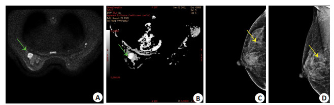
图 1 乳腺癌MRI图像及DBT图像
A~B: 左侧乳腺外下象限乳腺癌(橙色箭头); DWI表现为高信号, ADC表现为低信号; ADC值: 1.06×10⁻³ mm2/ s; C~D: 乳腺癌DBT钙化特征(蓝色箭头); 钙化分数: 9.
Figure 1. breast cancer MRI and DBT images.

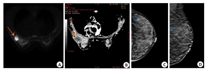
图 2 乳腺纤维瘤MRI图像及DBT图像
A~B: 左侧乳腺纤维瘤(绿色箭头); DWI表现为高信号, ADC表现为稍高信号; ADC值: 1.57×10⁻³ mm2/ s; C~D: 乳腺腺瘤DBT钙化特征(黄色箭头), 钙化分数: 4.
Figure 2. Breast fibroma of MRI and DBT images.

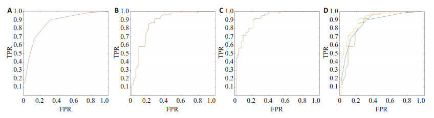
图 3 乳腺良恶性病变ADC、钙化分数各参数及双模式相结合ROC曲线图
A: 乳腺良恶性病变ADC值ROC曲线图, AUC=0.853; B: 乳腺良恶性病变DBT钙化分数ROC曲线图, AUC=0.855; C~D: 黄色: 钙化分数+ADC预测概率ROC曲线; 蓝色: 钙化分数ROC曲线; 橙色: ADC值ROC曲线; 其中, C: 乳腺良恶性病变双模式(DBT钙化分数和MRI-ADC)联合预测概率ROC曲线图, AUC=0.903; D: 对双模式(DBT钙化分数和MRI-ADC)联合预测概率ROC曲线、钙化分数ROC曲线、ADC值ROC曲线两两进行Delong检验.
Figure 3. ROC curve of ADC, calcification score and dual mode of benign and malignant breast lesions.
表 1 乳腺良恶性肿瘤钙化分数、ADC及两种方法相结合的AUC
Table 1. Calcification fraction, ADC and area under ROC curve of benign and malignant breast tumors combined with the two methods
分类 曲线下面积 敏感度(%) 特异性(%) 钙化分数 0.855 85.3 84.7 ADC 0.853 83.4 84.2 钙化分数+ADC 0.903 86.7 85.9 ADC: 表现扩散系数.  下载: 导出CSV
-
[1] Cen D, Xu L, Zhang S, et al. BI-RADS 3-5 microcalcifications: prediction of lymph node metastasis of breast cancer[J]. Oncotarget, 2017, 8(18): 30190-8. doi: 10.18632/oncotarget.16318 [2] 卢简言. 乳腺疾病X线诊断[M]. 福州: 福建科学技术出版社, 2012. [3] Farrokh D, Boloursaz S, Homai F. Relationship among mammographic findings with histopathologic type of breast cancer and human epidermal growth factor receptor 2 (HER2) in young women[J]. Electron Physician, 2017, 9(5): 4300-5. doi: 10.19082/4300 [4] 尤超, 顾雅佳, 彭卫军, 等. 采用数字乳腺断层结合合成二维图像对乳腺病变的鉴别诊断价值[J]. 中华放射学杂志, 2017, 51(11): 828-33. doi: 10.3760/cma.j.issn.1005-1201.2017.11.003 [5] Byun J, Lee JE, Cha ES, et al. Visualization of breast microcalcifications on digital breast tomosynthesis and 2-dimensional digital mammography using specimens[J]. Breast Cancer: Auckl, 2017, 11: 1178223417703388. http://la-press.com/redirect_file.php?fileId=8515&filename=10.1177_1178223417703388.pdf&fileType=pdf [6] 黄美铃, 蔡思清, 颜建湘, 等. 全数字化乳腺摄影与乳腺三维断层摄影对乳腺钙化的对比研究[J]. 中国医学物理学杂志, 2018, 35(7): 796- 800. doi: 10.3969/j.issn.1005-202X.2018.07.011 [7] 张冬雪, 段茜婷, 李卓琳, 等. 数字乳腺断层X线成像技术在乳腺癌筛查中的应用[J]. 放射学实践, 2020, 35(7): 938-40. https://www.cnki.com.cn/Article/CJFDTOTAL-FSXS202007029.htm [8] Bray F, Ferlay J, Soerjomataram I, et al. Global cancer statistics 2018: GLOBOCAN estimates of incidence and mortality worldwide for 36 cancers in 185 countries[J]. CA: A Cancer J Clin, 2018, 68(6): 394-424. doi: 10.3322/caac.21492 [9] 位寒, 刘鸿利, 王思奇, 等. 数字乳腺三维断层摄影及全数字化乳腺摄影对乳腺疾病诊断价值对比的初步研究[J]. 临床放射学杂志, 2019, 38(1): 171-6. https://www.cnki.com.cn/Article/CJFDTOTAL-LCFS201901041.htm [10] Spangler ML, Zuley ML, Sumkin JH, et al. Detection and classification of calcifications on digital breast tomosynthesis and 2D digital mammography: a comparison[J]. Am J Roentgenol, 2011, 196(2): 320-4. doi: 10.2214/AJR.10.4656 [11] You C, Zhang Y, Gu Y, et al. Comparison of the diagnostic performance of synthesized two-dimensional mammography and full-field digital mammography alone or in combination with digital breast tomosynthesis[J]. Breast Cancer, 2020, 27(1): 47-53. doi: 10.1007/s12282-019-00992-1 [12] 柳杰, 刘佩芳. 数字乳腺断层摄影在乳腺筛查中的应用进展[J]. 国际医学放射学杂志, 2018, 41(3): 319-23. https://www.cnki.com.cn/Article/CJFDTOTAL-GWLC201803016.htm [13] 马乐, 林晓佳, 曾辉, 等. 数字化断层融合(DBT)与全视野数字X线摄影(FFDM)引导乳腺病灶定位对比[J]. 放射学实践, 2021, 36(6): 742-6. https://www.cnki.com.cn/Article/CJFDTOTAL-FSXS202106014.htm [14] 王智慧, 卢国雄, 颜卓恒, 等. 全数字化乳腺摄影、数字乳腺断层摄影与DCE-MRI对乳腺肿物诊断效能的比较[J]. 中山大学学报: 医学科学版, 2020, 41(4): 603-10. https://www.cnki.com.cn/Article/CJFDTOTAL-ZSYK202004014.htm [15] Tang W, Hu FX, Zhu H, et al. Digital breast tomosynthesis plus mammography, magnetic resonance imaging plus mammography and mammography alone: a comparison of diagnostic performance in symptomatic women[J]. Clin Hemorheol Microcirc, 2017, 66(2): 105-16. doi: 10.3233/CH-16242 [16] Burton A, Byrnes G, Stone J, et al. Mammographic density assessed on paired raw and processed digital images and on paired screenfilm and digital images across three mammography systems[J]. Breast Cancer Res, 2016, 18(1): 130. doi: 10.1186/s13058-016-0787-0 [17] Cai SQ, Yan JX, Chen QS, et al. Significance and application of digital breast tomosynthesis for the BI-RADS classification of breast cancer[J]. Asian Pac J Cancer Prev, 2015, 16(9): 4109-14. doi: 10.7314/APJCP.2015.16.9.4109 [18] Seo M, Chang JM, Kim SA, et al. Addition of digital breast tomosynthesis to full-field digital mammography in the diagnostic setting: additional value and cancer detectability[J]. J Breast Cancer, 2016, 19(4): 438-46. doi: 10.4048/jbc.2016.19.4.438 [19] Li DC, Xu JJ, Zhang J, et al. Application of three-dimensional wire localization and orientation in the resection of non-palpable breast lesions[J]. Oncol Lett, 2017, 13(6): 4013-6. doi: 10.3892/ol.2017.6014 [20] 刘凤梅, 刘扬, 肖新华, 等. 3.0T MRI联合X线检查触诊阴性含有钙化灶乳腺癌诊断应用[J]. 放射学实践, 2020, 35(10): 1258-62. https://www.cnki.com.cn/Article/CJFDTOTAL-FSXS202010013.htm [21] 阳君, 赵欣, 苏丹柯, 等. 钼靶和超声及MRI对乳腺癌的诊断价值多中心研究及卫生经济学评价[J]. 放射学实践, 2018, 33(6): 579-81. https://www.cnki.com.cn/Article/CJFDTOTAL-FSXS201806011.htm [22] Zuley ML, Guo B, Catullo VJ, et al. Comparison of two-dimensional synthesized mammograms versus original digital mammograms alone and in combination with tomosynthesis images[J]. Radiology, 2014, 271(3): 664-71. doi: 10.1148/radiol.13131530 [23] Skaane P, Bandos AI, Gullien R, et al. Comparison of digital mammography alone and digital mammography plus tomosynthesis in a population-based screening program[J]. Radiology, 2013, 267 (1): 47-56. doi: 10.1148/radiol.12121373 [24] 马存文, 杨素梅, 杨丽, 等. 磁共振动态增强联合扩散加权成像对乳腺良恶性病变的诊断价值[J]. 放射学实践, 2019, 34(4): 440-4. https://www.cnki.com.cn/Article/CJFDTOTAL-FSXS201904017.htm [25] Taviani V, Alley MT, Banerjee S, et al. High-resolution diffusionweighted imaging of the breast with multiband 2D radiofrequency pulses and a generalized parallel imaging reconstruction[J]. Magn Reson Med, 2017, 77(1): 209-20. doi: 10.1002/mrm.26110 [26] Shi RY, Yao QY, Wu LM, et al. Breast lesions: diagnosis using diffusion weighted imaging at 1.5T and 3.0T-systematic review and meta-analysis[J]. Clin Breast Cancer, 2018, 18(3): e305-20. doi: 10.1016/j.clbc.2017.06.011 [27] Kwak MK, Lee NK, Kim S, et al. A case of epidermoid cyst in an intrapancreatic accessory spleen mimicking pancreas neoplasms: MRI with DWI[J]. Clin Imaging, 2016, 40(1): 164-6. doi: 10.1016/j.clinimag.2015.09.004 [28] 史旭波, 黄贵生, 苏欢欢, 等. 1.5T磁共振功能成像在女性乳腺癌检查中的临床价值及其与Her-2的相关性[J]. 中国医学物理学杂志, 2018, 35(12): 1441-6. doi: 10.3969/j.issn.1005-202X.2018.12.014 [29] 范文文, 欧阳汉, 周纯武, 等. 数字乳腺断层成像与磁共振成像对乳腺肿瘤的诊断价值[J]. 放射学实践, 2020, 35(3): 360-4. https://www.cnki.com.cn/Article/CJFDTOTAL-FSXS202003027.htm -

 点击查看大图
计量
- 文章访问数: 148
- HTML全文浏览量: 141
- PDF下载量: 7
- 被引次数: 0