

 Find Duplicates
 Check Document
 Submission(new)
 Experts Office
 Editorial Office
2023 Vol. 46, No. 3
Display Method:
2023, 46(3): 381-388.
doi: 10.12122/j.issn.1674-4500.2023.03.01
Abstract:
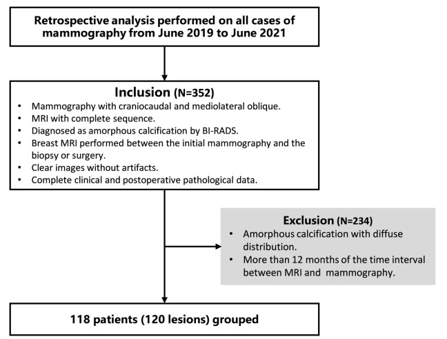
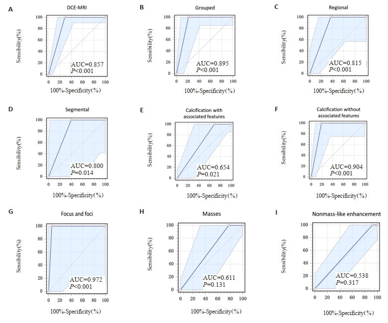
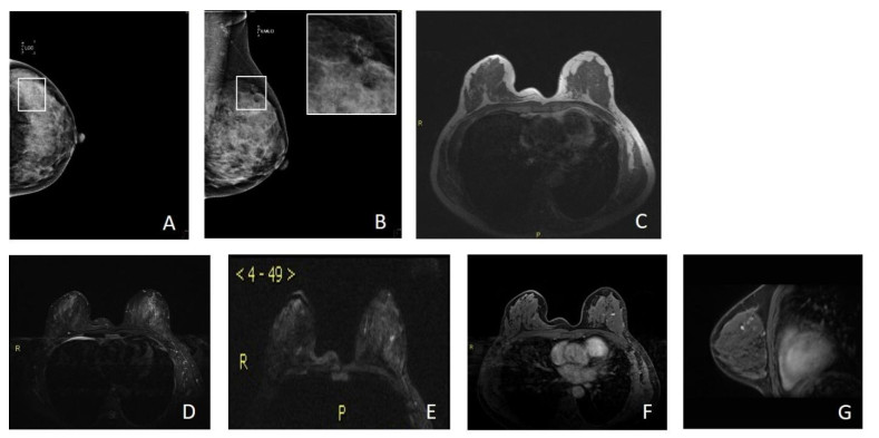
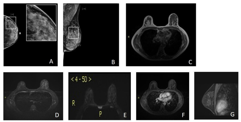
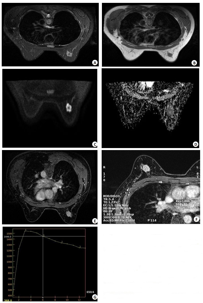
Objective To explore the factors that affect the accuracy of diagnosis of suspicious amorphous calcification and explore while the diagnostic performance of each factor is quantitatively evaluated. Methods A retrospective study was conducted on a selected group of 118 patients (120 lesions), who underwent mammography from June 1, 2019 to June 1, 2021. All patients were female, with an average age of 46.7±9.7 years old and a median age of 46 years old. A double-blind diagnosis of all cases was performed by two doctors who engaged in breast imaging diagnosis for more than 10 years. The patients were described and classified according to the 5th edition of breast imaging reporting and diagnostic system (BI-RADS) in 2013 as diagnostic criteria, while the pathological findings were chosen as the gold standard. The evaluation indexes of diagnostic tests and ROC curves were used to analyze and determine the diagnostic performance of each affecting factor on the benign and malignant results of suspicious amorphous calcification. Results The sensitivity, the specificity, the positive predictive value and the negative predictive value of breast dynamic contrast-enhanced magnetic resonance imaging (DCE-MRI) for suspicious amorphous calcification were 100%, 71.4%, 71.4% and 100% respectively. The area under curve was 0.857 (95%CI: 0.782-0.914). All the malignant lesions were correctly diagnosed. The diagnostic specificity of the focus and foci enhancement cases was 94.4% and the area under curve was 0.972 (95% CI: 0.785-1.000, P < 0.05), that were both largest among the three types of enhancement demonstrated by DCE-MRI. The malignancy rate of suspicious amorphous calcification was 41.7% in all patients, but decreased to 24% in the pure calcification group without associated features. When considering the four kinds of calcification distribution types, only the malignancy rate of suspicious amorphous calcification with grouped and regional distribution was in BI- RADS 4B, and the area under curve of them were 0.895 (95% CI: 0.798- 0.955) and 0.815 (95% CI: 0.650-0.924), respectively. The malignancy rate of the cases with regional distribution was the lowest as 25% among all distribution types. It could even drop to 8% in the pure calcification group of it. Conclusion Breast DCE- MRI proves sensitive enough to diagnose all malignant lesions correctly and has the highest diagnostic specificity for the focus and foci enhancement cases. Among the factors affecting the diagnosis of suspicious amorphous calcification, the diagnosis specificity of pure calcification group is significantly higher than that of the cases with associated features, which is of great value for judging the benign and malignant of suspicious amorphous calcification. The cases with pure calcification in regional distribution group have lowest rate of malignancy. The regular clinical follow-up can be chosen to avoid some unnecessary biopsy or surgery.
2023, 46(3): 389-397.
doi: 10.12122/j.issn.1674-4500.2023.03.02
Abstract:
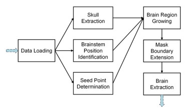
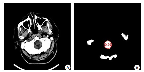
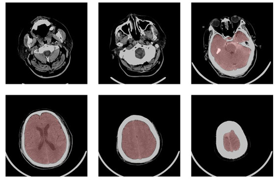
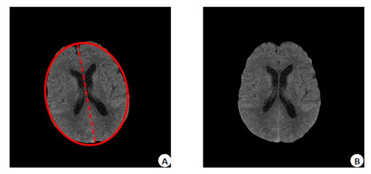
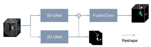
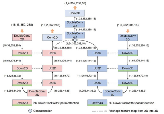
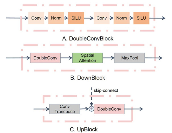
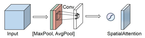
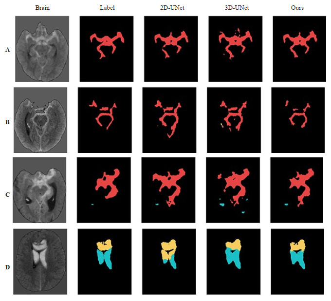
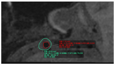
Objective To propose an end- to- end, fully automated segmentation method for multiple types of intracranial hematomas caused by the rupture of intracranial aneurysm. Methods A total of 644 brain CT images of intracranial hemorrhage caused by the rupture of intracranial aneurysms were selected and divided into train and test datasets by the ratio of 8:2. Brain extraction was firstly acquired by region growing, then multi- type segmentation was performed on the hemorrhage regions using deep learning. Results The dice coefficients of subarachnoid hemorrhage, intracerebral parenchymal hemorrhage, intraventricular hemorrhage, and intracerebral hemorrhage were 62.13%, 68.64%, 50.08% and 71.10% on the test dataset. Conclusion The segmentation network proposed in this paper can effectively complete multi-type hematoma segmentation for intracranial hemorrhage. The end-to-end processing process can be automatically completed with the brain extraction algorithm on the clinical data. The method effectively improves the diagnosis and treatment efficiency of intracranial hemorrhage caused by the rupture of intracranial aneurysms and possesses good clinical application value.
2023, 46(3): 398-406.
doi: 10.12122/j.issn.1674-4500.2023.03.03
Abstract:
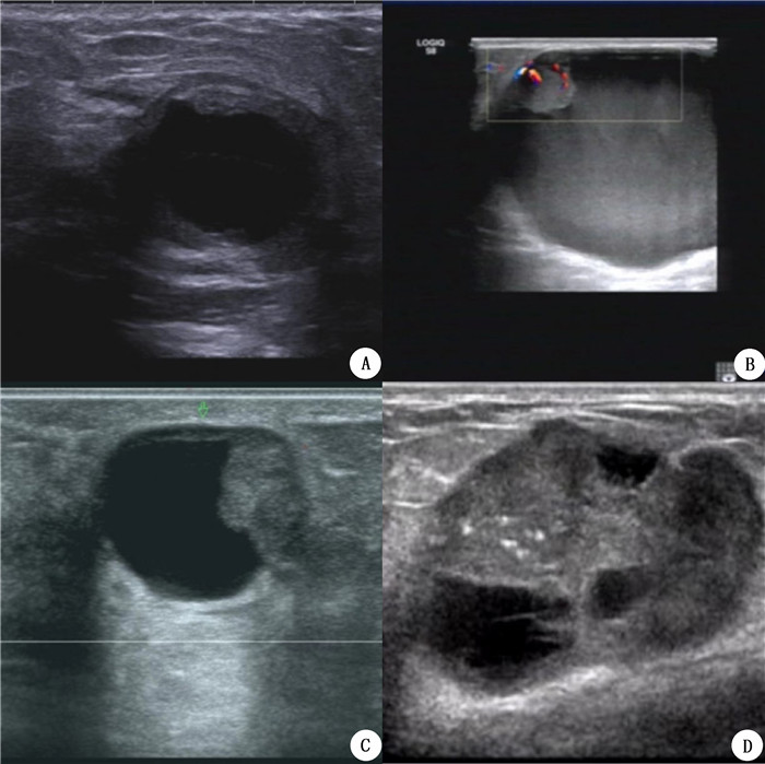
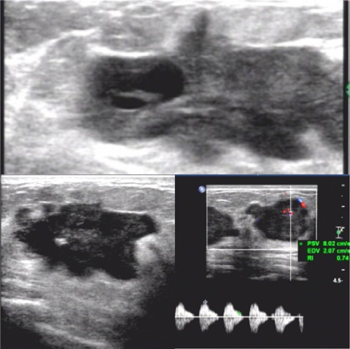
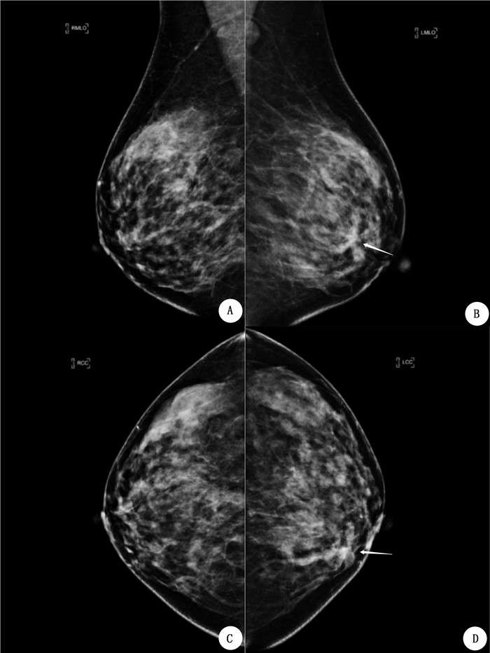
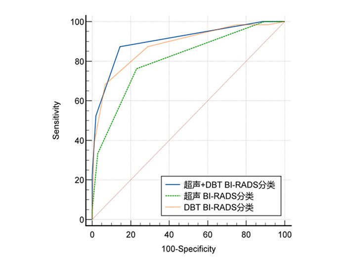
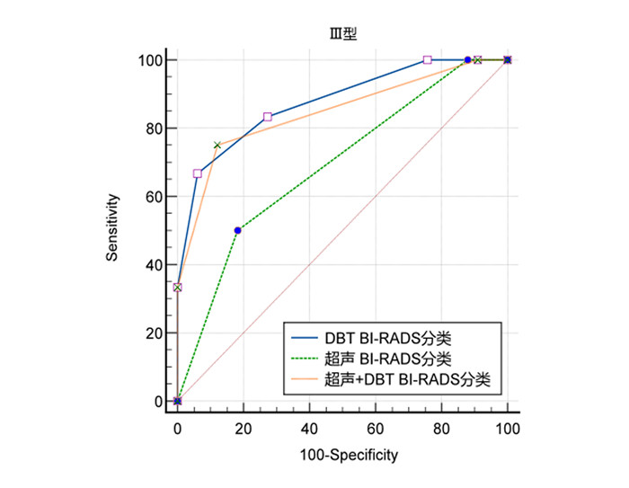
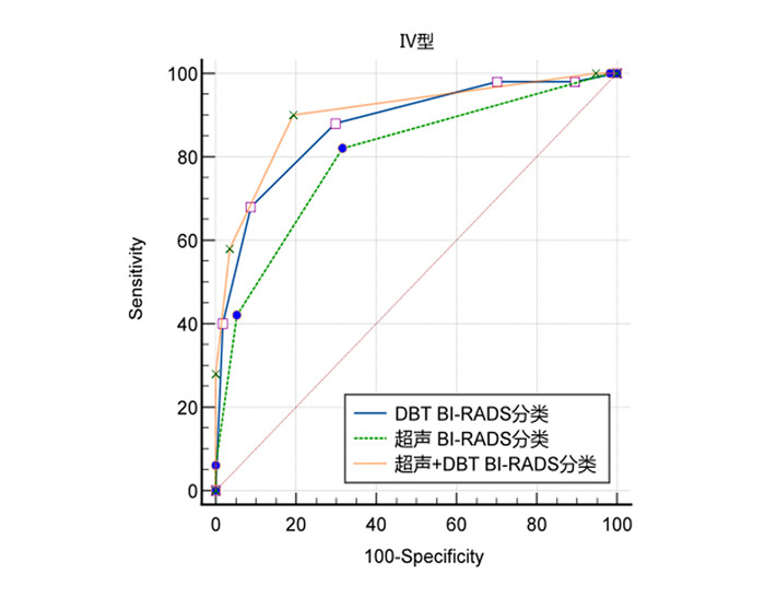
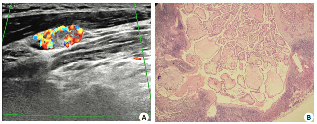
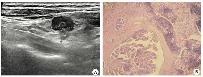
Objective To analyze the ultrasonographic and DBT features of breast complex cystic and solid mass, and to explore the diagnostic value of ultrasound (US), digital breast tomosynthesis (DBT) and their combination in differentiating benign and malignant breast complex cystic and solid mass. Methods A total of 257 masses in 251 patients with breast complex cystic and solid mass were analyzed retrospectively. According to the sonographic features, the masses were divided into 4 types, and the positive predictive values of each type were calculated. The correlation between the features of sonogram, DBT and malignancy was analyzed. The diagnostic efficacy of US, DBT and US + DBT was compared according to the pathological results as the gold standard. Results The malignant rate of breast complex cystic and solid mass was 30.4%. The positive predictive values of Ⅰ-Ⅳ type complex cystic and solid mass were 0, 22.2%, 19.3% and 38.0%, respectively(P < 0.001). There were significant differences in age, tumor size, classification, morphology, growth mode, margin, calcification, blood flow in the tumor, abnormal ALN in ultrasonic characteristics, and tumor size, morphology, margin, density, calcification, trabecular structure and gland classification in DBT characteristics (P < 0.05). The AUC values for US, DBT and US+DBT were 0.806, 0.880 and 0.903, respectively. There was significant difference in AUC between US and DBT, US + DBT (P < 0.05), but there was no significant difference in AUC between US+DBT and DBT (P < 0.05). Conclusion The diagnostic efficacy of DBT in differentiating benign and malignant breast complex cystic and solid mass is better than that of US. The combination of DBT and US can significantly improve the diagnostic accuracy, sensitivity and specificity.
2023, 46(3): 407-412.
doi: 10.12122/j.issn.1674-4500.2023.03.04
Abstract:
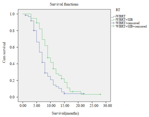
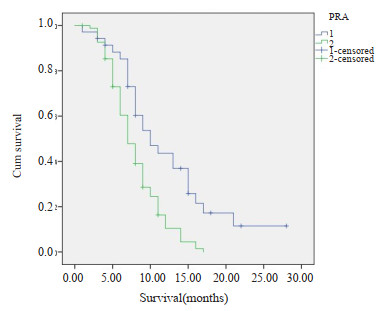
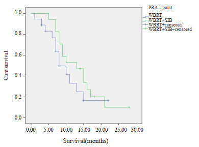
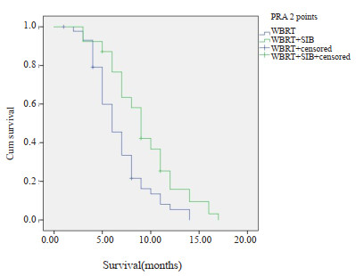
Objective To compare the therapy effect and toxic side effects of whole brain radiotherapy and whole brain with or without simultaneous integrated boost in patients with more than 3 brain metastatic lesions. Methods Patients with more than 3 brain metastatic lesions undergoing radiotherapy in the Department of Radiation Oncology of Shenzhen People's Hospital from July 2011 to July 2020 were analyzed retrospectively, which grouped to whole brain radiotherapy (WBRT group, n=62) and whole brain radiotherapy plus simultaneous integrated boost radiotherapy (WBRT + SIB group, n=56). We observed the survival rate and toxic side effects of the two groups. Results The median survival time of WBRT+SIB group and the WBRT group were 10.7 months and 7.8 months, respectively. The 1-year survival rate were 32% and 13% (P=0.02), respectively. The median survival time of WBRT+SIB and WBRT for those with RPA 1 point were 13.6 months and 10.5 months, and the 1-year survival rates were 51% and 28% (P=0.01). The median survival time of WBRT + SIB and WBRT for those with RPA 2 points were 9.3 months and 6.8 months, and the 1- year survival rates were 18% and 11% (P=0.39). There were no significant differences of the early and late toxic side reactions between the two groups(P>0.05). Conclusion For patients with more than 3 brain metastases, the survival rate of WBRT+SIB was better than whole brain radiotherapy, and there were no difference in side effects.
2023, 46(3): 413-420.
doi: 10.12122/j.issn.1674-4500.2023.03.05
Abstract:
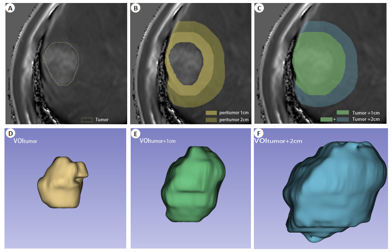
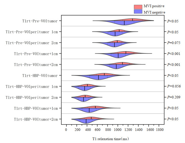
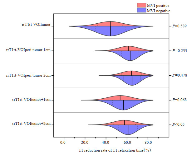
Objective To investigate the value of dual-region T1 mapping quantitative parameters of whole tumor and peritumoral volume of interest (VOI) combined with GD-EOB-DTPA enhanced MRI in evaluating microvascular invasion (MVI) of hepatocellular carcinoma. Methods From January 2019 to September 2022, 72 patients with single hepatocellular carcinoma diagnosed by postoperative pathology in our hospital were retrospectively analyzed. According to the expression status of MVI, it was divided into MVI positive group (n=23) and MVI negative group (n=49). GD-EOB-DTPA enhanced MRI T1 mapping scan was performed before operation, and T1 mapping images of pre-enhancement phase (Pre) and hepatobiliary phase (HBP) were obtained. The VOI of tumor, peritumoral and tumor plus peritumor were drawn layer by layer by using 3D Slicer software and VOItumor, VOIperitumor 1 cm, VOIperitumor 2 cm, VOItumor + 1 cm, VOItumor + 2 cm were obtained. The quantitative parameters of each VOI, T1 relaxation time (T1rt) and T1 relaxation time reduction rate (rrT1rt) were measured. We compared the differences of quantitative parameters between the two groups. ROC curve and net reclassification index (NRI) were used to analyze the diagnostic efficacy of each quantitative parameter. Results T1rt-Pre-VOItumor, T1rt-Pre-VOIperitumor 1 cm, T1rt-Pre- VOItumor + 1 cm, T1rt-Pre-VOItumor+2 cm, T1rt-HBP-VOItumor, T1rt-HBP-VOItumor+1 cm, T1rt-HBP-VOItumor+2 cm, rrT1rt-VOItumor+2 cm between the two groups were statistically significant (P < 0.05). The AUC were 0.720, 0.689, 0.748, 0.730, 0.727, 0.726, 0.717, 0.639. The AUC of multiphase quantitative parameter, T1rt-Pre-HBP-VOItumor, T1rt- Pre- HBP- VOItumor + 1 cm, T1rt-Pre-HBP-VOItumor + 2 cm, were 0.740, 0.756, 0.743, among which T1rt-Pre-HBP-VOItumor+ 1 cm had the highest diagnostic efficiency. NRI analysis showed that T1rt-Pre- VOItumor + 1 cm, T1rt- HBP- VOItumor + 1 cm had positive improvement compared with T1rt-Pre-VOItumor + 2 cm, T1rt-HBP-VOItumor + 2 cm. NRI values were 0.6158 and 0.4011. T1rt-Pre-HBP-VOItumor + 1 cm showed positive improvement compared with T1rt-Pre-VOItumo1 cm and T1rt-HBP-VOItumor + 1 cm, and the NRI values were 0.0692 and 0.5643, respectively. Conclusion The T1 relaxation time of tumor in Pre and HBP has good diagnostic efficacy for microvascular invasion in hepatocellular carcinoma. Combined with peritumoral 1 cm has higher diagnostic efficacy than peritumoral 2 cm. The diagnostic efficacy of multiphase quantitative parameter is higher than single-phase quantitative parameter and T1rt-Pre-HBP-VOItumor+1 cm is the highest.
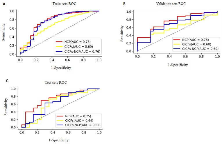
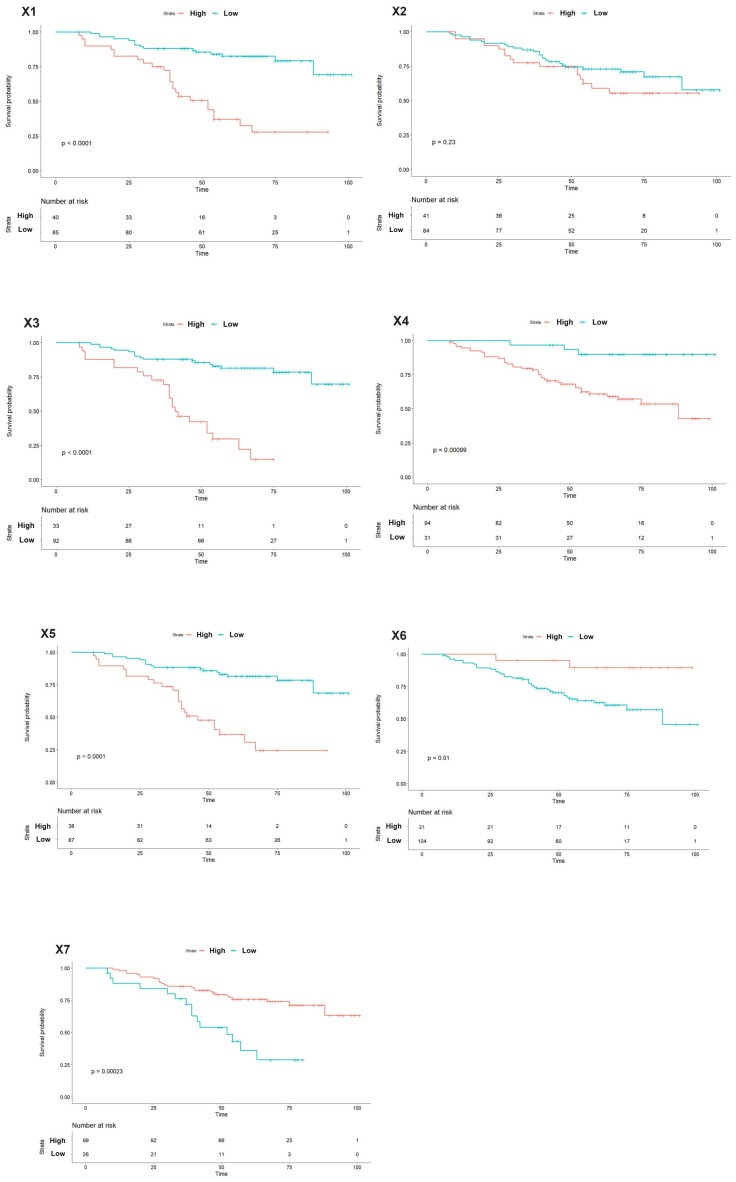
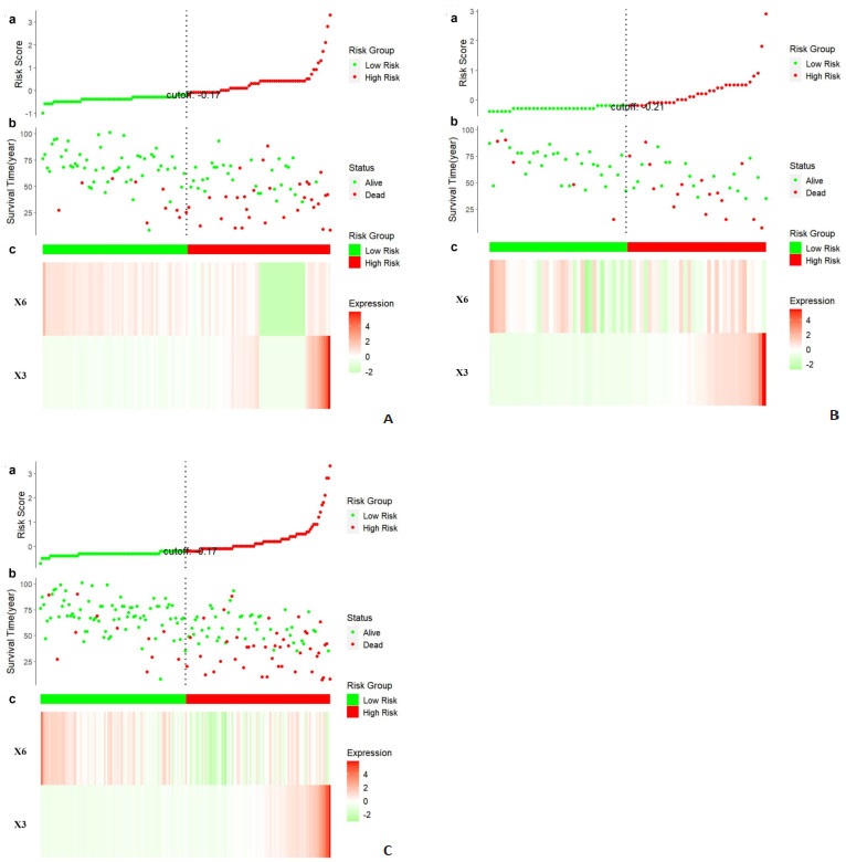
2023, 46(3): 421-430.
doi: 10.12122/j.issn.1674-4500.2023.03.06
Abstract:
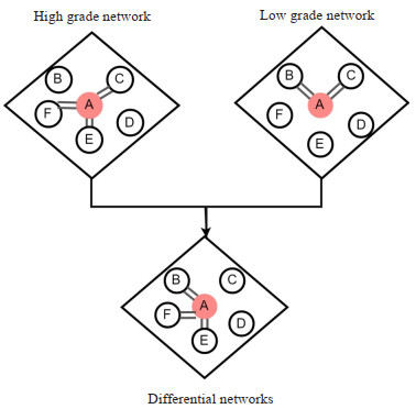
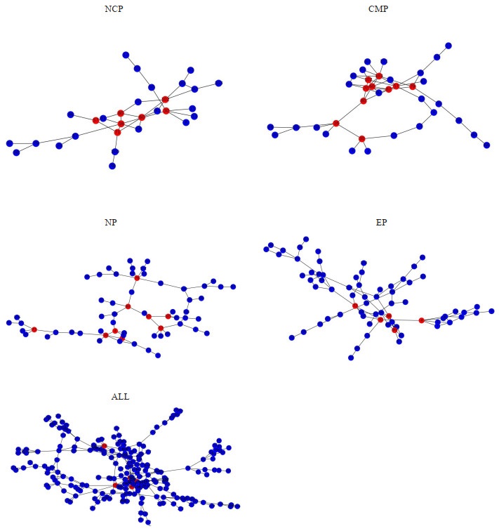
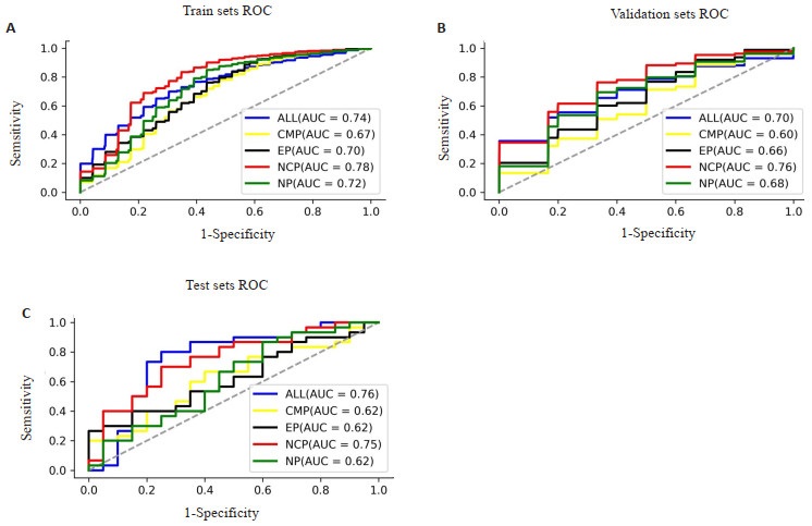
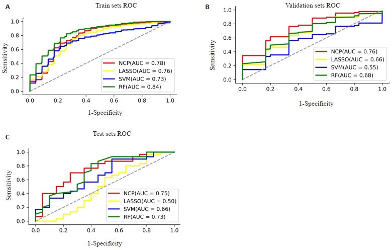
Objective To predict the World Health Organization/International Society of Urological Pathology (WHO/ISUP) grade of clear cell renal cell carcinoma (ccRCC) using a CT radiomics model based on differential network analysis feature selection and to explore the interpretability of features. Methods A total of 175 ccRCC patients with CT and clinicopathologic data were enrolled in the study, which included a training set of 105 patients and a test set of 70 patients. Segmentation images and feature extraction were performed using ITK-SNAP and PyRadiomics software. The WHO/ISUP grade prediction model was constructed using differential network analysis feature selection with different phases in the training set. The best phase model was then compared with other machine learning models and clinical models. The importance of features in the best phase model was evaluated using K-M survival analysis, Cox regression analysis and risk scores to verify their interpretability. Results The non-contrast phase model was identified as the best phase model, with an AUC of 0.76 in the validation set, performing better than other machine learning models and clinical models (P < 0.05). Kaplan-Meier survival analysis, Cox regression analysis and risk score analyses revealed that the non-contrast phase model could predict progression-free survival well. Conclusion The differential network analysis feature selection was used to select the non- contrast phase model, which could accurately predict the WHO/ISUP grade of ccRCC. The characteristics of this model could predict progression- free survival with strong interpretability.
2023, 46(3): 431-435.
doi: 10.12122/j.issn.1674-4500.2023.03.07
Abstract:
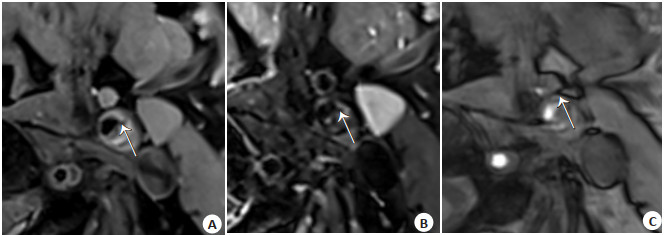
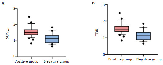
Objective To investigate the correlation between 18F-NaF uptake of plaques and fibrous cap rupture (FCR) of plaques, and the correlation between 18F-NaF uptake positivity and FCR. Methods Thirty-one patients with confirmed atherosclerosis at the carotid bifurcation who were admitted to our hospital within a year of diagnosis were selected. The patients underwent high-resolution magnetic resonance vessel wall imaging with MR-VPD software. The plaque FCR conditions was analyzed. The 18F-NaF PET / CT was performed with in 1 week after high-resolution magnetic resonance vessel wall imaging examination. Based on the extent of 18F-NaF uptake in carotid arteries, patients were divided into 18F-NaF uptake positive and 18F-NaF uptake negative groups. Differences of plaque FCR between groups were analyzed. Results There were 23 patients with both plaques in bilateral carotid bifurcations out of 31 patients and the remaining 8 patients with unilateral carotid plaques were included in this study. Fifty-four plaques were collected. Among 54 atherosclerotic carotid arteries, 33 carotid arteries were positive by 18F-NaF imaging and 21 carotid arteries were negative by 18F-NaF imaging, showing 61.11% positive and 38.89% negative carotid artery images. The proportion of plaques with FCR present was 58% in the 18F-NaF positive group and 19% in the 18F-NaF negative group. The difference in the FCR situation of plaques between the two groups was statistically significant, with a higher proportion of plaques positive for 18F-NaF uptake having FCR than plaques negative for 18F-NaF uptake.(t=2.034, P=0.045). Conclusion Plaques positive for 18F-NaF uptake has greater odds of FCR than plaques negative for 18F-NaF uptake. 18F-NaF uptake to some extent reflects plaques with FCR, which in turn assesses plaque vulnerability.
2023, 46(3): 436-441.
doi: 10.12122/j.issn.1674-4500.2023.03.08
Abstract:
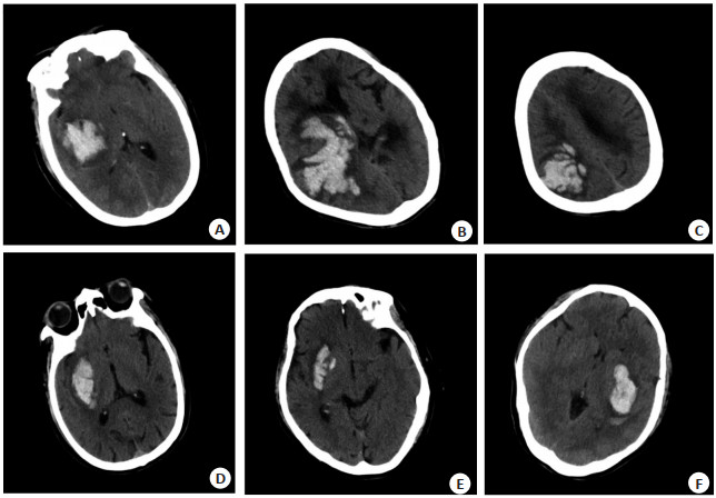
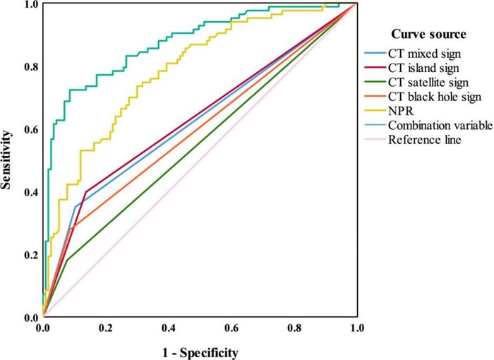
Objective To analyze the predictive value of CT mixed sign, island sign combined with neutrophil to platelet ratio (NPR) for the hematoma enlargement of spontaneous basal ganglia hemorrhage (sBGICH). Methods We retrospectively reviewed the data of 200 patients with sBGICH admitted to the Department of Emergency and Department of Neurosurgery of our hospital from February 2020 to November 2022. The patients were grouped as bleeding hematoma enlargement group (n=43) and no bleeding hematoma enlargement group (n=157) according to whether the bleeding had enlarged within 24 hours of onset. The CT mixed sign, island sign, satellite sign, swirl sign, black hole sign, NPR and prognosis scores were compared between the two groups. Results There were no statistically significant differences in gender, age, onset time, CT swirl sign, and intraventricular rupture between the two groups (P > 0.05). The CT mixed sign, island sign, satellite sign, black hole sign, and NPR were significantly higher in the bleeding hematoma enlargement group than in the no bleeding hematoma enlargement group, while the prognosis score was significantly lower (P < 0.05). Multivariate logistic regression analysis showed that CT mixed sign, island sign, satellite sign, black hole sign, and NPR > 5 were risk factors for bleeding hematoma enlargement within 24 hours of sBGICH onset (P < 0.05). The ROC curve was drawn according to whether bleeding hematoma enlargement occurred within 24 h of sBGICH onset, and the area under the curve of the joint diagnosis and prediction of CT mixed sign, island sign, satellite sign, black hole sign and NPR was 0.873, with a sensitivity and specificity of 72.30% and 91.50%, respectively. Conclusion CT mixed sign, island sign combined with NPR have a high predictive value for the hematoma enlargement of sBGICH and it can be flexibly used according to the actual clinical situation.
2023, 46(3): 442-447.
doi: 10.12122/j.issn.1674-4500.2023.03.09
Abstract:
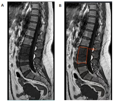
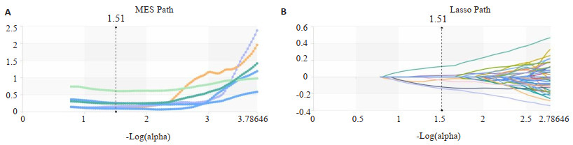
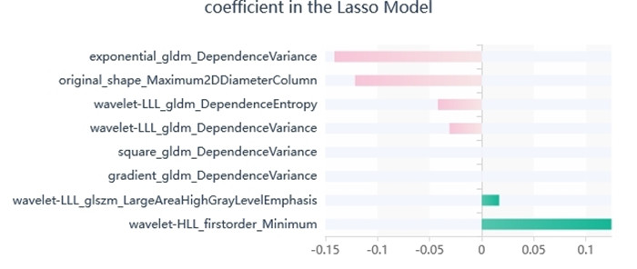
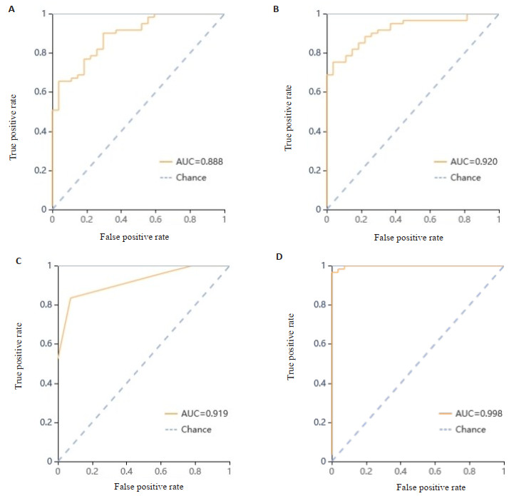
Objective To investigate the value of machine learning of MR T1WI radiomics in distinguishing tuberculous spondylitis (TS) from brucellar spondylitis (BS). Methods The clinical data of 77 TS patients and 34 BS patients diagnosed in our hospital were retrospectively collected. The patients were divided into training set (n=88) and verification set (n=23) according to the ratio of 0.8:0.2. First, two experienced diagnostic radiologists delineated the ROI on MR T1WI and extracted their imaging features. Then, we used the variance selection method, univariate feature selection method, LASSO for the feature information of radiomics to select and dimensionally reduce, and selected the 8 most characteristic values. Four machine learning algorithms, including XGBoost method, Logistic regression method, support vector machine method, and K-nearest neighbor method, were used to construct the MR T1WI imaging model in distinguishing TS from BS. The ROC curve and AUC were used to evaluate the diagnostic the effectiveness of these models. Results The AUC of XGBoost model was 0.80 (95% CI: 0.59-1.00), and the accuracy was 0.83. The AUC of Logistic regression model was 0.85 (95% CI: 0.65-1.00) and the accuracy was 0.78. The AUC of support vector machine model was 0.79 (95% CI: 0.62- 0.97), and the accuracy was 0.78. The AUC of K-nearest neighbor model was 0.75 (95% CI: 0.53-0.98), and the accuracy was 0.83. In the validation, Logistic regression model was the highest diagnostic efficiency in four machine learning algorithms. Conclusion The machine learning of MR T1WI radiomics has a good diagnostic efficiency in differentiating TS from BS. Logistic regression model is the best diagnostic efficiency in four machine learning algorithms.
2023, 46(3): 448-452.
doi: 10.12122/j.issn.1674-4500.2023.03.10
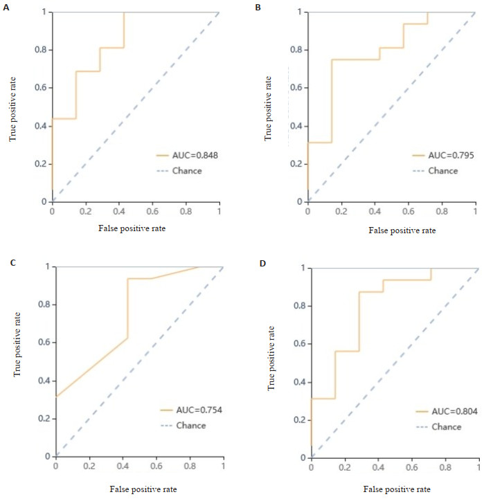
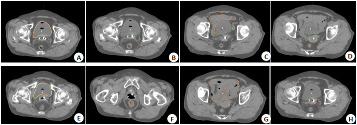
Abstract:
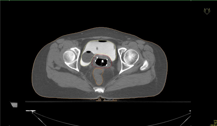
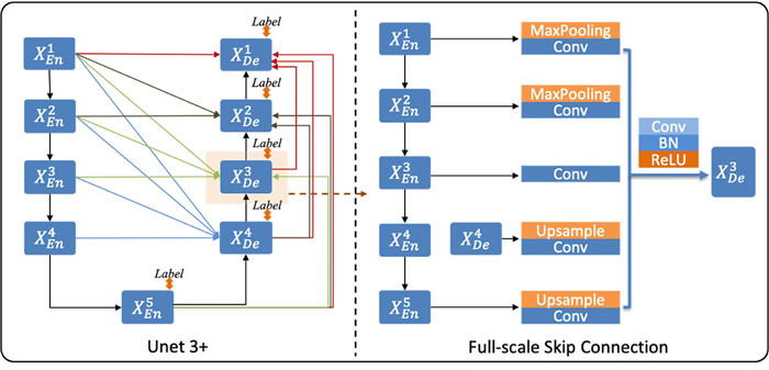
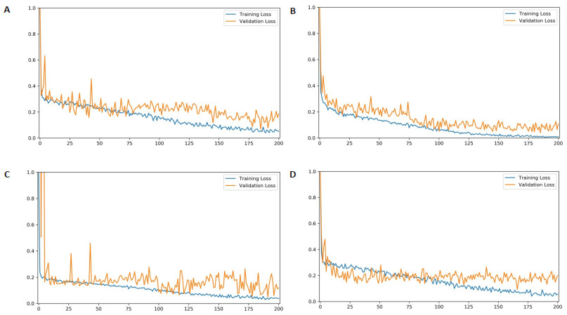
Objective To conduct a model to predicate high-risk target areas and dangerous organ locations in after-loading therapy for cervical cancer based on deep learning. Methods An end-to-end automatic segmentation framework based on U-net3+ was constructed, and 213 cervical cancer patients who had undergone after-loading high-dose-rate therapy in the two centers were delineated. The patients were divided into training set, validation set and test set according to the ratio of 7:2:1. The high-risk clinical target, bladder, rectum, and small intestine were delineated, and the accuracy of the predictive model was assessed using the Hausdorff distance and the Dice similarity coefficient, respectively. Results The Dice similarity coefficient values of automatic bladder delineation were 0.953, rectum and small intestine were 0.885 and 0.857, respectively. The mean value of organ risk was 0.898, and the average Hausdorff distance value was 5.4 mm. The Dice similarity coefficient value of the high-risk clinical target was 0.869 and the Hausdorff distance value was 8.1 mm. Conclusion The U-net3+based model for predicting the location of target and dangerous organs in after-loading therapy for cervical cancer has a high accuracy rate, while the training is less time-consuming. It is expected to be promoted for clinical application.
2023, 46(3): 453-457.
doi: 10.12122/j.issn.1674-4500.2023.03.11
Abstract:
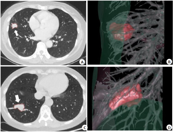
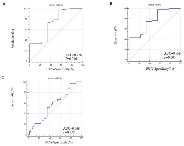
Objective To explore the correlation between CT signs and quantitative parameters of lung cancer and the expression level of Ki-67 antigen. Methods Clinical and imaging data of 93 patients with lung cancer confirmed by pathology who underwent CT scan in our hospital were retrospectively analyzed. Routine CT scan was performed using spectral CT, and tumor specimens were subjected to immunohistochemical staining to detect the expression level of Ki-67 antigen. CT signs of lung cancer were observed. Computer-aided diagnosis software was used to delineate and segment the primary lung cancer, and CT quantitative parameters were calculated. The correlation between Ki-67 expression level and CT signs and quantitative parameters was analyzed. ROC curve was drawn to analyze the predictive efficacy of CT signs and quantitative parameters on Ki-67 expression level. Results The expression level of Ki-67 was related to gender and pathological type(P < 0.05), with higher expression index in males and lung squamous cell carcinoma. There was no correlation between the expression level of Ki-67 and various CT signs(P > 0.05). The difference in tumor vascular volume between different Ki-67 expression groups was statistically significant (P < 0.05). Spearman non-parametric correlation analysis showed a positive correlation between vascular volume and Ki-67 expression level (r=0.224, P=0.03). The area under curves were 0.726, 0.759, and 0.569 in the low-medium level, low-high level, and medium-high level, respectively. Conclusion CT quantitative parameters (vascular volume) can be used to evaluate the expression level of Ki-67 in lung cancer. They are positively correlated with Ki-67 expression level. They can assist in the clinical evaluation of tumor cell proliferation activity in patients with lung cancer to a certain extent.
2023, 46(3): 458-463.
doi: 10.12122/j.issn.1674-4500.2023.03.12
Abstract:
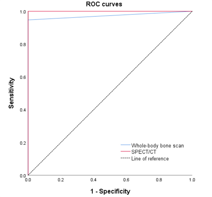
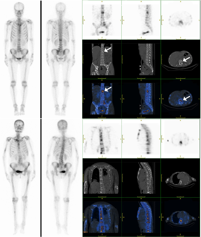
Objective To investigate the imaging characteristics and diagnostic value of whole-body bone scan and SPECT/CT in nontuberculous mycobacterial-infected bone destruction in HIV-negative patients. Methods We retrospectively analyzed 25 patients with nontuberculous mycobacterial (NTM) who underwent whole-body bone scan and SPECT/CT in our hospital from November 2020 to July 2022. The clinical and imaging characteristics of NTM-infected bone destruction were summarized, and the diagnostic efficacy of the two imaging methods was compared. Results The sensitivity of the whole-body bone scan for the diagnosis of NTM-infected bone destruction was 94.7%, the specificity was 100%, and the diagnostic coincidence rate was 96%. The sensitivity, specificity, and diagnostic compliance rate of SPECT/CT were 100%. There was a significant difference in the diagnostic compliance rate of the two methods in diagnosing NTM-infected bone destruction (P < 0.05). The lesions were mainly distributed in the ribs, long bone of extremities, and spinal column, manifested as intense radioactive uptake on whole-body bone scan, and mostly showed osteolytic changes on SPECT/CT. Conclusion Whole- body bone scan combined with SPECT/CT imaging can monitor lesions in multiple parts of the body and better show the metabolic situation and imaging characteristics of NTM-infected bone destruction, which can help clinical diagnosis and treatment.
2023, 46(3): 464-469.
doi: 10.12122/j.issn.1674-4500.2023.03.13
Abstract:
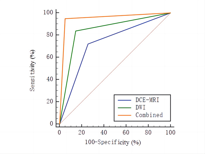
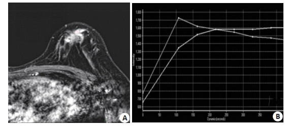
Objective To analyze the imaging characteristics of dynamic contrast-enhanced MRI (DCE- MRI) and diffusion-weighted imaging (DWI) of breast cancer and the efficacy of combined diagnosis. Methods The clinical data of 230 patients with breast cancer admitted to our hospital from June 2018 to August 2022 were retrospectively collected as the malignant group, and the clinical data of 252 patients with benign breast lesions who were examined and treated in our hospital during the same period were selected as the benign group. All patients were diagnosed by surgery or pathological puncture biopsy. The imaging characteristics of DCE-MRI and DWI in patients with benign and malignant breast lesions were counted, and the diagnostic results of DCE- MRI and DWI alone and in combination were counted. ROC curve was used to analyze the diagnostic efficacy of DCE- MRI and DWI alone and in combination for breast cancer. Results In the malignant group, the lesions were lobulated and irregular (43.04%, 45.65%), with marginal burrs (48.70%), internal heterogeneous enhancement, cluster enhancement and branching enhancement (20.87%, 38.70%, 36.09%), and the proportion of patients with TIC type Ⅲ (43.04%) was higher than that in benign group (33.33%, 15.48%, 12.70%, 8.33%, 14.29%, 19.05%, 3.57%, P < 0.05). DCE-MRI and DWI alone and jointly diagnosed benign and malignant breast lesions in 187 cases, 215 cases, 239 cases, 166 cases, 193 cases and 218 cases respectively, and the detection rates were 74.21%, 85.32%, 94.84% and 72.17%, 83.91%, 94.78%, respectively. The detection rate of benign and malignant breast lesions diagnosed by DWI alone and DCE-MRI combined with DWI was higher than that by DCE- MRI alone, and that by DCE- MRI combined with DWI was higher than that by DWI alone (P < 0.05).The sensitivity (83.91%, 94.78%), specificity (85.32%, 94.84%) and area under the curve (0.846, 0.948) of DWI alone and DCE-MRI combined with DWI in diagnosing benign and malignant breast lesions were higher than those of DCE- MRI alone (72.17%, 74.21%, 0.732), the combined diagnosis of DCE-MRI and DWI was higher than that of DWI alone (P < 0.05). Conclusion DCE-MRI and DWI have specific imaging characteristics in the diagnosis of benign and malignant breast lesions. The combination of DCE-MRI and DWI can effectively improve the diagnostic efficiency of benign and malignant breast lesions and had good application value.
2023, 46(3): 470-474.
doi: 10.12122/j.issn.1674-4500.2023.03.14
Abstract:
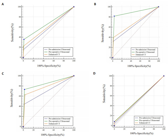
Objective To investigate the diagnostic value of cervical ultrasonography in the cervical lymph node metastasis of thyroid carcinoma. Methods A total of 932 patients who underwent radical thyroidectomy in our hospital from August 2020 to November 2021 were analyzed retrospectively. All patients were examined at least twice by experienced sonographers (pre-admission and pre-operation respectively). The imaging data of cervical ultrasound and cervical enhanced CT were collected. According to the pathological results of cervical lymph nodes as the gold standard. SPSS 26.0 and MedCacl 19.0.4 were used statistical analysis and compare the diagnostic efficacy index and AUC value of cervical ultrasound and cervical enhanced CT in cervical lymph node metastasis. Results The diagnostic accuracy, sensitivity, specificity, false-positive rate and false-negative rate of pre-operation ultrasound in cervical lateral Ⅲ and Ⅳ lymph nodes were superior to those of pre-admission ultrasound and cervical enhanced CT. The AUC value of pre-operation ultrasound in cervical lateral lymph nodes was better than that of pre-admission ultrasound and cervical enhanced CT. The AUC values of pre-operation ultrasound in cervical lateral Ⅲ and Ⅳ lymph nodes were above 0.80 (0.89 and 0.85, respectively), and the difference with the AUC values of pre-admission ultrasound and cervical enhanced CT was significant (P < 0.05). The diagnostic value of pre- operation ultrasound in cervical lateral Ⅲ and Ⅳ lymph nodes was better than that in cervical lateral Ⅱ and Ⅴ lymph nodes (AUC value was 0.67 and 0.54, respectively). Conclusion It is important and necessary for patients with thyroid cancer to undergo cervical lateral lymph nodes exploration again by experienced sonographers before operation.
2023, 46(3): 475-478.
doi: 10.12122/j.issn.1674-4500.2023.03.15
Abstract:
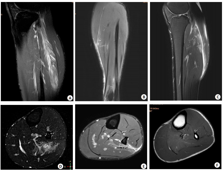
Objective To explore the diagnostic value of MR in male professional football players with calf muscle injuries. Methods Sixteen male professional football players, with an average age of 29.3±3.4 years, underwent MR examinations within 3 days after injury in the Third Affiliated Hospital of Southern Medical University from December 2010 to September 2022. MR data of 28 cases of calf muscle injuries were obtained. Two experienced radiologists read the MR images together to evaluate muscle injuries, including the location and degree of injured muscle, the shape of abnormal signal intensity, and whether there was muscle fascia injury, superficial fascia exudation, intermuscular bundle exudation, intermuscular effusion, effusion of broken muscles, or hemorrhage. Results A total of 40 muscles affected, mainly including soleus muscle injury in 21 cases, gastrocnemius muscle injury in 10 cases. The injury mainly involved the tendon belly transition zone (36 muscles), including 35 muscles in grade Ⅰ injuries and 5 muscles in grade Ⅱ injury. The injured muscles demonstrated contour feathery or downy feathery like and lamellar patterns. There were 7 cases of muscle aponeurosis injury, 18 cases of superficial fascia exudation, 21 cases of intramuscular effusion and 3 cases of hemorrhage. Conclusion Calf muscle injuries in male professional football players often involve the gastrocnemius muscle, and MR can show the imaging features of the injured muscles, clarify the location and degree of injury, and provide imaging information for treatment for athletic injury.
2023, 46(3): 479-482.
doi: 10.12122/j.issn.1674-4500.2023.03.16
Abstract:
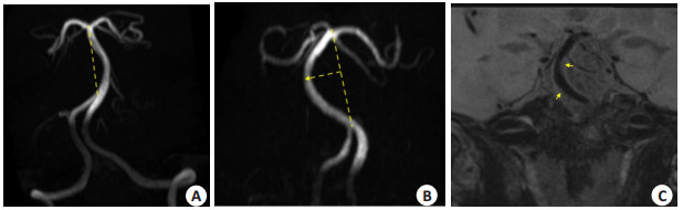
Objective To analyze the longitudinal distribution characteristics of atherosclerotic plaques in the tortuous basilar artery using high-resolution magnetic resonance imaging, for predicting the occurrence of posterior circulation ischemic stroke. Methods We involved the retrospective analysis of 60 patients who underwent high-resolution magnetic resonance imaging exams at our hospital's neurology department, and 90 plaques were detected. All patients were divided into three groups based on the relationship between the plaque's location and the tortuous arc top: bifurcation (n=19), proximal (n=43) and distal (n=28). The characteristics of the vessel wall, plaque, and general data were compared between the three groups. All patients in the proximal group and the distal group were divided into the right tortuosity group (n=52) and the left tortuosity group (n=19) based on the direction of basilar artery convexity. The relationship between the distribution of plaque and the direction of basilar artery convexity was compared. Results Among 60 patients, plaques were mostly located in the proximal group. The vessel area in the proximal group was larger than that in the distal group (P=0.049), and the vessel wall area in the proximal group was larger than that in the distal group (P=0.010). There was a statistically significant difference in the distribution of plaques among the three groups (P=0.001). The plaques at the proximal and the distal groups were mostly located in the lateral wall, and the plaques at the bifurcation group were mostly located in the anterior wall. For both the proximal and the distal groups, the plaque distribution in the left wall was more frequent in patients with right convex basilar artery(10.5% vs 42.3%, P=0.012), the plaque distribution in the right wall was more frequent in patients with left convex basilar arter (25.0% vs 57.9%, P=0.009). Conclusion Plaques in the tortuous basilar artery were mostly located at the proximal of the arc top, and plaques at the arc top's proximal and distal ends were mostly located in the tortuous arc's lateral and inner walls.
2023, 46(3): 483-488.
doi: 10.12122/j.issn.1674-4500.2023.03.17
Abstract:
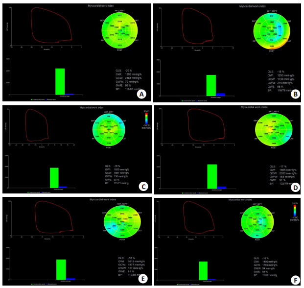
Objective To evaluate left ventricular work in patients with different rheumatic immune diseases using pressure-strain loop technique. Methods We prospectively selected 171 patients with rheumatic immune diseases diagnosed in the Affiliated Hospital of Yangzhou University from April 2021 to November 2022. The patients were divided into systemic lupus erythematosus group (n=30), ankylosing spondylitis group (n=33), Sjogren's syndrome group (n=35), rheumatoid arthritis group (n=43) and gout group (n=30). The course of disease was ≤ 5 years. Another 32 cases of physical examination during the same period were selected as the control group. All participants underwent conventional echocardiography to collect images, and obtained pressure strain loop parameters through offline ECHOPAC analysis. Statistical analysis of the correlation between routine parameters and myocardial work among groups was conducted to assess the left ventricular myocardial work status. Results Compared with the control group, the A peak of rheumatic immune disease groups increased and the E/A ratio decreased (P < 0.05). The E peak of systemic lupus erythematosus group and Sjogren's syndrome group decreased significantly (P < 0.05).The global wasted work of all rheumatic immune disease groups was higher, and the global work efficiency was lower (P < 0.05). The global longitudinal strain, global work index, and global constructive work in the systemic lupus erythematosus group and ankylosing spondylitis group and the global longitudinal strain, global work index, and global constructive work in the rheumatoid arthritis group and the global work index in the gout group were all decreased, with a statistically significant difference (P < 0.05). The absolute value of global longitudinal strain, left ventricular ejection fraction, and E/A ratio in the rheumatic immune disease group were positively correlated with global work index, global constructive work, and global work efficiency, while they were negatively correlated with global wasted work (P < 0.01). Conclusion The pressure strain loop can effectively assess the left ventricular myocardial work status in patients with rheumatic immune disease.It will objectively provide a more objective new diagnostic basis for clinical practice.
2023, 46(3): 489-493.
doi: 10.12122/j.issn.1674-4500.2023.03.18
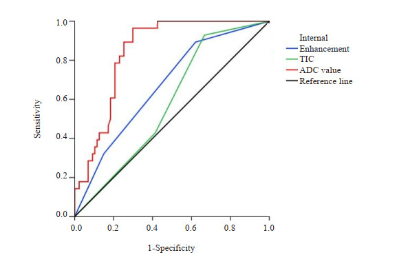
Abstract:
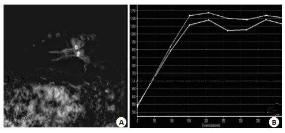
Objective To analyze the application value of MRI in growing BI-RADS class 3 breast lesions. Methods A total of 115 patients who diagnosed with growing BI-RADS class 3 lesions in the hospital from November 2021 to June 2022 were selected as the study subjects. The MRI features and their predictive value for condition changes were analyzed. Results Breast ultrasound follow- up found that among the 115 patients, there were 87 patients with decreased grade of BI-RADS class 3 lesions or stable BI-RADS class 3 lesions, and 28 patients with increased grade of BI-RADS 3 lesions. There was statistically significant differences in MRI findings such as morphology, boundary, internal enhancement, time-signal intensity curve (TIC) and apparent diffusion coefficient (ADC) value between the decreased grade or stable group and the increased grade group (P < 0.05). Multivariate logistic regression analysis found that internal enhancement, TIC curve and ADC value were influencing factors of changes in growing BI-RADS class 3 lesions (P < 0.05). ROC curve showed that the area under the curve values of internal enhancement, TIC curve and ADC value to predict changes in growing BI-RADS class 3 lesions were 0.669, 0.582 and 0.844, respectively (P < 0.05). Conclusion MRI features of patients with growing BI-RADS class 3 breast lesions are closely related to the grading and upgrading of lesions. Features such as internal enhancement, tic curve and ADC value can be used as important indicators to predict the upgrading of BI-RADS class 3 lesions, it provides a reliable basis for the diagnosis of benign and malignant breast lesions and the prediction of disease progression.
2023, 46(3): 494-499.
doi: 10.12122/j.issn.1674-4500.2023.03.19
Abstract:
Objective To evaluate the clinical value of multimodal MRI assessment of the response to apatinib mesylate therapy in advanced cervical cancer. Methods Sixty inpatients with advanced cervical cancer receiving apatinib mesylate therapy in our hospital from March 2017 to January 2022 were retrospectively enrolled. Patients were assigned into group A (complete response, n=48) and group B (partial response, n=12) according to their treatment response. The general data of patients and multimodal MRI findings were collected. Tumor signal intensity on diffusion-weighted imaging, T1WI, T2WI, and enhanced T2 star-weighted angiography scans were compared. Then ROC curve was plotted to assess the predictive efficacy of multimodal MRI for treatment response of advanced cervical cancer patients to apatinib mesylate therapy. Results The lesions of both groups displayed iso-signal intensity on T1WI sequence, and mixed signal intensity on T2WI sequence, with no statistical difference on the overall signal intensity of the lesions between two groups (P > 0.05). Lesions appeared as mixed signal intensity on diffusion-weighted imaging sequence in both groups. The amplitude map was all dominated by low signals, and the phase and R2* maps were dominated by mixed signals, with statistical difference in the signal intensity (P < 0.05). The apparent diffusion coefficient (ADC), phase value and R2* value demonstrated statistical difference between two groups (P < 0.05). ROC curve analysis denoted that the area under the curve of ADC value, phase value and R2* value in predicting the treatment response to apatinib mesylate therapy were 0.613, 0.656 and 0.759. Conclusion The signs in multimodal MRI of advanced cervical cancer patients with and without response to apatinib mesylate therapy have overlap and are non-specific, but patients with response have more complex performance on diffusion- weighted imaging and enhanced T2 star-weighted angiography images compared with non-response patients. ADC values, phase values and R2* values can be used as quantitative indicators to determine the presence or absence of treatment response after apatinib mesylate therapy, among which R2* value is more valuable.

2023, 46(3): 500-503.
doi: 10.12122/j.issn.1674-4500.2023.03.20
Abstract:
Objective To investigate the value of gastric contrast-enhanced ultrasound combined with intravenous contrast-enhanced ultrasound in evaluating the invasion and angiogenesis of gastric cancer. Methods Eighty-two patients with gastric cancer admitted to the hospital from January 2020 to Decembe 2021 were enrolled. All patients received gastric with intravenous contrast-enhanced ultrasound (double contrast-enhanced ultrasound) examinations one week before operation. The pathological tissues were collected during surgery, tumor microvessel density was detected by immunohistochemistry. Taking the pathological examination results as "gold standard", the value of double contrast-enhanced ultrasound in evaluating the T-stage of gastric cancer was analyzed. The relationship between time intensity curve parameters and microvessel density of gastric cancer lesions and normal gastric wall tissues was discussed by Pearson correlation analysis. Results Surgical pathological examination confirmed that 82 gastric cancer patients included 23 patients at stage T1, 26 at stage T2, 18 at stage T3, 9 at stage T4a and 6 at stage T4b. Taking the results of surgical pathological examination as the "gold standard", the total accuracy of dual-contrast ultrasound in the preoperative diagnosis of T-stage of gastric cancer was 78.05%. The baseline intensity, time to peak and infusion time of gastric cancer lesions were significantly lower than those of normal gastric wall tissues, while the peak intensity and enhanced intensity were significantly higher than those of normal gastric wall tissues (P < 0.05). No significant difference was found in arrival time between two tissues (P > 0.05). Correlation analysis showed that microvessel density in gastric cancer tissue was negatively correlated with the ultrasound contrast parameters baseline intensity, time to peak and infusion time (P < 0.05), and positively correlated with peak intensity and enhanced intensity (P < 0.05). Conclusion Double contrast-enhanced ultrasound has a good application value in the preoperative staging of gastric cancer. Its time intensity curve parameters are also of certain value in the indication of tumor angiogenesis, which is expected to be an effective tool in the preoperative assessment of gastric cancer.
2023, 46(3): 504-507.
doi: 10.12122/j.issn.1674-4500.2023.03.21
Abstract:
Objective To observe the effect of myringoplasty under otoendoscope in the treatment of traumatic tympanic membrane perforation and its effect on the poor air- bone conductivity of patients. Methods A total of 40 patients with traumatic tympanic membrane perforation were included from January 2019 to February 2021. After fully informing the design ideas of this study, all included patients underwent otoscopic myringoplasty. The treatment effect, hearing condition (comparison of patients' air conduction hearing threshold and air- bone conduction difference before operation, one month after operation, and three months after operation) and pain severity were observed [visual analogue scale (VAS) was used before operation, 1d after operation, and at discharge]. Results Among the 40 patients with traumatic large perforation of tympanic membrane treated by endoscopic myringoplasty, 28 cases were markedly improved, 10 cases were effective, and 2 cases were ineffective, the total effective rate was 95.00%. After treatment with otoscopic myringoplasty, the air conduction hearing threshold and air-bone conduction difference were lower one month after operation and three months after operation compared with those before operation, and the difference was statistically significant (P < 0.001). After treatment with endoscopic myringoplasty, their VAS scores were decreased 1 d after operation and at the time of discharge compared with those before operation, the VAS score of the patients before operation, 1 d after operation and during hospitalization showed statistical significance (P < 0.001); There was no significant difference in VAS score between pre-surgery and 1 d post-surgery (P > 0.05); Compared with pre-surgery, VAS score of patients at discharge was lower and the difference was statistically significant (P < 0.05). Conclusion Endoscopic myringoplasty is effective for patients with traumatic tympanic membrane perforation, which is beneficial to postoperative hearing recovery and pain relief. It is recommended to widely carry out used in clinical patients with traumatic tympanic membrane perforation.
2023, 46(3): 508-512.
doi: 10.12122/j.issn.1674-4500.2023.03.22
Abstract:
Objective To explore the value of medical thoracoscopy and CT-guided needle biopsy in the diagnosis of malignant pleural effusion. Methods A total of 104 patients with unexplained pleural effusion who were treated in the hospital were selected from January 2019 to February 2022. Among the patients, 49 cases who received medical thoracoscopy biopsy under local anesthesia were included in thoracoscopy group, and 55 cases who underwent CT-guided percutaneous puncture biopsy of thoracic lesions were inclded in CT-guided group. The number of biopsies, success rate of one-time biopsy and success rate of material sampling in the two groups of patients by different diagnostic methods were counted, and the accuracy rates of the two methods for diagnosing malignant pleural effusion were compared. The lesion signs under medical thoracoscopy and chest CT scan were observed, and the tolerance of examination and incidence rates of complications were compared between the two groups. Results The one-time success rate and success rate of material sampling in thoracoscopy group were higher than those in CT-guided group (P < 0.05). There were no significant differences in the accuracy rates of the two methods in the diagnosis of malignant pleural effusion and pathological types (malignant mesothelioma, lung adenocarcinoma, lung squamous cancer, lung small cell carcinoma, breast cancer, tuberculosis) (P > 0.05). Among the 49 patients who underwent medical thoracoscopy, pleural adhesion was seen in 36 cases (73.47%), pleural thickening was seen in 2 cases (4.08%) and nodular lesion was seen in 44 cases (89.80%). Among the 55 patients receiving chest CT scan, 34 cases (61.82%) of pleural adhesion, 3 cases (5.45%) of pleural thickening and 51 cases (92.73%) of nodular lesion were seen. The differences in surgical tolerance and total incidence rate of complications between the two groups were not significant (P > 0.05). Conclusion The value of medical thoracoscopy and CT- guided needle biopsy in the diagnosis of malignant pleural effusion is comparable. There are no differences in postoperative complications and comfort. Medical thoracoscopy has higher success rates of biopsy and material sampling and greater clinical promotion value.
2023, 46(3): 513-517.
doi: 10.12122/j.issn.1674-4500.2023.03.23
Abstract:
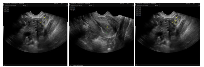
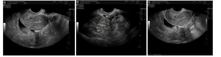
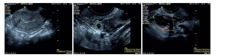
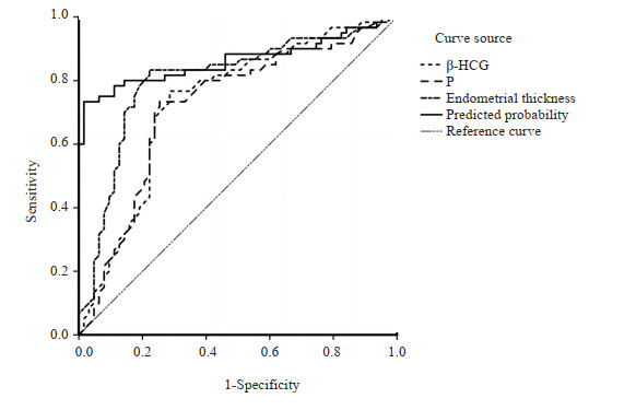
Objective To evaluate the clinical value of transvaginal and abdominal ultrasound combined with serum β-human chorionic gonadotropin (β-HCG) and progesterone detection in the diagnosis of ectopic pregnancy. Methods A retrospective analysis was performed. Sixty-three patients with ectopic pregnancy admitted to obstetrics and gynecology department of our hospital from January 2019 to October 2022 were included in ectopic pregnancy group, and 60 normal early pregnant women with similar gestational weeks were enrolled as control group. The data including transvaginal and abdominal ultrasound results, and serum levels of β-HCG and progesterone were compared between two groups. Then the value of transvaginal ultrasound, abdominal ultrasound, β-HCG and progesterone levels in the diagnosis of ectopic pregnancy were evaluated. Results Compared with control group, ectopic pregnancy group had lower levels of β-HCG and progesterone in peripheral blood and thinner endometrial thickness, with statistical difference (P < 0.05). The detection rate of pelvic adnexal mass of transvaginal ultrasound was higher than that of abdominal ultrasound (P < 0.05), while no statistical difference was found in the detection rates of embryo bud, fetal heart beat, pelvic effusion, and the display in both ipsilateral and contralateral ovarian (P > 0.05). The efficacy of β- HCG, progesterone and endometrial thickness alone in the diagnosis of ectopic pregnancy was as follow: AUC=0.732, 95% CI: 0.642- 0.823; AUC=0.719, 95% CI: 0.626- 0.812; AUC=0.803, 95% CI: 0.720- 0.885, and the combined detection of three indexes in diagnosis of ectopic pregnancy had the highest value than that of separate detection (AUC=0.862, 95% CI: 0.790-0.934). Conclusion The combined application of transvaginal and abdominal ultrasound combined with serum β-HCG and progesterone detection can effectively improve the diagnostic efficacy of ectopic pregnancy.
2023, 46(3): 518-521.
doi: 10.12122/j.issn.1674-4500.2023.03.24
Abstract:
Objective To investigate the diagnostic value of abnormal bronchial and vascular CT images for early lung cancer, and compare them with pathology. Methods A total of 100 patients with pathologically confirmed early lung cancer who were admitted to the hospital from October 2019 to December 2021 were selected as the research subjects. All patients underwent CT examination during hospitalization. The CT data were analyzed and compared with pathology. Results There were 15 cases of vacuole sign (15.00%), 13 cases of spicule sign (13.00%), 39 cases of abnormal air bronchogram (39.00%) and 33 cases of intratumoral vascular changes (33.00%). Among the 100 patients with early lung cancer, there were 45 patients with small cell lung cancer (45.00%), 17 patients with squamous cell carcinoma (17.00%), 29 patients with adenocarcinoma (29.00%) and 9 patients with alveolar carcinoma (9.00%). The incidence of abnormal air bronchogram in squamous cell carcinoma and the incidence of intratumoral vascular changes in adenocarcinoma were significantly higher than those in the other types of early lung cancer (P < 0.05). Conclusion Clinical manifestations of early lung cancer lack of specificity, but its CT images are characteristic, which is closely related to pathological types. Abnormal bronchial and vascular CT images are helpful for diagnosing early lung cancer.
2023, 46(3): 522-526.
doi: 10.12122/j.issn.1674-4500.2023.03.25
Abstract:
Objective To investigate the efficacy of high-intensity focused ultrasound (HIFU) in ablation of adenomyosis, and its influence on serum CA125. Methods From December 2020 to December 2021, 73 patients with adenomyosis were treated with HIFU. The effectiveness of HIFU treatment according to the postoperative condition of patients was evaluated. The dysmenorrhea score and menstrual flow score were compared before operation and 1, 3, 6 months after operation. The volume of uterine and glandular muscle lesions was measured, and the change of serum CA125 level was observed. All patients were followed up for 6 months, and the occurrence and recurrence of adverse reactions were recorded. Results Among the 73 patients of adenomyosis, 22 patients (30.14%) were markedly effective and 47 patients (64.38%) were effective, with a total effective rate of 94.52%. After 1, 3 and 6 months, the dysmenorrhea score, menstrual flow score, lesion volume and uterus volume were lower than those before operation (P < 0.05). After 1, 3 and 6 months follow-up, there were statistical differences in dysmenorrhea score, menstrual flow score and lesion volume (P < 0.05), but there was no statistical difference in uterine volume (P > 0.05), and the level of serum CA125 gradually decreased and became normal (P < 0.05). After operation, IgA, IgM, complement C3, C4 and Th17/Treg ratio were lower than those before operation, but IgG level was higher than that before operation (P < 0.05). After 6 months of follow- up, the total incidence of adverse reactions was 10.96%, and 2 patients (2.74%) recurred. Conclusion HIFU ablation can effectively relieve the clinical symptoms such as dysmenorrhea in patients with adenomyosis, reduce the volume of the lesion, and reduce the adverse reaction rate and recurrence rate. It is a safe and effective method to treat adenomyosis. When HIFU is used, the change of CA125 level can be detected, which can provide reference for evaluating the curative effect of patients with adenomyosis.
2023, 46(3): 527-531.
doi: 10.12122/j.issn.1674-4500.2023.03.26
Abstract:
Objective To analyze the evaluation value of neoadjuvant chemotherapy in patients with non-small cell lung cancer (NSCLC) based on tumor volume measured by CT. Methods Seventy- four patients with NSCLC who received neoadjuvant chemotherapy in our hospital from May 2019 to May 2020 were selected. After chemotherapy, they were divided into effective group (n=51) and ineffective group (n=23) based on pathological results. Tumor volume of all patients was measured by CT. Its influencing factors were analyzed by univariate and Logistic multivariate regression analysis. Results After chemotherapy, the tumor volume of 2 groups was significantly smaller than before chemotherapy (P < 0.05). The tumor volume of effective group was significantly smaller than before ineffective (P < 0.05). There was no significant difference in 6-month survival rate between 2 groups after chemotherapy (P > 0.05). One year after chemotherapy, the survival rate of effective group was significantly higher than that of ineffective group (P < 0.05). Univariate analysis showed that clinical stage, degree of differentiation and chemotherapy cycle were influencing factors of tumor volume (P < 0.05). Logistic regression analysis showed that clinical stage, differentiation degree and chemotherapy cycle were the influencing factors of tumor volume in patients with NSCLC (P < 0.05). ROC curve analysis showed that the sensitivity and specificity of tumor volume in predicting the efficacy of neoadjuvant chemotherapy in NSCLC patients were 0.732 and 0.800. Conclusion Tumor volume measured by CT has certain value in evaluating the efficacy of neoadjuvant chemotherapy in patients with NSCLC.
2023, 46(3): 532-536.
doi: 10.12122/j.issn.1674-4500.2023.03.27
Abstract:
Objective To observe the relationship between cognitive dysfunction and brain structural network changes in patients with white matter lesions based on magnetic resonance diffusion tensor imaging. Methods A total of 120 patients with white matter lesions in our hospital from January 2010 to February 2020 were enrolled as the research objects. According to the neuropsychological evaluation results, they were divided into normal cognitive function group (n=48), mild cognitive dysfunction group (n=44) and dementia group (n=28). All patients underwent 3.0T magnetic resonance examination to measure white matter high- signal volume and diffusion tensor imaging imaging to construct brain structural network. The para-ventricular, deep and total white matter high-signal volume, brain structure network parameters [global efficiency, local efficiency, shortest path, clustering coefficient, normalized clustering coefficient (γ), approximate standard characteristic path length (λ), small-world attribute value (σ)] were compared among the three groups. Pearson correlation was used to analyze the correlation between brain structural network parameters and MoCA score. Results The hyperintensity volume and total volume of paraventricular and deep white matter in normal cognitive function group, mild cognitive dysfunction group and dementia group were increased successively, and the differences among groups were statistically significant (P < 0.05). The global efficiency, local efficiency, agglomeration coefficient, γ, λ and σ of dementia group were smaller than those of normal cognitive function group and mild cognitive dysfunction group, and the shortest path of dementia group was larger than those of normal cognitive function group and mild cognitive dysfunction group, and the differences were statistically significant (P < 0.05). Pearson correlation analysis showed that the MoCA score was negatively correlated with the shortest path, but positively correlated with the global efficiency, local efficiency, agglomeration coefficient, γ, λ and σ (P < 0.05). Conclusion Cognitive dysfunction in white matter lesions patients is associated with white matter high signal volume and changes in brain structural network properties.
2023, 46(3): 537-542.
doi: 10.12122/j.issn.1674-4500.2023.03.28
Abstract:
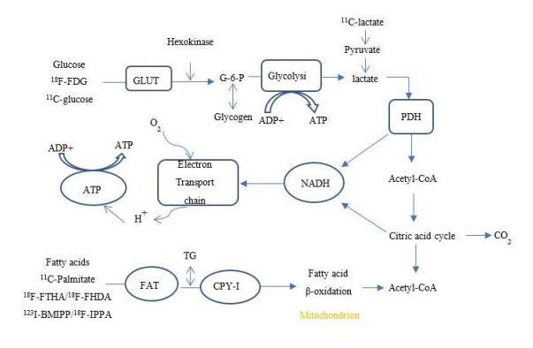
Positron emission tomography/computed tomography (PET/CT), due to its sensitivity and noninvasiveness, has occupied an important role in the diagnosis of cardiovascular diseases. Different positron imaging agents are guaranteed to improve the specificity and sensitivity of the examination. In myocardial metabolic disease, the change of metabolic substrate often occurs before the anatomical structure, and it is important to estimate the myocardial damage by judging the change of metabolic way, so that the choice of metabolic substrate by myocardium provides ideas and ideas for the design of positron imaging agents. In this paper, we divided them into three categories, including oxygen metabolism pathway, glucose metabolism pathway and fatty acid metabolism according to different metabolic pathways, and its advantages and disadvantages are categorically discussed from different nuclides and their applicable diagnostic directions, with a view to combining more technologies in the future to obtain more performance metabolic imaging agents.
Positron emission tomography/computed tomography (PET/CT), due to its sensitivity and noninvasiveness, has occupied an important role in the diagnosis of cardiovascular diseases. Different positron imaging agents are guaranteed to improve the specificity and sensitivity of the examination. In myocardial metabolic disease, the change of metabolic substrate often occurs before the anatomical structure, and it is important to estimate the myocardial damage by judging the change of metabolic way, so that the choice of metabolic substrate by myocardium provides ideas and ideas for the design of positron imaging agents. In this paper, we divided them into three categories, including oxygen metabolism pathway, glucose metabolism pathway and fatty acid metabolism according to different metabolic pathways, and its advantages and disadvantages are categorically discussed from different nuclides and their applicable diagnostic directions, with a view to combining more technologies in the future to obtain more performance metabolic imaging agents.
2023, 46(3): 543-547.
doi: 10.12122/j.issn.1674-4500.2023.03.29
Abstract:
The early diagnosis and treatment of liver cancer has always been a difficult problem in the medical field, and the development of nanotechnology brings new opportunities for the early diagnosis and treatment of liver cancer. At present, fluorescent nanoprobes have been widely used in the imaging, diagnosis and treatment of liver cancer due to their advantages of good targeting and high sensitivity. In this paper, the research progress of fluorescent nanoprobes in early diagnosis and treatment of liver cancer will be summarized from three aspects: specific targeted liver cancer probe, near- infrared fluorescence imaging and multimodal fluorescence imaging, and the integration of diagnosis and treatment. The problems to be solved urgently in the development of nanoprobes will be put forward, and the application of theranostic nano-system in diagnosis and treatment of liver cancer will be predicted.
The early diagnosis and treatment of liver cancer has always been a difficult problem in the medical field, and the development of nanotechnology brings new opportunities for the early diagnosis and treatment of liver cancer. At present, fluorescent nanoprobes have been widely used in the imaging, diagnosis and treatment of liver cancer due to their advantages of good targeting and high sensitivity. In this paper, the research progress of fluorescent nanoprobes in early diagnosis and treatment of liver cancer will be summarized from three aspects: specific targeted liver cancer probe, near- infrared fluorescence imaging and multimodal fluorescence imaging, and the integration of diagnosis and treatment. The problems to be solved urgently in the development of nanoprobes will be put forward, and the application of theranostic nano-system in diagnosis and treatment of liver cancer will be predicted.
2023, 46(3): 548-553.
doi: 10.12122/j.issn.1674-4500.2023.03.30
Abstract:
In recent years, photoacoustic imaging technology with high resolution, high contrast and high imaging depth has been paid more and more attention in the diagnosis and treatment of liver cancer. Photothermal therapy has also attracted more and more attention because of its high specificity and good therapeutic effect. This paper mainly introduces the basic principle of photoacoustic imaging technology and photothermal therapy, as well as nano- diagnostic agents with high photoacoustic signal and good photothermal conversion efficiency, and shows the application of photoacoustic imaging technology combined with photothermal therapy in the integration of diagnosis and treatment of liver cancer in the form of nano-diagnostic agents. Photoacoustic imaging is used to monitor the enrichment of nano-diagnostic agents in tumors, and laser irradiation is used to kill tumor cells, regulate tumor microenvironment and inhibit tumor metastasis and recurrence. The review describes the feasibility and advantages of combination technique in the diagnosis and treatment of hepatocellular carcinoma, and promote the practical application of combined technology clinically.
In recent years, photoacoustic imaging technology with high resolution, high contrast and high imaging depth has been paid more and more attention in the diagnosis and treatment of liver cancer. Photothermal therapy has also attracted more and more attention because of its high specificity and good therapeutic effect. This paper mainly introduces the basic principle of photoacoustic imaging technology and photothermal therapy, as well as nano- diagnostic agents with high photoacoustic signal and good photothermal conversion efficiency, and shows the application of photoacoustic imaging technology combined with photothermal therapy in the integration of diagnosis and treatment of liver cancer in the form of nano-diagnostic agents. Photoacoustic imaging is used to monitor the enrichment of nano-diagnostic agents in tumors, and laser irradiation is used to kill tumor cells, regulate tumor microenvironment and inhibit tumor metastasis and recurrence. The review describes the feasibility and advantages of combination technique in the diagnosis and treatment of hepatocellular carcinoma, and promote the practical application of combined technology clinically.
2023, 46(3): 554-559.
doi: 10.12122/j.issn.1674-4500.2023.03.31
Abstract:
Major depressive disorder is a kind of complex psychiatric disorder with poor prognosis, but its pathogenesis has not been fully elucidated. The development of magnetic resonance imaging has laid the foundation for revealing its neuropathological mechanism and clarifying its objective diagnosis basis. On the other hand, there is growing evidence of a strong association between major depressive disorder and dysfunction of the microbiota- gut- brain axis. Advances in neuroimaging and sequencing technologies are making it increasingly feasible to explore the interplay between the brain, gut, and microbiome. Studies on neuroimaging and gut-brain interaction will provide a more precise perspective for revealing the pathological mechanism behind major depressive disorder and constructing a gut- mediated therapy for major depressive disorder. This review will summarize the current state of brain imaging research on major depressive disorder, the relationship between gut microbiota and major depressive disorder, and the research progress on brain imaging and gut-brain interaction.
Major depressive disorder is a kind of complex psychiatric disorder with poor prognosis, but its pathogenesis has not been fully elucidated. The development of magnetic resonance imaging has laid the foundation for revealing its neuropathological mechanism and clarifying its objective diagnosis basis. On the other hand, there is growing evidence of a strong association between major depressive disorder and dysfunction of the microbiota- gut- brain axis. Advances in neuroimaging and sequencing technologies are making it increasingly feasible to explore the interplay between the brain, gut, and microbiome. Studies on neuroimaging and gut-brain interaction will provide a more precise perspective for revealing the pathological mechanism behind major depressive disorder and constructing a gut- mediated therapy for major depressive disorder. This review will summarize the current state of brain imaging research on major depressive disorder, the relationship between gut microbiota and major depressive disorder, and the research progress on brain imaging and gut-brain interaction.
2023, 46(3): 560-565.
doi: 10.12122/j.issn.1674-4500.2023.03.32
Abstract:
In recent years, the aging of our population is accelerating. The risk of spinal diseases is on the rise. With the cross integration of artificial intelligence and spinal medicine, deep learning has gradually become a popular research method in the field of spinal imaging and diagnosis and treatment. However, the research of deep learning in spine is relatively few and still in the initial stage, which has roomily great development potential and progress space in the future. This article reviews the application and research progress of deep learning in spinal image recognition, segmentation and measurement, spinal disease diagnosis and prognosis, and spinal surgery evaluation, to power deeper and higher level development of spine imaging and spine clinical research.
In recent years, the aging of our population is accelerating. The risk of spinal diseases is on the rise. With the cross integration of artificial intelligence and spinal medicine, deep learning has gradually become a popular research method in the field of spinal imaging and diagnosis and treatment. However, the research of deep learning in spine is relatively few and still in the initial stage, which has roomily great development potential and progress space in the future. This article reviews the application and research progress of deep learning in spinal image recognition, segmentation and measurement, spinal disease diagnosis and prognosis, and spinal surgery evaluation, to power deeper and higher level development of spine imaging and spine clinical research.
2023, 46(3): 566-570.
doi: 10.12122/j.issn.1674-4500.2023.03.33
Abstract:
Intracranial atherosclerosis is one of the most common causes of global ischemic stroke, and it has a high risk of recurrence. In terms of imaging diagnosis, risk stratification, treatment selection, and prognosis evaluation of stroke, multimodal MRI technology has advantages such as spatial resolution and cerebrospinal fluid suppression that other imaging technologies cannot. This article will review the application value of multimodal MRI in assessing stroke risk from multiple dimensions such as the imaging characteristics of plaques, hemodynamics and collateral compensation status, aiming to provide reference for research on precise risk stratification and prognosis evaluation of stroke.
Intracranial atherosclerosis is one of the most common causes of global ischemic stroke, and it has a high risk of recurrence. In terms of imaging diagnosis, risk stratification, treatment selection, and prognosis evaluation of stroke, multimodal MRI technology has advantages such as spatial resolution and cerebrospinal fluid suppression that other imaging technologies cannot. This article will review the application value of multimodal MRI in assessing stroke risk from multiple dimensions such as the imaging characteristics of plaques, hemodynamics and collateral compensation status, aiming to provide reference for research on precise risk stratification and prognosis evaluation of stroke.
2023, 46(3): 571-574.
doi: 10.12122/j.issn.1674-4500.2023.03.34
Abstract:
Imaging medicine is one of the main application directions of artificial intelligence in the medical field. In daily diagnosis and treatment work, the clinical demand for imaging examination is huge, but the growth of the number of imaging physicians and the accumulation of clinical experience is far from the growth rate of imaging data. The cross fusion of artificial intelligence and imaging data can reduce the pressure of imaging physicians to process massive image data. At present, based on deep learning technology and ultrasound, X-ray, CT and MRI data, various artificial intelligence assisted imaging quantitative analysis algorithms have been developed and widely applied in clinical practice, achieving early diagnosis, precise treatment, efficacy evaluation, and prediction of diseases, significantly improving the efficiency and accuracy of imaging physicians in processing image information, and providing quantitative basis for clinical diagnosis and treatment.
Imaging medicine is one of the main application directions of artificial intelligence in the medical field. In daily diagnosis and treatment work, the clinical demand for imaging examination is huge, but the growth of the number of imaging physicians and the accumulation of clinical experience is far from the growth rate of imaging data. The cross fusion of artificial intelligence and imaging data can reduce the pressure of imaging physicians to process massive image data. At present, based on deep learning technology and ultrasound, X-ray, CT and MRI data, various artificial intelligence assisted imaging quantitative analysis algorithms have been developed and widely applied in clinical practice, achieving early diagnosis, precise treatment, efficacy evaluation, and prediction of diseases, significantly improving the efficiency and accuracy of imaging physicians in processing image information, and providing quantitative basis for clinical diagnosis and treatment.