


Automated detection and segmentation method for aneurysmal intracranial hemorrhage
-
摘要:
目的 提出一种端到端的颅内动脉瘤破裂引起的颅内出血多类型血肿全自动分割方法。 方法 选择颅内动脉瘤破裂引起的颅内出血644例CT影像数据,按8:2的比例分为训练集和测试集。首先通过区域生长的方式获取脑组织区域,然后利用深度学习对出血区域进行多类型血肿分割。 结果 测试集上的结果表明,蛛网膜下腔出血、脑实质出血、脑室内出血和颅内出血的Dice系数分别为62.13%、68.64%、50.08%、71.10%。 结论 本文提出的分割网络可以有效地对颅内动脉瘤破裂引起的颅内出血完成多类型血肿分割,配合脑组织提取算法可以在未处理的临床数据上自动完成端到端的处理流程。该方法有效提升颅内动脉瘤破裂引起的颅内出血的诊治效率,有较好的临床应用价值。 Abstract:Objective To propose an end- to- end, fully automated segmentation method for multiple types of intracranial hematomas caused by the rupture of intracranial aneurysm. Methods A total of 644 brain CT images of intracranial hemorrhage caused by the rupture of intracranial aneurysms were selected and divided into train and test datasets by the ratio of 8:2. Brain extraction was firstly acquired by region growing, then multi- type segmentation was performed on the hemorrhage regions using deep learning. Results The dice coefficients of subarachnoid hemorrhage, intracerebral parenchymal hemorrhage, intraventricular hemorrhage, and intracerebral hemorrhage were 62.13%, 68.64%, 50.08% and 71.10% on the test dataset. Conclusion The segmentation network proposed in this paper can effectively complete multi-type hematoma segmentation for intracranial hemorrhage. The end-to-end processing process can be automatically completed with the brain extraction algorithm on the clinical data. The method effectively improves the diagnosis and treatment efficiency of intracranial hemorrhage caused by the rupture of intracranial aneurysms and possesses good clinical application value. -

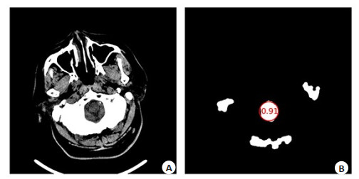
图 2 脑干识别示意图
Figure 2. Diagram for brainstem recognition. Image B could be done from image A, which performed tissue extraction, erosion and removal of too-large or too-small ar-eas. The IOU of brainstem position identification was 0.91.

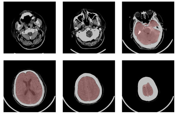
图 3 脑组织提取结果
Figure 3. Brain extraction results. The area under the red masked is the brain tissue. There were 28 slices in this case, and the currently selected slice was 2, 6, 11, 18, 23, 26. The 6th slice was the last brainstem position after identification.

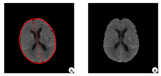
图 4 基于椭圆拟合的旋转矫正
Figure 4. Rotation correction. A: Result of ellipse fitting; B: Result of rotation.

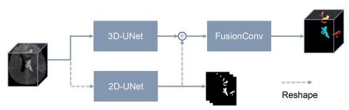
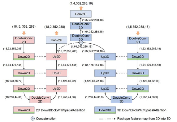
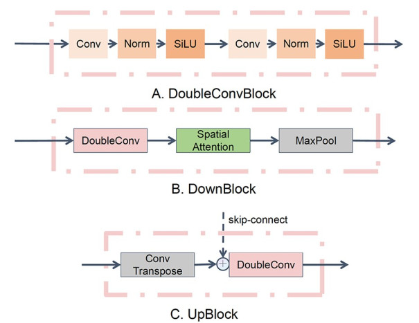
图 5 混合2D/3D特征融合分割网络框架图
Figure 5. The framework of 2D/3D segmentation network with feature fusion.

图 6 混合2D/3D特征融合分割网络设计图
Figure 6. The design of 2D/3D segmentation network with feature fusion.

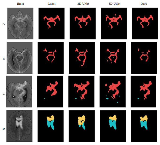
图 10 分割结果图
Figure 10. Segmentation result. Red, yellow and blue represent the hematoma of SAH, IPH and IVH. The images from left to right are brain tissue, ground truths, prediction of 2D-UNet, prediction of 3D-UNet and prediction of the model in this article.
表 1 血肿分割的表现
Table 1. The results of hematoma segmentation (%, Dice)
Model ICH SAH IPH IVH 2D-UNet 70.54 62.21 66.44 45.91 3D-UNet 68.09 60.83 72.90 49.73 Ours 71.10 62.13 68.64 50.08 ICH: Intracranial hemorrhage; SAH: Subarachnoid hemorrhage; IPH: Intracerebral parenchymal hemorrhage; IVH: Intraventricu-lar hemorrhage.  下载: 导出CSV
-
[1] Wang N, Tong F, Tu YC, et al. Extraction of cerebral hemorrhage and calculation of its volume on CT image using automatic segmentation algorithm[J]. J Phys: Conf Ser, 2019, 1187(4): 042088. doi: 10.1088/1742-6596/1187/4/042088 [2] 张天麒, 康波, 孟祥飞, 等. 基于U-Net的颅内出血识别算法[J]. 北京邮电大学学报, 2020, 43(3): 92-8. https://www.cnki.com.cn/Article/CJFDTOTAL-BJYD202003014.htm [3] 汪亮, 金福江, 陈峻严. 基于区域生长和FCM模糊聚类的颅内出血CT图像分割[J]. 系统仿真学报, 2014, 26(2): 231-5. doi: 10.16182/j.cnki.joss.2014.02.027 [4] Gautam A, Sadhya D, Raman B. A Modified FCM-Based Brain Lesion Segmentation Scheme for Medical Images[C]. Proceedings of 3rd International Conference on Computer Vision and Image Processing. Singapore: Springer, 2020: 149-159. [5] 赵杰祎, 周正松, 王晓宇, 等. 基于分水岭及区域增长算法建立一种测量自发性脑出血血肿体积的分割方法[J]. 四川大学学报: 医学版, 2022, 53(3): 511-6. https://www.cnki.com.cn/Article/CJFDTOTAL-HXYK202203025.htm [6] K rkk inen K, Fazeli S, Sarrafzadeh M. Unsupervised acute intracranial hemorrhage segmentation with mixture models[C]. 2021 IEEE 9th International Conference on Healthcare Informatics (ICHI), Victoria, BC, Canada. IEEE, 2021: 120-9. [7] Kuang HL, Najm M, Menon BK, et al. Joint Segmentation of Intracerebral Hemorrhage and Infarct from Non-Contrast CT Images of Post- treatment Acute Ischemic Stroke Patients[C]. International Conference on Medical Image Computing and Computer-Assisted Intervention. Cham: Springer, 2018: 681-688. [8] Ji ZY, Han X, Lin T, et al. A dense-gated U-net for brain lesion segmentation[C]. 2020 IEEE International Conference on Visual Communications and Image Processing (VCIP), Macau, China. IEEE, 2020: 104-7. [9] Shijitha R, Karthigaikumar P, Paul A. Efficient morphological segmentation of brain hemorrhage stroke lesion through MultiResUNet[J]. Comput Mater Continua, 2022, 70(3): 5233-49. doi: 10.32604/cmc.2022.020227 [10] Rangaraj S, Islam M, Vs V, et al. Identifying risk factors of intracerebral hemorrhage stability using explainable attention model [J]. Med Biol Eng Comput, 2022, 60(2): 337-48. doi: 10.1007/s11517-021-02459-y [11] 周正松, 陈旭淼, 张皞宇, 等. 改进型Unet网络在脑CT图像出血区域识别与分割中的应用[J]. 四川大学学报: 医学版, 2022, 53(1): 114-20. https://www.cnki.com.cn/Article/CJFDTOTAL-HXYK202201019.htm [12] 于金扣, 余南南, 于贺, 等. 基于多尺度层级化注意力模型的脑血肿分割算法[J]. 航天医学与医学工程, 2021, 34(1): 44-51. https://www.cnki.com.cn/Article/CJFDTOTAL-HYXB202101009.htm [13] Ronneberger O, Fischer P, Brox T. U-Net: Convolutional Networks for Biomedical Image Segmentation[C]. International Conference on Medical Image Computing and Computer-Assisted Intervention. Cham: Springer, 2015: 234-241. [14] Woo S, Park J, Lee JY, et al. CBAM: Convolutional Block Attention Module[C]. European Conference on Computer Vision. Cham: Springer, 2018: 3-19. [15] Ma J, Chen JN, Ng M, et al. Loss odyssey in medical image segmentation[J]. Med Image Anal, 2021, 71: 102035. [16] Sato T, Sasaki T, Sakuma J, et al. Quantification of subarachnoid hemorrhage by three-dimensional computed tomography: correlation between hematoma volume and symptomatic vasospasm[J]. Neurol Med Chir (Tokyo), 2011, 51(3): 187-94. [17] Muschelli J, Ullman NL, Mould WA, et al. Validated automatic brain extraction of head CT images[J]. NeuroImage, 2015, 114: 379-85. [18] Smith-Bindman R, Kwan ML, Marlow EC, et al. Trends in use of medical imaging in US health care systems and in Ontario, Canada, 2000-2016[J]. JAMA, 2019, 322(9): 843-56. [19] Bruls RM, Kwee RM. Workload for radiologists during on-call hours: dramatic increase in the past 15 years[J]. Insights Imaging, 2020, 11(1): 121. [20] Strub WM, Leach JL, Tomsick T, et al. Overnight preliminary head CT interpretations provided by residents: locations of misidentified intracranial hemorrhage[J]. AJNR Am J Neuroradiol, 2007, 28(9): 1679-82. [21] Ironside N, Chen CJ, Mutasa S, et al. Fully automated segmentation algorithm for hematoma volumetric analysis in spontaneous intracerebral hemorrhage[J]. Stroke, 2019, 50(12): 3416-23. [22] Yu NN, Yu H, Li HN, et al. A robust deep learning segmentation method for hematoma volumetric detection in intracerebral hemorrhage[J]. Stroke, 2022, 53(1): 167-76. [23] Li XY, Luo GN, Wang W, et al. Hematoma expansion context guided intracranial hemorrhage segmentation and uncertainty estimation[J]. IEEE J Biomed Health Inform, 2022, 26(3): 1140-51. [24] 苗政, 李明洋, 陈忠萍, 等. 基于深度学习分割模型的脑出血CT图像自动分割研究[J]. 中国医疗设备, 2022, 37(8): 46-50, 86. https://www.cnki.com.cn/Article/CJFDTOTAL-YLSX202208011.htm [25] Shang F, Wang S, Wang X, et al. An Effective Transformer-based Solution for RSNA Intracranial Hemorrhage Detection Competition [J/OL].arXiv, 2022. https://arxiv.org/abs/2205.07556 [26] Thanellas A, Peura H, Lavinto M, et al. Development and external validation of a deep learning algorithm to identify and localize subarachnoid hemorrhage on CT scans[J]. Neurology, 2023, 100 (12): e1257-e1266. [27] Inkeaw P, Angkurawaranon S, Khumrin P, et al. Automatic hemorrhage segmentation on head CT scan for traumatic brain injury using 3D deep learning model[J]. Comput Biol Med, 2022, 146: 105530. [28] Nijiati M, Tuersun A, Zhang Y, et al. A symmetric prior knowledge based deep learning model for intracerebral hemorrhage lesion segmentation[J]. Front Physiol, 2022, 13: 977427. [29] Chang PD, Kuoy E, Grinband J, et al. Hybrid 3D/2D convolutional neural network for hemorrhage evaluation on head CT[J]. Am J Neuroradiology, 2018, 39(9): 1609-1616. [30] Nag MK, Chatterjee S, Sadhu AK, et al. Computer-assisted delineation of hematoma from CT volume using autoencoder and Chan Vese model[J]. Int J Comput Assist Radiol Surg, 2019, 14(2): 259-69. [31] Bauer S, Fejes T, Reyes M. A skull-stripping filter for ITK[J]. Insight J, 2012, 1. doi: https://doi.org/ 10.54294/dp4mfp. -

 点击查看大图
计量
- 文章访问数: 161
- HTML全文浏览量: 120
- PDF下载量: 24
- 被引次数: 0