


Comparative study between abnormal bronchial and vascular CT images and pathology of early lung cancer
-
摘要:
目的 探讨肺癌组织内支气管、血管CT异常影像在早期肺癌中的诊断价值,并与病理学进行对照分析。 方法 选取2019年10月~2021年12月本院收治的100例经病理诊断为早期肺癌患者为研究对象,患者住院期间均进行CT检查,分析CT资料并与病理学对照。 结果 患者CT征象包括空泡征15例(15.00%)、毛刺征13例(13.00%)、异常空气支气管征39例(39.00%)、肿瘤内血管改变33例(33.00%);100例早期肺癌患者中小细胞肺癌45例(45.00%)、鳞状细胞癌17例(17.00%)、腺癌29例(29.00%)、肺泡癌型9例(9.00%);上述各类型早期肺癌支气管、血管CT异常影像比较,鳞状细胞癌异常空气支气管征及腺癌中的肿瘤内血管改变征象高于其他类型(P < 0.05)。 结论 早期肺癌临床表现缺乏特异性,但其CT影像具有特征性,与病理学类型密切相关,早期肺癌组织内支气管、血管CT异常影像可对早期肺癌的诊断提供价值。 Abstract:Objective To investigate the diagnostic value of abnormal bronchial and vascular CT images for early lung cancer, and compare them with pathology. Methods A total of 100 patients with pathologically confirmed early lung cancer who were admitted to the hospital from October 2019 to December 2021 were selected as the research subjects. All patients underwent CT examination during hospitalization. The CT data were analyzed and compared with pathology. Results There were 15 cases of vacuole sign (15.00%), 13 cases of spicule sign (13.00%), 39 cases of abnormal air bronchogram (39.00%) and 33 cases of intratumoral vascular changes (33.00%). Among the 100 patients with early lung cancer, there were 45 patients with small cell lung cancer (45.00%), 17 patients with squamous cell carcinoma (17.00%), 29 patients with adenocarcinoma (29.00%) and 9 patients with alveolar carcinoma (9.00%). The incidence of abnormal air bronchogram in squamous cell carcinoma and the incidence of intratumoral vascular changes in adenocarcinoma were significantly higher than those in the other types of early lung cancer (P < 0.05). Conclusion Clinical manifestations of early lung cancer lack of specificity, but its CT images are characteristic, which is closely related to pathological types. Abnormal bronchial and vascular CT images are helpful for diagnosing early lung cancer. -
Key words:
- early lung cancer /
- bronchial CT image /
- vascular CT image /
- pathological finding
-

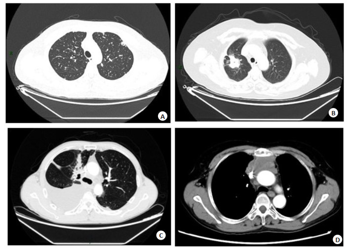
图 1 早期肺癌CT征象
Figure 1. CT signs of early lung cancer. A: Small diffuse nodules in both lungs, with partial vacuolar changes; B: Partial bronchial obstruction in upper lobe of the right lung, spicule on the edge, thickening and stretching of adjacent pleu-ra, heterogeneous and significant enhancement after enhanced scan; C: Irregular soft tissue shadows near hilar re-gion of the right upper lobe, with blurry boundaries and heterogeneous enhancement after enhanced scan; Local ob-struction of bronchial branch in corresponding upper lobe of the right lung and obstructive pneumonia of corre-sponding distal segment, with bronchial air bronchogram inside; D: An irregular mass shadow was observed in su-perior mediastinal region, with a larger cross-section of approximately 7.5 cm×3.6 cm, with blurry boundaries. Part breaks through the mediastinal pleural and dashes forward to the upper right chest area. The contact surface be-tween the lesion and the right upper lobe is rough. Enhanced scan of the mass shows heterogeneous enhancement and a large region with relatively weak enhancement inside. The boundaries between the mass and the brachioce-phalic trunk, ascending aorta, bilateral brachiocephalic veins and superior vena cava are blurry, accompanied by compression and narrowing of superior vena cava lumen. The left brachiocephalic vein is partially enveloped and the lumen is not clearly displayed. On above basis, malignant tumor with invasion of surrounding structures is con-sidered.
表 1 早期肺癌组织内支气管、血管CT异常影像与病理对照
Table 1. Comparison between abnormal bronchial and vascular CT images and pathology of early lung cancer (n)
Pathological type Abnormal air bronchogram Intratumoral vascular changes Other signs Small cell lung cancer (n=45) 10 7 28 Squamous cell carcinoma (n=17) 14 3 0 Adenocarcinoma (n=29) 10 19 0 Alveolar carcinoma(n=9) 5 4 0 Total 39 33 28  下载: 导出CSV
-
[1] Nishio M, Barlesi F, West H, et al. Atezolizumab plus chemothera-py for first-line treatment of nonsquamous NSCLC: results from the randomized phase 3 IMpower132 trial[J]. J Thorac Oncol, 2021, 16 (4): 653-64. doi: 10.1016/j.jtho.2020.11.025 [2] Camidge DR, Kim HR, Ahn MJ, et al. Brigatinib Versus Crizotinib in Advanced ALK Inhibitor-Naive ALK-Positive Non-Small Cell Lung Cancer: Second Interim Analysis of the Phase Ⅲ ALTA-1L Trial[J]. J Clin Oncol, 2020, 38(31): 3592-603. doi: 10.1200/JCO.20.00505 [3] 肖蓉, 潘频华. 老年肺癌CT影像学特征与特异性标记物的相关性研究及联合诊断[J]. 国际老年医学杂志, 2020, 41(6): 49-52. https://www.cnki.com.cn/Article/CJFDTOTAL-GWLL202006013.htm [4] Infante M, Sestini S, Galeone C, et al. Lung cancer screening with low-dose spiral computed tomography: evidence from a pooled analysis of two Italian randomized trials[J]. Eur J Cancer Prev, 2017, 26(4): 324-9. doi: 10.1097/CEJ.0000000000000264 [5] 陈琦, 朱全新, 郁义星, 等. 肺部单发微小磨玻璃结节(< 10mm)MSCT特征对肺腺癌病理亚型的诊断价值[J]. 放射学实践, 2019, 34(7): 778-83. [6] 金梅, 吴重重, 方瑞, 等. 纯磨玻璃密度肺腺癌的危险因素CT量化研究[J]. 中华放射学杂志, 2018, 52(11): 836-41. [7] 杨龙海, 叶波, 魏星, 等. 最新国际肺癌TNM分期标准(第8版)修订稿解读[J]. 中国医刊, 2016, 51(9): 22-5. https://www.cnki.com.cn/Article/CJFDTOTAL-ZGYI201609007.htm [8] Forde PM, Spicer J, Lu S, et al. Neoadjuvant nivolumab plus chemotherapy in resectable lung cancer[J]. N Engl J Med, 2022, 386(21): 1973-85. doi: 10.1056/NEJMoa2202170 [9] 贾永军, 于楠, 杨创勃, 等. 基于模型的迭代重建在改善胸部低剂量CT评价早期周围型肺癌中的价值[J]. 中国医学影像学杂志, 2019, 27(9): 682-6. https://www.cnki.com.cn/Article/CJFDTOTAL-ZYYZ201909015.htm [10] 谢惠康, 谢冬, 陈昶, 等. 磨玻璃结节早期肺腺癌病理学诊断中的问题与探讨[J]. 中华外科杂志, 2019, 57(1): 63-7. [11] 田笑如, 张毅. 拉曼光谱检测技术在早期肺癌诊断方面的研究进展[J]. 中国肺癌杂志, 2018, 21(7): 560-4. https://www.cnki.com.cn/Article/CJFDTOTAL-FAIZ201807013.htm [12] 蔡荣, 王骥, 张新红, 等. 肺癌病理特征及CT征象与EGFR的相关性分析[J]. 医学影像学杂志, 2022, 32(10): 1716-21. https://www.cnki.com.cn/Article/CJFDTOTAL-XYXZ202210015.htm [13] 李倩倩, 王志刚, 刘剑辉, 等. 多层螺旋CT多征象联合肿瘤标志物检测对于磨玻璃结节肺癌的诊断价值研究[J]. 中国医学装备, 2022, 19 (8): 37-42. https://www.cnki.com.cn/Article/CJFDTOTAL-YXZB202208009.htm [14] 张文超, 李靖煦, 关玉宝, 等. 多原发肺癌的CT表现与患者预后相关性研究[J]. CT理论与应用研究, 2019, 28(1): 29-38. https://www.cnki.com.cn/Article/CJFDTOTAL-CTLL201901004.htm [15] Gadgeel S, Rodríguez-Abreu D, Speranza G, et al. Updated Analysis From KEYNOTE-189: Pembrolizumab or Placebo Plus Pemetrexed and Platinum for Previously Untreated Metastatic Nonsquamous Non-Small-Cell Lung Cancer[J]. J Clin Oncol, 2020, 38(14): 1505-17. [16] 张红娟, 武志峰, 鄂林宁, 等. 小细胞肺癌与非小细胞肺癌早晚期CT征象对比分析[J]. 山西医科大学学报, 2019, 50(1): 54-8. https://www.cnki.com.cn/Article/CJFDTOTAL-SXYX201901011.htm [17] 张亚涛, 王鑫, 孙腾月, 等. 探讨周围型肺癌MSCT影像学表现与临床组织病理学的相关性研究[J]. 中国CT和MRI杂志, 2021, 19 (10): 46-8. https://www.cnki.com.cn/Article/CJFDTOTAL-CTMR202110014.htm [18] 杨国才, 柴振达, 张善华. 周围型小肺癌2年以上CT随访观察研究[J]. 肿瘤学杂志, 2020, 26(4): 323-8. https://www.cnki.com.cn/Article/CJFDTOTAL-XHON202004010.htm [19] 黄定品, 傅钢泽, 项益岚, 等. 纯磨玻璃肺小腺癌内异常空气支气管征与病理亚型的相关性[J]. 医学影像学杂志, 2019, 29(12): 2047-50. https://www.cnki.com.cn/Article/CJFDTOTAL-XYXZ201912015.htm [20] 耿云平, 李真真, 任悠悠, 等. 高分辨率CT对pGGN肺腺癌病理学亚型的诊断价值[J]. 中国实用医刊, 2021(16): 92-4. [21] 程娟, 李江, 汪田田, 等. 41例孤立型细支气管肺泡癌患者CT影像表现及其不同病理基础对照研究[J]. 中国CT和MRI杂志, 2022, 20 (2): 45-7. https://www.cnki.com.cn/Article/CJFDTOTAL-CTMR202202012.htm [22] 段秀杰, 李福元, 付玉存. 周围型非小细胞肺癌CT征象与临床病理分型的关系[J]. 现代肿瘤医学, 2020, 28(15): 2622-26. https://www.cnki.com.cn/Article/CJFDTOTAL-SXZL202015016.htm -

 点击查看大图
计量
- 文章访问数: 163
- HTML全文浏览量: 104
- PDF下载量: 8
- 被引次数: 0