


Comparation of pathological features and CT features of pulmonary space occupying in AIDS patients with lung cancer, tuberculosis patients with lung cancer and simple lung cancer
-
摘要:
目的 分析艾滋病合并肺癌、肺结核合并肺癌与单纯肺癌的病理特征及肺内占位CT特征。 方法 选择2021年11月~2022年6月我院收治的36例艾滋病合并肺癌、36例肺结核合并肺癌及36例单纯肺癌患者为研究对象, 收集其一般资料、临床症状、病理特征、肺内占位CT特征等临床资料, 并将3组患者上述资料进行对比分析。 结果 艾滋病合并肺癌组、肺结核合并肺癌组发热、纳差比例均高于单纯肺癌组(P < 0.05), 艾滋病合并肺癌组贫血、真菌感染比例高于肺结核合并肺癌、单纯肺癌组(P < 0.05), 3组其他临床症状差异无统计学意义(P>0.05)。艾滋病合并肺癌组腺癌比例高于肺结核合并肺癌组、单纯肺癌组(P < 0.05), 鳞癌比例低于肺结核合并肺癌组、单纯肺癌组(P < 0.05), 肺结核合并肺癌组病理分型与单纯肺癌组的差异均无统计学意义(P>0.05), 3组TNM分期差异均无统计学意义(P>0.05)。艾滋病合并肺癌组肺内占位CT表现中纤维条索影比例均高于肺结核合并肺癌组和单纯肺癌组(P < 0.05);肺结核合并肺癌患者肺内占位CT表现中长毛刺征、空洞、纵膈淋巴结增大、胸腔积液均高于单纯肺癌组(P < 0.05), 粟粒性阴影、卫星灶比例高于艾滋病合并肺癌组和单纯肺癌组(P < 0.05)。 结论 艾滋病合并肺癌、肺结核合并肺癌与单纯肺癌的病理特征及肺内占位CT特征存在不同之处, 临床可结合患者病理特征及肺内占位CT特征对不同类型肺癌患者病情予以评估。 Abstract:Objective To compare the pathological features and CT features of pulmonary space occupying in AIDS complicated with lung cancer, tuberculosis complicated with lung cancer and simple lung cancer. Methods Thirty-six patients with AIDS combined with lung cancer, 36 patients with tuberculosis combined with lung cancer and 36 patients with simple lung cancer admitted to our hospital from November 2021 to June 2022 were selected as the research objects.General data, clinical symptoms, pathological characteristics, and CT features of lung space occupying lesions were collected, and the above data of the three groups of patients were compared. Results The proportions of fever and poor appetite in the AIDS combined lung cancer group and the tuberculosis combined lung cancer group were higher than those in the simple lung cancer group (P < 0.05).The proportion of anemia and fungal infection in the group of AIDS with lung cancer was higher than that in the group of tuberculosis with lung cancer and the group of simple lung cancer (P < 0.05).There was no significant difference in other clinical symptoms among the three groups (P>0.05).The proportion of adenocarcinoma in the AIDS combined lung cancer group was higher than that in the tuberculosis combined lung cancer group and the simple lung cancer group (P < 0.05).The proportion of squamous cell carcinoma was lower than that in the tuberculosis combined lung cancer group and the simple lung cancer group (P < 0.05).There were no significant difference in pulmonary pathological classification between the tuberculosis combined lung cancer group and the simple lung cancer group (P>0.05).There were no significant difference in TNM staging among the three groups (P>0.05).The proportion of fibrous shadow in CT manifestations of HIV/AIDS patients with lung cancer was higher than that in pulmonary tuberculosis patients with lung cancer and lung cancer alone (P < 0.05).The CT findings of pulmonary space occupying in patients with pulmonary tuberculosis combined with lung cancer were higher than those in patients with simple lung cancer (P < 0.05).The percentages of miliary shadow and satellite focus were higher than those in patients with AIDS combined with lung cancer and patients with simple lung cancer (P < 0.05). Conclusion There are differences in pathological features and CT features of pulmonary space occupying between AIDS combined with lung cancer, tuberculosis combined with lung cancer and simple lung cancer.Clinical evaluation of patients with different types of lung cancer can be made according to the pathological features and CT features of pulmonary space occupying. -

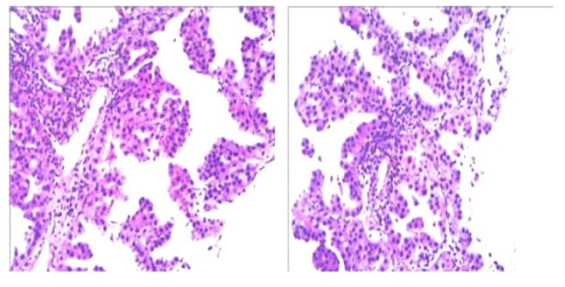
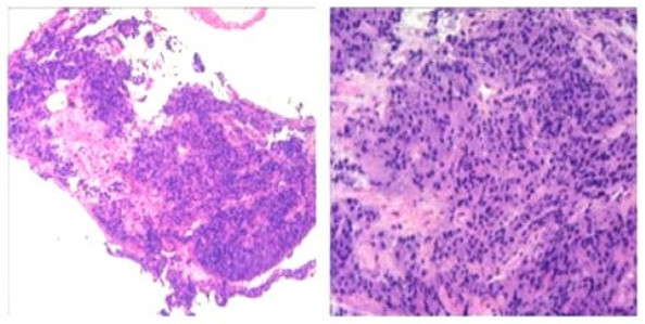
图 1 肺结核合并肺腺癌病理图像,镜下形态符合肺腺癌(HE染色,40×10)
Figure 1. Tuberculosis was complicated with lung adenocarcinoma pathological images, and the microscopic morphology was consistent with lung adenocarcinoma(HE staining, 40×10).

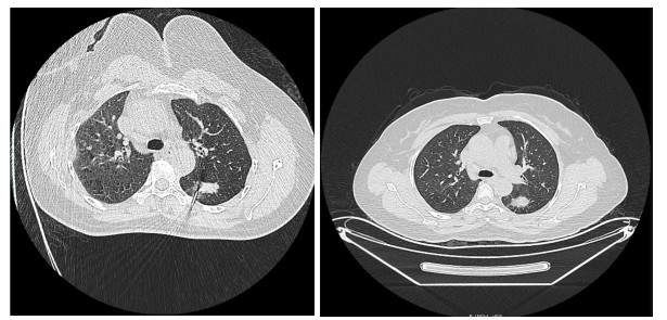
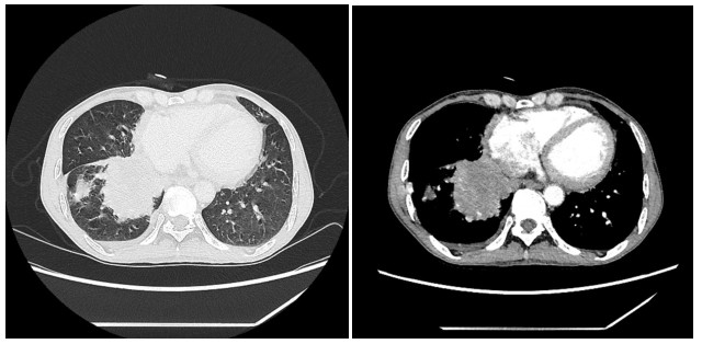
图 2 肺结核合并肺腺癌肺内占位CT征象
Figure 2. CT sign of space occupying in the lung of tuberculosis complicated with lung adenocarcinoma.

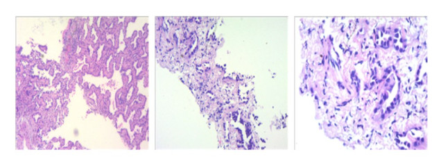
图 3 肺结核合并肺腺癌病理图像,镜下形态符合肺腺癌(HE染色,40×10)
Figure 3. Tuberculosis was complicated with lung adenocarcinoma pathological images, and the microscopic morphology was consistent with lung adenocarcinoma(HE staining, 40×10).

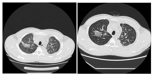
图 4 肺结核合并肺腺癌肺内占位CT征象
Figure 4. CT sign of space occupying in the lung of tuberculosis complicated with lung adenocarcinoma.

图 5 单纯肺腺癌病理图像,镜下形态符合肺腺癌(HE染色,40×10)
Figure 5. Pathological images of simple lung adenocarcinoma, with microscopic morphology consistent with lung adenocarcinoma(HE staining, 40×10).

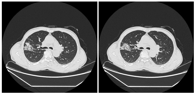
图 6 单纯肺腺癌肺内占位CT征象
Figure 6. CT signs of occupying space in the lung in simple lung adenocarcinoma.

图 7 艾滋病合并肺小细胞癌病理图像,镜下形态符合肺小细胞癌(HE染色,40×10)
Figure 7. Pathological images of AIDS combined with lung small cell carcinoma, with microscopic morphology consistent with lung small cell carcinoma (HE staining, 40×10).

图 8 艾滋病合并肺小细胞癌肺内占位CT征象
Figure 8. CT signs of lung occupancy in AIDS complicated with pulmonary small cell carcinoma.
表 1 一般资料比较
Table 1. Comparison of general demographic characteristics (n, n=36)
资料 艾滋病合并肺癌组 肺结核合并肺癌组 单纯肺癌组 χ2 P 性别 0.568 0.753 男 23 25 22 女 13 11 14 年龄(岁) 1.408 0.495 < 60 20 18 15 ≥60 16 18 21 文化程度 0.869 0.929 初中及以下 18 16 19 高中及专科 11 14 11 本科及以上 7 6 6 婚姻状况 0.727 0.695 已婚 32 33 34 未婚 4 3 2  下载: 导出CSV
表 2 三组患者临床症状比较
Table 2. Comparison of the clinical symptoms among the three groups (n, n=36)
临床症状 艾滋病合并肺癌组 肺结核合并肺癌组 单纯肺癌组 χ2 P 咳嗽、咳痰 21 25 18 2.838 0.242 咯血 6 4 3 1.224 0.542 发热 16* 17* 7 7.227 0.027 胸痛 14 15 10 1.686 0.430 胸闷 19 22 16 2.006 0.367 纳差 18* 20* 9 7.760 0.021 消瘦 17 18 9 5.599 0.060 贫血 16*# 5 4 13.845 0.001 盗汗 16 19 12 2.788 0.248 真菌感染 12*# 4 3 9.325 0.009 *P < 0.05 vs单纯肺癌组; #P < 0.05 vs肺结核合并肺癌组.
下载: 导出CSV
表 3 三组患者病理特征比较
Table 3. Comparison of the pathological characteristics among the three groups (n, n=36)
病理特征 艾滋病合并肺癌组 肺结核合并肺癌组 单纯肺癌组 χ2 P 病理类型 17.902 0.001 鳞癌 6*# 15 23 腺癌 26*# 16 10 小细胞癌 4 5 3 TNM分期 0.971 0.615 Ⅰ~Ⅱ期 19 18 22 Ⅲ~Ⅳ期 17 18 14 *P < 0.05 vs单纯肺癌组; #P < 0.05 vs肺结核合并肺癌组.
下载: 导出CSV
表 4 患者肺内占位CT特征比较
Table 4. Comparison of CT characteristics of pulmonary occupancy (n=36)
CT特征 艾滋病合并肺癌组 肺结核合并肺癌组 单纯肺癌组 χ2/F P 肿瘤最大直径(cm, Mean±SD) 1.83±0.51 1.76±0.56 1.85±0.60 0.258 0.773 边界模糊 29 28 27 0.321 0.852 形态不规则 30 31 28 0.894 0.640 双肺受累 13 17 11 2.202 0.333 长毛刺征 6 13* 4 7.403 0.025 毛刺征 20 18 22 0.900 0.638 分叶征 13 11 12 0.250 0.882 粟粒性阴影 10# 19* 6 11.244 0.004 纤维条索影 19*# 10 8 8.469 0.014 钙化 9 10 15 2.661 0.264 空洞 21 26* 15 6.892 0.032 纵膈淋巴结增大 16 20* 9 7.086 0.029 胸腔积液 14 21* 10 7.086 0.029 卫星灶 4# 22* 2 35.100 < 0.001 *P < 0.05 vs单纯肺癌组; #P < 0.05 vs肺结核合并肺癌组.
下载: 导出CSV
-
[1] 丁晓娟, 刘啸峰, 胡磊. 不同类型肺磨玻璃结节的CT表现及其与肺腺癌病理特征的关系研究[J]. 川北医学院学报, 2021, 36(6): 766-9. doi: 10.3969/j.issn.1005-3697.2021.06.021 [2] 籍强, 吕骏卿, 赵鑫, 等. 晚期NSCLC组织巨噬细胞表面Tim-3表达与临床特征和预后的关系[J]. 中南医学科学杂志, 2021, 49(6): 668-71, 702. https://www.cnki.com.cn/Article/CJFDTOTAL-HYYY202106012.htm [3] 吴尊友. 艾滋病预防技术进展与防治策略[J]. 中华预防医学杂志, 2018, 52(12): 1204-9. doi: 10.3760/cma.j.issn.0253-9624.2018.12.002 [4] 王琳, 宋言峥. HIV合并肺癌的个体化综合治疗[J]. 中国肺癌杂志, 2018, 21(4): 327-32. https://www.cnki.com.cn/Article/CJFDTOTAL-FAIZ201804021.htm [5] 张维, 李奇穗, 邓长刚, 等. 合并与未合并HIV感染的肺癌患者临床特征分析[J]. 中国艾滋病性病, 2022, 28(9): 1005-8. https://www.cnki.com.cn/Article/CJFDTOTAL-XBYA202209002.htm [6] 程增辉, 施裕新, 袁敏, 等. 艾滋病合并肺癌的CT表现[J]. 放射学实践, 2015, 30(9): 901-4. https://www.cnki.com.cn/Article/CJFDTOTAL-FSXS201509008.htm [7] 李杰, 李铁丰. CT在肺结核合并肺癌患者中的诊断价值分析[J]. 中国地方病防治杂志, 2020, 35(5). https://www.cnki.com.cn/Article/CJFDTOTAL-DYBF202005039.htm [8] 本刊编辑部. 2018年版原发性肺癌诊疗规范[J]. 实用心脑肺血管病杂志, 2018, 26(S2): 217. https://www.cnki.com.cn/Article/CJFDTOTAL-SYXL2018S2093.htm [9] 中华医学会感染病学分会艾滋病丙型肝炎学组, 中国疾病预防控制中心. 中国艾滋病诊疗指南(2021年版)[J]. 协和医学杂志, 2022, 13(2): 203-26. https://www.cnki.com.cn/Article/CJFDTOTAL-XHYX202202008.htm [10] 中华人民共和国国家卫生和计划生育委员会. 肺结核诊断[J]. 传染病信息, 2017, 30(6): 12. https://www.cnki.com.cn/Article/CJFDTOTAL-DDYI201228007.htm [11] 邓莉平, 熊勇, Sesay IZ, 等. 人类免疫缺陷病毒感染/艾滋病者恶性肿瘤的类型及其标化发生率分析[J]. 中华全科医师杂志, 2020, 19(8): 737-40. [12] 卢培培, 张楠, 王家林. 中国恶性肿瘤健康管理现状研究[J]. 中国公共卫生管理, 2019, 35(6): 760-3. https://www.cnki.com.cn/Article/CJFDTOTAL-GGWS201906014.htm [13] 全斌, 喻艳林. 肺结核合并肺癌的发生机制研究进展[J]. 山东医药, 2015, 55(24): 104-6. https://www.cnki.com.cn/Article/CJFDTOTAL-SDYY201524051.htm [14] 李成海, 邱万成, 周新华, 等. 71例肺结核并发肺癌患者的CT表现特征及临床病理分析[J]. 中国防痨杂志, 2017, 39(6): 576-80. [15] 韦菊临, 梁桂录, 刘军, 等. 艾滋病合并肺结核的影像学检查和特征[J]. 实用放射学杂志, 2018, 34(9): 1351-3. https://www.cnki.com.cn/Article/CJFDTOTAL-GJWJ201605022.htm [16] Park SK, Cho LY, Yang JJ, et al. Lung cancer risk and cigarette smoking, lung tuberculosis according to histologic type and gender in a population based case-control study[J]. Lung Cancer, 2010, 68(1): 20-6. [17] 曹敏, 方伟军, 黎惠如, 等. 肺结核合并肺癌的CT表现[J]. 放射学实践, 2020, 35(1): 40-4. https://www.cnki.com.cn/Article/CJFDTOTAL-FSXS202001012.htm [18] 黄德扬, 刘晋新, 张烈光, 等. 艾滋病合并肺癌的CT表现分析[J]. 中国CT和MRI杂志, 2019, 17(1): 81-3. https://www.cnki.com.cn/Article/CJFDTOTAL-CTMR201901025.htm [19] Sigel KM, Stone K, Wisnivesky JP, et al. Short-term outcomes for lung cancer resection surgery in HIV infection[J]. AIDS, 2019, 33(8): 1353-60. [20] Makinson A, Eymard-Duvernay S, Raffi F, et al. Feasibility and efficacy of early lung cancer diagnosis with chest computed tomography in HIV-infected smokers[J]. AIDS, 2016, 30(4): 573-82. [21] 马超豪, 李琦, 陈辉月. 对比观察炎性肺癌与渗出为主型肺结核CT表现[J]. 中国医学影像技术, 2020, 36(6): 849-52. https://www.cnki.com.cn/Article/CJFDTOTAL-ZYXX202006014.htm [22] 刘超, 孟庆军, 刘立红, 等. 胸腺瘤患者螺旋CT影像特征与CKP、VEGF表达的相关性研究[J]. 中南医学科学杂志, 2018, 46(4): 373-6. https://www.cnki.com.cn/Article/CJFDTOTAL-HYYY201804010.htm [23] 陈龙兰, 游金辉. 肺癌多模态医学影像学研究现状与进展[J]. 川北医学院学报, 2017, 32(5): 806-10. https://www.cnki.com.cn/Article/CJFDTOTAL-NOTH201705045.htm [24] 张彦, 胡国启, 司丽, 等. 艾滋病合并肺结核与单纯肺结核患者的临床特点对比分析研究[J]. 传染病信息, 2018, 31(6): 552-4. https://www.cnki.com.cn/Article/CJFDTOTAL-CRBX201806014.htm [25] 贾守勤, 张旭, 王武章, 等. 肺结核合并肺癌的临床特点及MSCT影像学分析[J]. 医学影像学杂志, 2021, 31(10): 1682-5. https://www.cnki.com.cn/Article/CJFDTOTAL-XYXZ202110017.htm [26] 田素升, 张炜, 范海涛, 等. 多样化CT征象鉴别周围性肺癌与肺结核[J]. 分子影像学杂志, 2018, 41(1): 11-5. doi: 10.3969/j.issn.1674-4500.2018.01.03 -


 点击查看大图
计量
- 文章访问数: 129
- HTML全文浏览量: 106
- PDF下载量: 8
- 被引次数: 0