


Value of C-TIRADS in differential diagnosis of benign and malignant thyroid nodules
-
摘要:
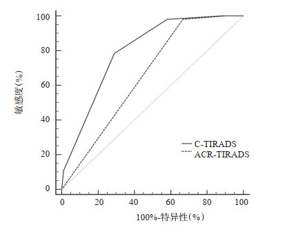
目的 通过对比美国放射学会(ACR)-甲状腺影像报告和数据系统(TIRADS),探讨中国版TIRADS(C-TIRADS)对甲状腺结节良恶性的鉴别诊断价值。 方法 回顾性收集2021年8月~2022年7月在本院接受甲状腺超声检查并得到明确病理诊断的甲状腺结节患者166例(195枚结节);以病理结果为金标准,分别评估C-TIRADS和TIRADS不同分类的恶性占比,绘制两种系统诊断甲状腺结节良恶性的ROC曲线,并对比二者诊断效能。 结果 两种TIRADS的风险分层级别下的实际恶性占比与指南推荐的恶性率相符。C-TIRADS诊断甲状腺结节良恶性ROC曲线的AUC为0.796(95% CI:0.741~0.852),相比ACR-TIRADS的0.658(95% CI:0.587~0.724)增大(P < 0.05)。C-TIRADS、ACR-TIRADS的最佳临界点分别为4C类、5类,两种分类系统的敏感度差异无统计学意义(94.12% vs 95.10%,P > 0.05),但C-TIRADS的特异性高于ACR-TIRADS(56.99% vs 35.48%,P < 0.05)。 结论 对于甲状腺结节的鉴别,C-TIRADS相比ACR-TIRADS诊断效能更高,提高了特异性。 -
关键词:
- 甲状腺癌 /
- 甲状腺结节 /
- 超声检查 /
- 甲状腺影像报告和数据系统
Abstract:Objective To explore the value of Chinese version of thyroid imaging reporting and data system (C-TIRADS) by comparing with the American College of Radiology (ACR) TIRADS in the differential diagnosis between benign and malignant thyroid nodules. Methods A total of 166 patients with thyroid nodules (195 nodules) who underwent thyroid ultrasonography and confirmed by pathology from August 2021 to July 2022 were retrospectively collected. The pathological results were used as the gold standard to evaluate the malignant proportion of different classifications of C‑TIRADS and TIRADS respectively. The ROC curves of the two systems for diagnosing benign and malignant thyroid nodules were drawn, and the diagnostic efficacy of the two systems were compared. Results The actual malignant proportion of the two TIRADS risk stratification levels were consistent with the malignant rate recommended by the guidelines. The AUC of C-TIRADS in the diagnosis between benign and malignant thyroid nodules was 0.796 (95% CI: 0.741-0.852), which was significantly higher than 0.658 (95% CI: 0.587-0.724) of ACR-TIRADS (P < 0.05). The optimal critical points of C‑TIRADS and ACR‑TIRADS were 4C and 5, respectively. There was no significant difference in sensitivity between the two classification systems (94.12% vs 95.10 %, P > 0.05), but the specificity of C‑TIRADS was significantly higher than that of ACR‑TIRADS (56.99% vs 35.48%, P < 0.05). Conclusion For the identification of thyroid nodules, C-TIRADS has higher diagnostic efficiency and higher specificity than ACR-TIRADS. -

图 1 C-TIRADS与ACR-TIRADS诊断甲状腺结节良恶性的ROC曲线
Figure 1. ROC curves of C-TIRADS and ACR-TIRADS in the diagnosis of benign and malignant thyroid nodules.

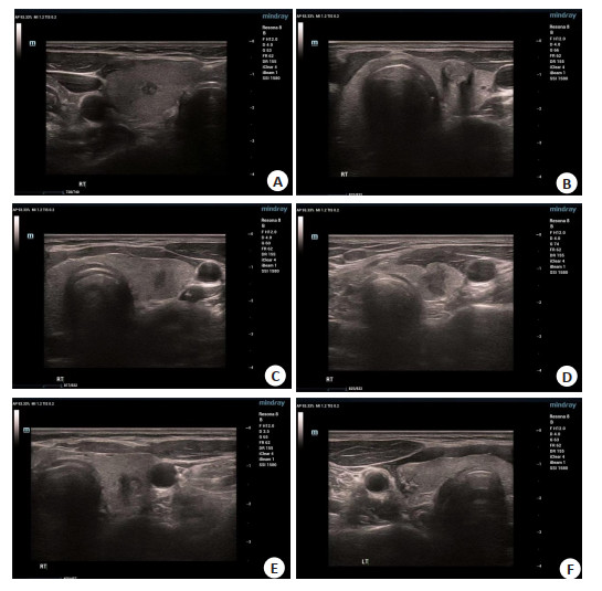
图 2 甲状腺结节的超声图像
A: 超声分类C-TR 4B类, A-TR 5级, FNA证实为甲状腺乳头状癌, Bethesda Ⅵ类; B: 超声分类C-TR 4C类, A-TR 5级, FNA证实为甲状腺乳头状癌, Bethesda Ⅵ类; C: 超声分类CTR 4C类,A-TR 5级, FNA证实为甲状腺乳头状癌, Bethesda Ⅵ类; D: 超声分类C-TR 4C类,A-TR 5级. FNA: 见淋巴细胞、嗜酸性细胞及滤泡上皮细胞,Bethesda Ⅱ类; E: 超声分类C-TR 5类. A-TR 5级, FNA见淋巴细胞及滤泡上皮细胞, 未见异型细胞, Bethesda Ⅱ类; F: 超声分类C-TR 5类. A-TR 5级, FNA证实为甲状腺乳头状癌,Bethesda Ⅵ类.
Figure 2. Ultrasound images of thyroid nodules.
表 1 195枚甲状腺结节不同分类方法与诊断效果对比
Table 1. Comparison of different classification methods and diagnostic effects of 195 thyroid nodules
不同分类 风险分层 良性(枚) 恶性(枚) 总数(枚) 实际恶性占比(%) 推荐恶性率(%) P C-TIRADS 2 0 0 0 0 0 < 0.001 3 13 0 13 0 < 2 4A 10 1 11 9.09 2~10 4B 28 4 32 12.50 10~50 4C 40 74 114 64.91 50~90 5 1 23 24 95.83 > 90 ACR-TIRADS 1 0 0 0 0 ≤2 < 0.001 2 3 0 3 0 ≤2 3 6 0 6 0 < 5 4 22 2 24 8.33 5~20 5 62 100 162 61.73 > 20 C-TIRADS: 中国甲状腺影像报告和数据系统; ACR-TIRADS: 美国放射学会甲状腺影像报告和数据系统.  下载: 导出CSV
表 2 C-TIRADS与ACR-TIRADS对甲状腺结节的诊断效能比较
Table 2. Comparison of diagnostic efficacy between C-TIRADS and ACR-TIRADS for thyroid nodules
不同分类系统 AUC(95% CI) 敏感度(%) 特异性(%) 准确度(%) 阳性预测值(%) 阴性预测值(%) C-TIRADS 0.796(0.741~0.852) 94.12(96/102) 56.99(53/93) 76.41(149/195) 70.59(96/136) 89.83(53/59) ACR-TIRADS 0.658(0.587~0.724) 95.10(97/102) 35.48(33/93) 30.58(130/195) 61.78(97/157) 96.84(33/38) Z/χ2 5.296 0.096 8.651 4.546 2.513 0.205 P < 0.001 0.757 0.003 0.033 0.113 0.650
下载: 导出CSV
-
[1] 尹丽萍, 万梦, 王晓华, 等. 某三甲医院健康体检人群甲状腺结节检出率及影响因素分析[J]. 安徽医学, 2021, 42(8): 941-4. doi: 10.3969/j.issn.1000-0399.2021.08.028 [2] Horvath E, Majlis S, Rossi R, et al. An ultrasonogram reporting system for thyroid nodules stratifying cancer risk for clinical management[J]. J Clin Endocrinol Metab, 2009, 94(5): 1748-51. doi: 10.1210/jc.2008-1724 [3] Kwak JY, Han KH, Yoon JH, et al. Thyroid imaging reporting and data system for US features of nodules: a step in establishing better stratification of cancer risk[J]. Radiology, 2011, 260(3): 892-9. doi: 10.1148/radiol.11110206 [4] Russ G, Bonnema SJ, Erdogan MF, et al. European thyroid association guidelines for ultrasound malignancy risk stratification of thyroid nodules in adults: the EU‑TIRADS[J]. Eur Thyroid J, 2017, 6(5): 225-37. doi: 10.1159/000478927 [5] Tessler FN, Middleton WD, Grant EG, et al. ACR thyroid imaging, reporting and data system (TI-RADS): white paper of the ACR TI-RADS committee[J]. J Am Coll Radiol, 2017, 14(5): 587-95. doi: 10.1016/j.jacr.2017.01.046 [6] 中华医学会超声医学分会浅表器官和血管学组, 中国甲状腺与乳腺超声人工智能联盟. 2020甲状腺结节超声恶性危险分层中国指南: C-TIRADS[J]. 中华超声影像学杂志, 2021, 30(3): 185-200. doi: 10.3760/cma.j.cn131148-20210205-00092 [7] 李晓宇, 刘静静, 刘利平, 等. 计算机辅助检测和诊断中K-TIRADS、ACR-TIRADS、ATA的诊断效能比较以及辅助超声医师诊断甲状腺结节的研究[J]. 中华超声影像学杂志, 2019, 28(10): 888-92. [8] Lauria Pantano A, Maddaloni E, Briganti SI, et al. Differences between ATA, AACE/ACE/AME and ACR TI-RADS ultrasound classifications performance in identifying cytological high-risk thyroid nodules[J]. Eur J Endocrinol, 2018, 178(6): 595-603. doi: 10.1530/EJE-18-0083 [9] 史宜鑫, 夏蜀珺, 陈林, 等. ACR 2017版甲状腺超声影像与数据报告系统在中国人群中的应用价值[J]. 中国超声医学杂志, 2020, 36(5): 394-7. doi: 10.3969/j.issn.1002-0101.2020.05.004 [10] Zhou JQ, Yin LX, Wei X, et al. 2020 Chinese guidelines for ultrasound malignancy risk stratification of thyroid nodules: the C-TIRADS[J]. Endocrine, 2020, 70(2): 256-79. doi: 10.1007/s12020-020-02441-y [11] Basha MAA, Alnaggar AA, Refaat R, et al. The validity and reproducibility of the thyroid imaging reporting and data system (TI‑RADS) in categorization of thyroid nodules: Multicentre prospective study[J]. Eur J Radiol, 2019, 117: 184-92. doi: 10.1016/j.ejrad.2019.06.015 [12] 李健, 殷延华, 戚建国, 等. 甲状腺结节超声恶性风险分层方法对甲状腺结节良恶性的鉴别诊断价值: 中美指南对比分析[J]. 中国全科医学, 2022, 25(9): 1077-81. doi: 10.12114/j.issn.1007-9572.2021.02.026 [13] 陈洁, 崔明祥, 刘淼. 甲状腺结节恶性分层系统在社区医院的应用价值研究[J]. 中国全科医学, 2020, 23(17): 2147-51. doi: 10.12114/j.issn.1007-9572.2019.00.648 [14] 李潜, 丁思悦, 郭兰伟, 等. 甲状腺结节超声恶性危险分层中国指南(C-TIRADS)联合人工智能辅助诊断对甲状腺结节鉴别诊断的效能评估[J]. 中华超声影像学杂志, 2021, 30(3): 231-5. doi: 10.3760/cma.j.cn131148-20201106-00858 [15] 王佳讯, 陈毓菁, 梁展鹏, 等. 超声造影结合细针穿刺活检诊断甲状腺结节[J]. 分子影像学杂志, 2017, 40(4): 397-400. doi: 10.3969/j.issn.1674-4500.2017.04.04 [16] 李朝喜, 温德惠, 陆海永, 等. ACR-TIRADS和C-TIRADS对桥本甲状腺炎背景下的桥本结节和甲状腺乳头状癌的诊断价值[J]. 临床耳鼻咽喉头颈外科杂志, 2022, 36(6): 447-52. https://www.cnki.com.cn/Article/CJFDTOTAL-LCEH202206008.htm [17] 田猛, 吴秀艳, 蔡雪珍, 等. 甲状腺结节超声恶性危险分层中国指南(C-TIRADS)对甲状腺乳头状癌的诊断价值[J]. 解放军医学院学报, 2022, 43(8): 823-9. doi: 10.3969/j.issn.2095-5227.2022.08.002 [18] 刘奕博, 李丽, 叶玉泉. 超声引导下细针穿刺活检在甲状腺中的应用研究进展[J]. 中国超声医学杂志, 2020, 36(10): 957-9. doi: 10.3969/j.issn.1002-0101.2020.10.030 [19] Migda B, Migda M, Migda MS, et al. Use of the Kwak Thyroid Image Reporting and Data System (K-TIRADS) in differential diagnosis of thyroid nodules: systematic review and meta-analysis[J]. Eur Radiol, 2018, 28(6): 2380-8. doi: 10.1007/s00330-017-5230-0 [20] 张秋霞, 张占超. 超声引导下细针穿刺活检甲状腺结节的临床应用分析[J]. 医学影像学杂志, 2022, 32(11): 1986-8. https://www.cnki.com.cn/Article/CJFDTOTAL-XYXZ202211032.htm [21] 王伟镇, 李颖嘉, 刘志, 等. 超声与细针穿刺诊在甲状腺结节诊断中的价值比较[J]. 分子影像学杂志, 2019, 42(2): 214-6. doi: 10.12122/j.issn.1674-4500.2019.02.17 -

 点击查看大图
计量
- 文章访问数: 193
- HTML全文浏览量: 133
- PDF下载量: 5
- 被引次数: 0