


Diagnostic value of musculoskeletal ultrasound and MRI in degenerative osteoarthritis of knee joint
-
摘要:
目的 评估肌骨超声及MRI对膝关节退行性骨关节炎(KOA)的诊断价值。 方法 选择2017年1月~2019年12月在我院接受治疗并全部接受关节镜检查的单侧KOA患者126例为研究对象。对所有纳入研究的患者行超声检查、MRI检查及关节镜检查,对肌骨超声图像及MRI图像行质量评价。以关节镜检查结果为金标准,分别评估超声、MRI检查对关节积液、滑膜增生、髌上囊积液、关节软骨损害、骨质增生、半月板损伤、韧带损伤等病变的诊断价值。 结果 126例患者成功接受肌骨超声、MRI及关节镜检查。以关节镜结果为金标准,超声检查及MRI检查在KOA患者滑膜增生、髌上囊积液、半月板损害、韧带损伤、肌腱断裂及腘窝囊肿的检出中与关节镜结果一致性好(超声Kappa值:0.81、0.85、0.79、0.79、1.00、1.00,MRI Kappa值:0.76、0.93、0.86、0.89、1.00、1.00,Kappa值均 > 0.75);在KOA滑膜增生和骨质增生的诊断中,超声检查的诊断效能好于MRI检查(AUC=0.978、0.972,P < 0.05);在关节积液、髌上囊积液、关节软骨损害、韧带损伤的诊断中,MRI检查的诊断效能好于超声检查(AUC=0.991、0.995、0.969、0.961,P < 0.05);在韧带损伤的诊断中,超声检查与MRI检查的诊断效能差异无统计学意义(P > 0.05);126例患者中,超声检查认为有57例患者存在关节间隙狭窄,平均关节间隙为3.54±0.53 mm,MRI检查认为有52例患者存在关节间隙狭窄,平均关节间隙为3.62±0.61 mm,两者测量差值无统计学意义(P > 0.05)。 结论 肌骨超声及MRI检查能较好的识别膝关节退行性骨关节炎病变特征,具有较高的临床运用价值。 -
关键词:
- 肌骨超声 /
- 磁共振成像 /
- 膝关节退行性骨关节炎
Abstract:Objective To assess the diagnostic value of musculoskeletal ultrasound and MRI in the diagnosis degenerative osteoarthritis of the knee (KOA). Methods A total of 126 patients with unilateral KOA who were treated in our hospital from January 2017 to December 2019 were selected and all underwent arthroscopy. Musculoskeletal ultrasound, MRI and arthroscope imaging were performed on all patients included in the study. Using arthroscopic findings as the gold standard, the diagnostic value of ultrasound and MRI examinations was assessed for joint effusion, synovial hyperplasia, suprapatellar bursa effusion, articular cartilage damage, osteophytes, meniscal damage and ligament damage, respectively. Results A total of 126 patients underwent musculoskeletal ultrasound, MRI and arthroscopy successfully. With arthroscopic results as the gold standard, ultrasonography and MRI were in good agreement with arthroscopic results in the detection of synovial hyperplasia, suprapatellar sac effusion, meniscus injury, ligament injury, tendon rupture and popliteal cyst in patients with KOA (Kappa values of ultrasound: 0.81, 0.85, 0.79, 0.79, 1.00, 1.00; Kappa values of MRI: 0.76, 0.93, 0.86, 0.89, 1.00, 1.00, Kappa values > 0.75). In the diagnosis of synovial hyperplasia and hyperosteogeny in KOA, the diagnostic efficacyof ultrasound was better than that of MRI (AUC=0.978, 0.972, P < 0.05). In the diagnosis of joint effusion, suprapatellar sac effusion, articular cartilage damage and ligament injury, MRI had better diagnostic performance than ultrasound (AUC=0.991, 0.995, 0.969, 0.961, P < 0.05). In the diagnosis of ligament injury, there was no statistical difference between ultrasound and MRI (P > 0.05). Among 126 patients, 57 patients were found to have joint space stenosis by ultrasound examination, and the average joint space was about 3.54±0.53 mm, while 52 patients were found to have joint space stenosis by MRI examination, and the average joint space was about 3.62±0.61 mm. There was no significant difference between the two measurements (P > 0.05). Conclusion Musculoskeletal ultrasound and MRI examination can better identify the characteristics of KOA and have high clinical value. -

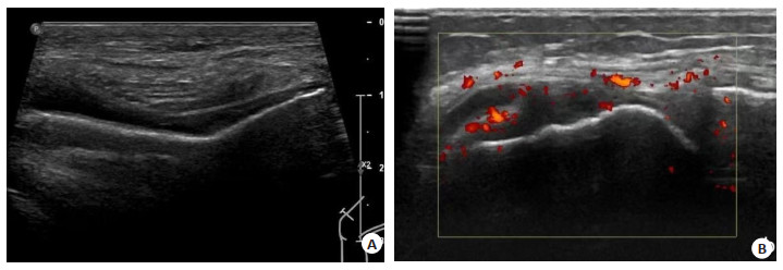
图 1 一例56岁女性KOA患者的肌骨超声图像
A: 股骨滑车处关节软骨部分变薄; B: 滑膜增生合并存在多普勒血流信号.
Figure 1. Musculoskeletal ultrasound image of a 56-year-old female KOA patient.

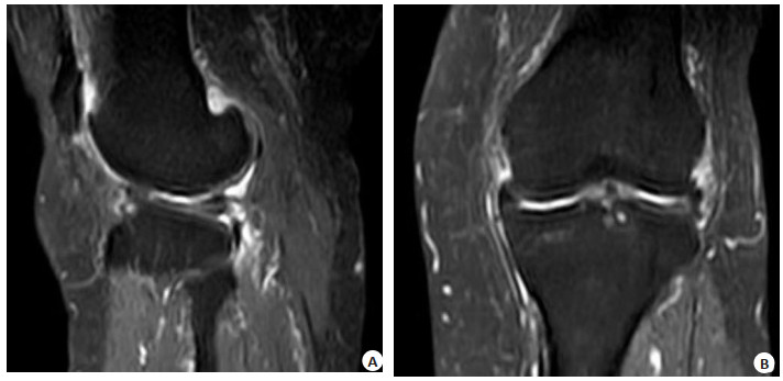
图 2 一例70岁男性KOA患者的MRI图像
A: 膝关节矢状位压脂序列显示股骨踝部骨髓水肿, 呈小囊状长T2信号(箭头处); B: 膝关节冠状位压脂序列显示胫骨平台骨髓水肿, 呈斑片状长T2信号(箭头处)
Figure 2. MRI image of a 70-year-old male KOA patient
表 1 超声、MRI对KOA患者具体病变的诊断价值
Table 1. Diagnostic value of ultrasound and MRI for specific lesions of KOA patients
病变 敏感度(%) 特异性(%) 阳性预测值(%) 阴性预测值(%) Kappa值 Youden指数 超声 MRI 超声 MRI 超声 MRI 超声 MRI 超声 MRI 超声 MRI 关节积液 92.5 95.3 89.4 84.2 98.0 97.1 68.0 76.2 0.73 0.72 0.82 0.80 滑膜增生 92.1 85.4 91.9 91.2 96.5 96.2 82.9 72.3 0.81 0.76 0.84 0.77 髌上囊积液 92.9 98.0 100.0 96.4 100.0 99.0 80.0 93.1 0.85 0.93 0.93 0.94 关节软骨损害 87.5 91.7 93.3 93.3 97.7 97.8 70.0 77.8 0.73 0.80 0.81 0.85 骨质增生 92.7 88.1 88.2 94.1 98.1 99.0 65.2 55.2 0.70 0.63 0.81 0.82 半月板损伤 81.3 89.1 98.4 96.8 98.1 96.6 83.6 90.0 0.79 0.86 0.80 0.86 韧带损伤 76.9 87.2 97.7 98.9 93.8 97.1 90.4 94.5 0.79 0.89 0.75 0.86 肌腱断裂 100.0 100.0 100.0 100.0 100.0 100.0 100.0 100.0 1.00 1.00 1.00 1.00 腘窝囊肿 100.0 100.0 100.0 100.0 100.0 100.0 100.0 100.0 1.00 1.00 1.00 1.00  下载: 导出CSV
表 2 超声、MRI对KOA患者具体病变的诊断结果比较
Table 2. Comparison of ultrasound and MRI diagnosis results of specific lesions in KOA patients
病变 AUC Z P 超声 MRI 关节积液 0.972 0.991 2.029 0.043 滑膜增生 0.978 0.944 2.522 0.012 髌上囊积液 0.964 0.995 2.515 0.012 关节软骨损害 0.948 0.969 2.032 0.042 骨质增生 0.972 0.945 2.508 0.012 半月板损伤 0.914 0.961 2.553 0.011 韧带损伤 0.910 0.949 1.780 0.075 肌腱断裂 1.000 1.000 - 1.000 腘窝囊肿 1.000 1.000 - 1.000
下载: 导出CSV
-
[1] Rathbun AM, Shardell MD, Ryan AS, et al. Association between disease progression and depression onset in persons with radiographic knee osteoarthritis[J]. Rheumatology (Oxford), 2020, 59(11): 3390-9. doi: 10.1093/rheumatology/keaa141 [2] Zampogna B, Papalia R, Papalia GF, et al. The role of physical activity as conservative treatment for hip and knee osteoarthritis in older people: a systematic review and meta-analysis[J]. J Clin Med, 2020, 9(4): 1167. doi: 10.3390/jcm9041167 [3] Murphy LB, Moss S, Do BT, et al. Annual incidence of knee symptoms and four knee osteoarthritis outcomes in the Johnston County osteoarthritis project[J]. Arthritis Care Res (Hoboken), 2016, 68(1): 55-65. doi: 10.1002/acr.22641 [4] 吕苏梅, 张瑞丽. 中老年膝骨关节炎的流行病学研究进展[J]. 中国老年学杂志, 2016, 36(16): 4133-5. doi: 10.3969/j.issn.1005-9202.2016.16.121 [5] 沈建成, 丁小方, 闫志刚, 等. 关节镜下清理术联合臭氧对膝骨关节炎患者膝关节功能和复发率的影响[J]. 中国医学装备, 2019, 16(4): 80-4. doi: 10.3969/J.ISSN.1672-8270.2019.04.022 [6] Bai X, Lv P, Liu K, et al. 3D black‑blood luminal angiography derived from high‑resolution MR vessel wall imaging in detecting MCA Stenosis: a preliminary study[J]. AJNR Am J Neuroradiol, 2018, 39(10): 1827-32. doi: 10.3174/ajnr.A5770 [7] 张晓慧, 邓雪蓉, 耿研, 等. 中国膝骨关节炎患者超声表现[J]. 中华风湿病学杂志, 2016, 20(9): 604-8. doi: 10.3760/cma.j.issn.1007-7480.2016.09.006 [8] Schmidt WA, Schmidt H, Schicke B, et al. Standard reference values for musculoskeletal ultrasonography[J]. Ann Rheum Dis, 2004, 63(8): 988-94. doi: 10.1136/ard.2003.015081 [9] Zhou XY, Zhang XX, Yu GY, et al. Effects of low-intensity pulsed ultrasound on knee osteoarthritis: a meta-analysis of randomized clinical trials[J]. Biomed Res Int, 2018, 2018: 7469197. [10] Draper DO, Klyve D, Ortiz R, et al. Effect of low-intensity long-duration ultrasound on the symptomatic relief of knee osteoarthritis: a randomized, placebo-controlled double-blind study[J]. J Orthop Surg Res, 2018, 13(1): 257. doi: 10.1186/s13018-018-0965-0 [11] Nasb M, Huang LJ, Gong CZ, et al. Human adipose-derived Mesenchymal stem cells, low-intensity pulsed ultrasound, or their combination for the treatment of knee osteoarthritis: study protocol for a first‑in‑man randomized controlled trial[J]. BMC Musculoskelet Disord, 2020, 21(1): 33. doi: 10.1186/s12891-020-3056-4 [12] Karakaş A, Dilek B, Şahin MA, et al. The effectiveness of pulsed ultrasound treatment on pain, function, synovial sac thickness and femoral cartilage thickness in patients with knee osteoarthritis: a randomized, double-blind clinical, controlled study[J]. Clin Rehabil, 2020, 34(12): 1474-84. doi: 10.1177/0269215520942953 [13] 林成才, 李丽娟, 陈吉波, 等. 三维快速自旋回波与常规序列诊断膝关节交叉韧带损伤的对比研究[J]. 中国医学装备, 2020, 17(2): 71-4. doi: 10.3969/J.ISSN.1672-8270.2020.02.019 [14] 曹峰, 张尹, 廖庆. 三种新型磁共振功能成像技术对早期膝关节软骨损伤的诊断价值分析[J]. 中国医学装备, 2020, 17(1): 76-80. doi: 10.3969/J.ISSN.1672-8270.2020.01.021 [15] 周雪添, 马勇, 郭杨, 等. 肌肉骨骼超声技术在骨科诊断治疗中的应用现状及机制[J]. 中国组织工程研究, 2019, 23(16): 2573-8. doi: 10.3969/j.issn.2095-4344.1167 [16] 王海, 陈小宇, 林千早, 等. 多层螺旋CT与超声诊断女性盆腔囊性肿块临床价值[J]. 医学影像学杂志, 2017, 27(2): 379-82. https://www.cnki.com.cn/Article/CJFDTOTAL-XYXZ201702058.htm [17] 刘琦, 吴长君. 超声在肌肉骨骼系统中应用的研究进展[J]. 医学综述, 2017, 23(12): 2433-7. doi: 10.3969/j.issn.1006-2084.2017.12.030 [18] 唐盛斐, 蓝常贡, 涂振阳. 肌骨超声在诊断肢体肌腱和韧带损伤中应用进展[J]. 中华实用诊断与治疗杂志, 2020, 34(2): 214-6. https://www.cnki.com.cn/Article/CJFDTOTAL-HNZD202002031.htm [19] Zhang X, Sun X, Chen G. Effect of the combinative use of acupotomy therapy and ultrasonic drug penetration in treating knee joint osteoarthritis[J]. QJM, 2022, 115(1): 12-6. doi: 10.1093/qjmed/hcaa278 [20] Wu Y, Zhu SB, Lv ZH, et al. Effects of therapeutic ultrasound for knee osteoarthritis: a systematic review and meta-analysis[J]. Clin Rehabil, 2019, 33(12): 1863-75. doi: 10.1177/0269215519866494 [21] Bevers K, Bijlsma JW, Vriezekolk JE, et al. Ultrasonographic features in symptomatic osteoarthritis of the knee and relation with pain[J]. Rheumatology (Oxford), 2014, 53(9): 1625-9. doi: 10.1093/rheumatology/keu030 -

 点击查看大图
计量
- 文章访问数: 166
- HTML全文浏览量: 156
- PDF下载量: 9
- 被引次数: 0