


In vivo fluorescence imaging of Evans Blue dye in zebrafish muscle necrosis model: a new platform for studying necrosis avid agents
-
摘要:
目的 探讨制作斑马鱼肌肉坏死模型的可行性,并应用此模型进行伊文思蓝(EBD)活体成像以研究其坏死亲和特性。 方法 采用显微注射泵注射10 nL无水酒精于3~7 d斑马鱼幼鱼卵黄囊背部肌肉处以制作斑马鱼肌肉坏死模型。肌肉坏死模型组和对照组于心脏大静脉处注射0.1% EBD 5 nL后0、4、24、48 h行荧光及激光共聚焦显微镜拍摄,动态观察两组斑马鱼体内EBD的分布情况,定量分析肌肉坏死区域和正常肌纤维区域EBD的荧光强度、范围及比值。 结果 斑马鱼肌肉坏死模型的制作成功率高。荧光及激光共聚焦显微镜显示,肌肉受损区域有大量EBD的红色荧光积聚,同明场下肌肉受损区域相一致;动态观察发现,随时间推移,红色荧光强度逐渐增强,于24 h达到峰值后逐渐减退;肌肉坏死区域同正常肌纤维区域之间荧光强度的差异具有统计学意义(P < 0.05),4 h和24 h时荧光强度差异最明显,分别为58.30±5.14、17.36±1.16和54.20±4.25、15.96±0.79。不同时间点的坏死亲和性比率的差异无统计学意义(P>0.05)。 结论 无水酒精诱导的斑马鱼肌肉坏死模型制作简捷经济,可作为一种新的研究坏死亲和性物质的实验平台。本研究初步证实EBD可选择性积聚于肌肉坏死区域,是一种潜在的坏死亲和性对比剂。 Abstract:Objective To explore the feasibility of making a muscle necrosis model in zebrafish, and to apply this model to investigate the necrosis affinity of Evans Blue dye (EBD) by in vivo fluorescence imaging. Methods In order to make a zebrafish muscle necrosis model, 10 nL of absolute alcohol was injected into the dorsal muscle of zebrafish larvae 3-7 d post fertilization by using the microinjection pump instrument. The distribution of EBD in the muscle necrosis model group and the control group were dynamically observed by fluorescence and laser confocal microscopy at 0, 4, 24 and 48 h after injection of 5 nL 0.1% EBD at the common cardinal vein. The fluorescence intensity, range and ratio of EBD in both the necrotic and normal muscle area were quantitative analyzed. Results Zebrafish model of muscle necrosis was successfully established with a high rate. Fluorescence and laser confocal microscopy showed that there was a large amount of EBD red fluorescence accumulation in the necrotic muscle area, which was consistent with the necrotic muscle in bright field. Dynamic observation showed that the red fluorescence intensity gradually increased, and gradually decreased after reaching the peak at 24 h. The difference in fluorescence intensity between necrotic and normal muscle was statistically significant (P < 0.05). At 4 h and 24 h, the difference was the most significant, which were 58.30±5.14, 17.36±1.16 and 54.20±4.25, 15.96±0.79, respectively. However, no significant differences were found in the necrosis affinity ratio among different time points (P>0.05). Conclusion The proposed model of muscle necrosis in zebrafish is simple and convenient, which can be used as a new experimental platform for studying necrosis avid agents. This study preliminary showed that EBD may selectively accumulate in the necrotic muscle of zebrafish, and it could be a potential necrosis avid contrast agent. -
Key words:
- Evans Blue dye /
- zebrafish /
- necrosis /
- necrosis avid agents /
- in vivo fluorescence imaging
-

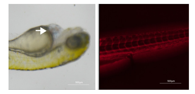
图 1 酒精诱导斑马鱼肌肉坏死模型的建立
Figure 1. Establishment of alcohol-induced muscle necrosis model in zebrafish.

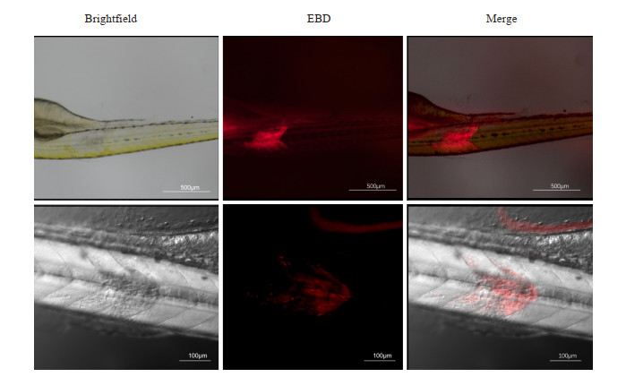
图 2 斑马鱼心脏大静脉注射EBD后的显微观察
Figure 2. Microscopic observation of the injection of EBD into the common cardinal vein of zebrafish.

图 3 斑马鱼肌肉坏死模型术后24 h荧光显微镜和激光共聚焦显微镜成像
Figure 3. Fluorescence and confocal laser imaging 24 h after establishment of zebrafish muscle necrosis model.

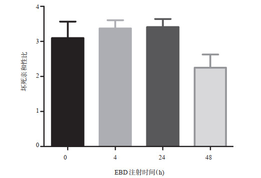
图 4 斑马鱼肌肉坏死模型的荧光显微镜动态观察及荧光强度定量分析
A: 斑马鱼肌肉坏死模型注射EBD术后0、4、24、48 h荧光显微镜成像; B: 不同时间点斑马鱼肌肉坏死区域的荧光强度定量分析; C: 不同时间点斑马鱼肌肉坏死区域面积定量分析(*P < 0.05).
Figure 4. Dynamic observation and quantitative analysis of fluorescence intensity of zebrafish muscle necrosis model by fluorescence microscope.
-
[1] Cona MM, Oyen R, Ni YC. Necrosis avidity of organic compounds: a natural phenomenon with exploitable theragnostic potentials[J]. Curr Med Chem, 2015, 22(15): 1829-1849. doi: 10.2174/0929867322666150227153550 [2] Galluzzi L, Vitale I, Aaronson SA, et al. Molecular mechanisms of cell death: recommendations of the Nomenclature Committee on Cell Death 2018[J]. Cell Death Differ, 2018, 25(3): 486-541. doi: 10.1038/s41418-017-0012-4 [3] Zhang DJ, Jiang CH, Feng YB, et al. Molecular imaging of myocardial necrosis: an updated mini-review[J]. J Drug Target, 2020, 28(6): 565-73. doi: 10.1080/1061186X.2020.1725769 [4] Chen F, Suzuki Y, Nagai N, et al. Rodent stroke induced by photochemical occlusion of proximal middle cerebral artery: evolution monitored with MR imaging and histopathology[J]. Eur J Radiol, 2007, 63(1): 68-75. doi: 10.1016/j.ejrad.2007.01.005 [5] Hernandez-Martinez JM, Sánchez-Reyes R, De la Garza-Salazar JG, et al. Onco-omics approaches and applications in clinical trials for cancer patients[J]. Adv Exp Med Biol, 2019, 1168: 79-90. [6] Smith BA, Smith BD. Biomarkers and molecular probes for cell death imaging and targeted therapeutics[J]. Bioconjug Chem, 2012, 23(10): 1989-2006. doi: 10.1021/bc3003309 [7] Silvestre-Roig C, de Winther MP, Weber C, et al. Atherosclerotic plaque destabilization: mechanisms, models, and therapeutic strategies[J]. Circ Res, 2014, 114(1): 214-26. doi: 10.1161/CIRCRESAHA.114.302355 [8] Xie BW, Stammes MA, van Driel PBAA, et al. Necrosis avid near infrared fluorescent cyanines for imaging cell death and their use to monitor therapeutic efficacy in mouse tumor models[J]. Oncotarget, 2015, 6(36): 39036-49. doi: 10.18632/oncotarget.5498 [9] Stammes MA, Maeda A, Bu JC, et al. The necrosis-avid small molecule HQ4-DTPA as a multimodal imaging agent for monitoring radiation therapy-induced tumor cell death[J]. Front Oncol, 2016, 6: 221. [10] Fang C, Wang K, Zeng CT, et al. Illuminating necrosis: from mechanistic exploration to preclinical application using fluorescence molecular imaging with indocyanine green[J]. Sci Rep, 2016, 6: 21013. doi: 10.1038/srep21013 [11] Yao LP, Xue X, Yu PP, et al. Evans blue dye: a revisit of its applications in biomedicine[J]. Contrast Media Mol Imaging, 2018, 2018: 7628037. [12] Miller D L, Li P, Dou C, et al. Evans blue staining of cardiomyocytes induced by myocardial contrast echocardiography in rats: evidence for necrosis instead of apoptosis[J]. Ultrasound Med Biol, 2007, 33(12): 1988-96. doi: 10.1016/j.ultrasmedbio.2007.06.008 [13] Hamer PW, McGeachie JM, Davies MJ, et al. Evans Blue Dye as an in vivo marker of myofibre damage: optimising parameters for detecting initial myofibre membrane permeability[J]. J Anat, 2002, 200(Pt 1): 69-79. [14] Smith SJ, Horstick EJ, Davidson AE, et al. Analysis of zebrafish larvae skeletal muscle integrity with Evans blue dye[J]. J Vis Exp, 2015(105): 53183. [15] Feng YB, Chen F, Ma ZL, et al. Towards stratifying ischemic components by cardiac MRI and multifunctional stainings in a rabbit model of myocardial infarction[J]. Theranostics, 2013, 4(1): 24-35. [16] Ye, L, Miao X, Chen T, et al. Zebrafish as a visual and dynamic model to study the transport of nanosized drug delivery systems across the biological barriers[J]. Colloids Surf B Biointerfaces, 2017, 156: 227-35. doi: 10.1016/j.colsurfb.2017.05.022 [17] Gaff DF, Okong'o-Ogola O. The use of non-permeating pigments for testing the survival of cells[J]. J Exp Bot, 1971, 22(3): 756-8. doi: 10.1093/jxb/22.3.756 [18] Matsuda R, Nishikawa A, Tanaka H. Visualization of dystrophic muscle fibers in mdx mouse by vital staining with Evans blue: evidence of apoptosis in dystrophin-deficient muscle[J]. J Biochem, 1995, 118(5): 959-64. doi: 10.1093/jb/118.5.959 [19] Li JJ, Cona MM, Chen F, et al. Exploring theranostic potentials of radioiodinated hypericin in rodent necrosis models[J]. Theranostics, 2012, 2(10): 1010-9. doi: 10.7150/thno.4924 [20] Klyen BR, Sampson DD, Shavlakadze T, et al. Identification of muscle necrosis in the mdx mouse model of Duchenne muscular dystrophy using three-dimensional optical coherence tomography[C]. /2011: 076013. [21] Zhang D, Gao M, Jin Q, et al. Updated developments on molecular imaging and therapeutic strategies directed against necrosis[J]. Acta Pharm Sin B, 2019, 9(3): 455-68. doi: 10.1016/j.apsb.2019.02.002 [22] Jin Q, Shan X, Luo Q, et al. 131I-Evans blue: evaluation of necrosis targeting property and preliminary assessment of the mechanism in animal models[J]. Acta Pharm Sin B, 2018, 8(3): 390-400. doi: 10.1016/j.apsb.2017.08.002 [23] Waugh TA, Horstick E, Hur J, et al. Fluoxetine prevents dystrophic changes in a zebrafish model of Duchenne muscular dystrophy[J]. Hum Mol Genet, 2014, 23(17): 4651-62. doi: 10.1093/hmg/ddu185 [24] Yasuda S, Ikuta K, Uwatoku T, et al. In vivo magnetic resonance imaging of atherosclerotic lesions with a newly developed Evans blue-DTPA-gadolinium contrast medium in apolipoprotein-E-deficient mice[J]. J Vasc Res, 2008, 45(2): 123-8. doi: 10.1159/000109930 [25] Tsopelas C, Bellon M, Bevington E, et al. Lymphatic mapping with 99mTc-Evans Blue dye in sheep[J]. Ann Nucl Med, 2008, 22(9): 777-85. doi: 10.1007/s12149-008-0171-y [26] Zang J, Liu QX, Sui HM, et al. Combined 68Ga-NOTA-Evans blue lymphoscintigraphy and 68Ga-NOTA-RM26 PET/CT evaluation of sentinel lymph node metastasis in breast cancer patients[J]. Bioconjug Chem, 2020, 31(2): 396-403. doi: 10.1021/acs.bioconjchem.9b00789 [27] Hou GZ, Li XQ, Hou B, et al. Lymphangioma on 68Ga-NOTA-Evans blue PET/MRI[J]. Clin Nucl Med, 2018, 43(7): 553-5. doi: 10.1097/RLU.0000000000002129 [28] Ehlerding EB, Lan XL, Cai WB. "albumin hitchhiking" with an Evans blue analog for cancer theranostics[J]. Theranostics, 2018, 8(3): 812-4. doi: 10.7150/thno.24183 [29] Zhang JJ, Lang LX, Zhu ZH, et al. Clinical translation of an albumin-binding PET radiotracer 68Ga NEB[J]. J Nucl Med, 2015, 56(10): 1609-14. doi: 10.2967/jnumed.115.159640 [30] Sutton R, Tsopelas C, Kollias J, et al. Sentinel node biopsy and lymphoscintigraphy with a technetium 99m labeled blue dye in a rabbit model[J]. Surgery, 2002, 131(1): 44-9. doi: 10.1067/msy.2002.118450 [31] Tsopelas C, Sutton R. Why certain dyes are useful for localizing the sentinel lymph node[J]. J Nucl Med, 2002, 43(10): 1377-82. [32] Chen F, Suzuki Y, Nagai N, et al. Rat cerebral ischemia induced with photochemical occlusion of proximal middle cerebral artery: a stroke model for MR imaging research[J]. MAGMA, 2004, 17(3): 103-8. -

 下载:
下载:

 点击查看大图
计量
- 文章访问数: 203
- HTML全文浏览量: 102
- PDF下载量: 8
- 被引次数: 0