


Clinical significance of serum STK1, ECM1 and sB7-H3 detection combined with multi-slice spiral CT on improving the differential diagnosis of hepatocellular carcinoma and hepatic hemangioma
-
摘要:
目的 探究血清胸腺苷激酶1(STK1)、可溶型B7-H3(sB7-H3)、细胞外基质蛋白-1(ECM1)联合多层螺旋CT对肝细胞癌与肝血管瘤的鉴别诊断。 方法 选取59例肝细胞癌及45例肝血管瘤患者,比较两种病灶的影像学特征及血清STK1、sB7-H3及ECM1水平;分析多层螺旋CT三期增强扫描对上述两种疾病的诊断效能。 结果 CT平扫显示,肝细胞癌及肝血管瘤病灶的形态特征、类型、边界清晰度的差异有统计学意义(P < 0.05);肝细胞癌患者的血清STK1、sB7-H3、ECM1水平高于肝血管瘤患者(P < 0.05),且两种患者的动脉期均匀高密度病灶的增强CT值的差异有统计学意义(P < 0.05);多层螺旋CT三期增强扫描诊断肝细胞癌、肝血管瘤,与病理结果一致性较好(Kappa=0.844)。 结论 多层螺旋CT三期增强扫描可有效鉴别肝细胞癌与肝血管瘤,动脉期均匀高密度病灶的增强CT值及血清STK1、sB7-H3、ECM1水平升高有利于疾病诊断。 Abstract:Objective To explore serum thymidine kinase 1 (STK1), soluble B7-H3 (sB7-H3) and extracellular matrix protein-1 (ECM1) combined with multi-slice spiral CT in the differential diagnosis of hepatocellular carcinoma and hepatic hemangioma. Methods Fifty-nine patients with hepatocellular carcinoma and 45 patients with hepatic hemangioma were selected. The imaging characteristics of the two lesions and levels of serum STK1, sB7-H3 and ECM1 were compared and the diagnostic efficiency of multi-slice spiral CT three-phase enhanced scan on the above two diseases was analyzed. Results CT plain scan results showed that there were significant differences in the morphology, type and boundary between hepatocellular carcinoma lesions and hepatic hemangioma lesions (P < 0.05). The levels of serum STK1, sB7-H3 and ECM1 in patients with hepatocellular carcinoma were higher than those in patients with hepatic hemangioma (P < 0.05). There was a statistical difference in the enhanced CT value of uniform high-density lesions in the arterial phase between two kinds of patients (P < 0.05). Multi-slice spiral CT three-phase enhanced scan in the diagnosis of hepatocellular carcinoma and hepatic hemangioma had good consistency with pathological results (Kappa=0.844). Conclusion Multi-slice spiral CT three-phase enhanced scan is effective in distinguishing hepatocellular carcinoma from hepatic hemangioma. Enhanced CT value of uniform high-density lesions in the arterial phase and increased levels of serum STK1, sB7-H3 and ECM1 are helpful for disease diagnosis. -
Key words:
- hepatocellular carcinoma /
- hepatic hemangioma /
- multi-slice spiral CT
-

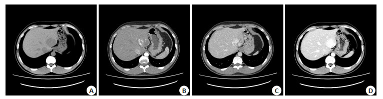
图 1 肝右叶巨块型肝细胞癌伴中心液化坏死图片
A: CT平扫病灶大小10.6 cm×12.0 cm×12.8 cm, CT值27~41 Hu, 密度不均, 内可见片状更低密度影; B~C: 增强后动脉期及门脉期病灶明显欠均匀强化, 强化程度高于肝实质; D: 延迟期强化程度略低于肝实质, 病灶中心低密度影强化不明显, 邻近肝实质密度欠均, 强化欠均.
Figure 1. Massive hepatocellular carcinoma of the right lobe with central liquefaction necrosis.

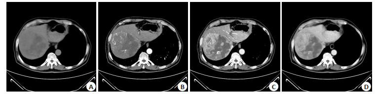
图 2 肝左外上段血管瘤图片
A: CT平扫较病灶大小约4.5 cm×3.9 cm, CT值47 Hu, 边界尚清; B: 渐进性强化增强, 动脉期病灶边缘结节状或斑片状强化明显; C~D: 门脉期及延迟期强化程度逐渐向中心扩展.
Figure 2. Left lateral upper hepatic hemangioma.
表 1 肝细胞癌和肝血管瘤病灶CT平扫影像学资料比较
Table 1. Comparison of CT plain scan imaging data of hepatocellular carcinoma and hepatic hemangioma [n(%)]
图像特征 肝细胞癌病灶(n=62) 肝血管瘤病灶(n=55) χ2 P 形态 30.717 < 0.001 类圆形 15(24.19) 14(25.45) 结节样 10(16.13) 33(60.00) 圆形 37(59.68) 8(14.55) 边界 5.699 0.017 清晰 33(53.23) 41(74.55) 模糊 29(46.77) 14(25.45) 类型 9.497 0.002 单发病灶 56(90.32) 37(67.27) 多发病灶 6(9.68) 18(32.73) 密度 0.857 0.651 高密度 1(1.61) 2(3.64) 等密度 8(12.90) 5(9.09) 低密度 53(85.48) 48(87.27)  下载: 导出CSV
表 2 动脉期均匀高密度病灶的增强CT值比较
Table 2. Comparison of enhanced CT value of uniform high-density lesions in arterial phase (Hu, Mean±SD)
疾病类型 增强CT值 阅片师1 阅片师2 肝细胞癌(n=40) 39.87±6.07 40.05±5.12 肝血管瘤(n=14) 96.45±19.33 99.23±20.46 t 16.561 17.093 P < 0.001 < 0.001
下载: 导出CSV
表 3 诊断效能分析
Table 3. Diagnostic efficiency analysis (n)
多层螺旋CT三期增强扫描 病理诊断 Kappa 肝细胞癌 肝血管瘤 合计 肝细胞癌 54 3 57 0.844 肝血管瘤 5 42 47 合计 59 45 104
下载: 导出CSV
表 4 血清STK1、sB7-H3、CEA水平比较
Table 4. Comparison of levels of serum STK1, sB7-H3 and CEA (Mean±SD)
指标 肝细胞癌(n=59) 肝血管瘤(n=45) t P STK1(pmol/L) 1.92±0.63 0.83±0.24 11.003 < 0.001 sB7-H3(pg/mL) 3968.53±564.15 2145.32±402.31 18.395 < 0.001 ECM1(pg/mL) 259.47±30.12 115.43±22.18 26.972 < 0.001 STK1: 血清胸腺苷激酶1; sB7-H3: 可溶型B7-H3; ECM1: 细胞外基质蛋白-1.
下载: 导出CSV
-
[1] Kulik L, El-Serag HB. Epidemiology and management of hepatocellular carcinoma[J]. Gastroenterology, 2019, 156(2): 477-91. e1. doi: 10.1053/j.gastro.2018.08.065 [2] Shabbir Z, Javaid A, Din IU. Infantile hepatic hemangioma[J]. J Pak MedAssoc, 2018, 68(12): 1846-7. [3] 陈茂东, 张静, 杨桂香, 等. 基于普美显增强磁共振的影像组学鉴别肝细胞癌与肝血管瘤[J]. 南方医科大学学报, 2018, 38(4): 428-33. doi: 10.3969/j.issn.1673-4254.2018.04.10 [4] 李卫侠, 林晓珠, 张静, 等. CT能谱成像在肝细胞肝癌与肝血管瘤鉴别诊断中的价值[J]. 放射学实践, 2018, 33(9): 882-7. https://www.cnki.com.cn/Article/CJFDTOTAL-FSXS201809005.htm [5] 中国抗癌协会肝癌专业委员会, 中国抗癌协会临床肿瘤学协作专业委员会, 中华医学会肝病学会肝癌学组, 等. 原发性肝癌规范化病理诊断方案专家共识[J]. 肿瘤, 2011, 31(4): 285-7. https://www.cnki.com.cn/Article/CJFDTOTAL-ZZLL201104004.htm [6] 陈孝平, 夏锋, 李雪松. 肝血管瘤诊断和治疗多学科专家共识(2019版)[J]. 临床肝胆病杂志, 2019, 35(9): 1928-32. doi: 10.3969/j.issn.1001-5256.2019.09.008 [7] Wang M, Wei CX, Shi ZJ, et al. Study on the diagnosis of small hepatocellular carcinoma caused by hepatitis B cirrhosis via multislice spiral CT and MRI[J]. Oncol Lett, 2018, 15(1): 503-8. [8] 吕杨, 季玲. 肝血管瘤超声、CT和MRI影像学特征及诊断价值比较[J]. 磁共振成像, 2020, 11(8): 672-4. https://www.cnki.com.cn/Article/CJFDTOTAL-CGZC202008020.htm [9] Zhang R, Liu ZW, Zhou CR, et al. Diagnostic value of multi-slice spiral CT perfusion imaging in primary hepatocellular carcinoma[J]. Minerva Med, 2021, 28(1): 4068. [10] 张晨彩, 王新文, 范凯, 等. 能谱CT平扫对原发性肝癌及肝血管瘤鉴别诊断的价值[J]. 实用放射学杂志, 2020, 36(9): 1426-9. doi: 10.3969/j.issn.1002-1671.2020.09.018 [11] Li WX, Li RK, Zhao XT, et al. Differentiation of hepatocellular carcinoma from hepatic hemangioma and focal nodular hyperplasia using computed tomographic spectral imaging[J]. J Clin Transl Hepatol, 2021, 9(3): 315-23. [12] 李茂胜, 谢微波. 肝细胞癌和局灶性结节增生CT定量参数差异及临床鉴别意义[J]. 肝脏, 2019, 24(8): 907-10. doi: 10.3969/j.issn.1008-1704.2019.08.020 [13] 胡菁, 夏茂鑫, 任艳. 超声及CT鉴别诊断肝硬化背景下肝血管瘤、肝细胞肝癌的临床价值探讨[J]. 中国CT和MRI杂志, 2020, 18(8): 80-2. doi: 10.3969/j.issn.1672-5131.2020.08.025 [14] Pang GD, Duan ZY, Shao CC, et al. Heterogeneity analysis of triphasic CT scan perfusion parameters in differential diagnosis of hepatocellular carcinoma and hemangioma[J]. Medicine, 2018, 97 (38): e12512. doi: 10.1097/MD.0000000000012512 [15] 罗江, 徐勤, 杨琛, 等. 肝脏不同肿瘤性病变中央瘢痕的CT及MRI表现与研究价值[J]. 医学影像学杂志, 2019, 29(12): 2063-7. https://www.cnki.com.cn/Article/CJFDTOTAL-XYXZ201912020.htm [16] Mamone G, Piazza AD, Carollo V, et al. Imaging of hepatic hemangioma: fromAto Z[J]. Abdom Radiol, 2020, 45(3): 672-91. [17] 唐鹏, 彭定辉, 陈薇, 等. 肺癌患者胸苷激酶1血清水平分析[J]. 微循环学杂志, 2020, 30(2): 60-2. https://www.cnki.com.cn/Article/CJFDTOTAL-WXHX202002017.htm [18] Huang LL, Zhou Y, Sun QW, et al. Evaluation of the role of soluble B7-H3 in association with membrane B7-H3 expression in gastric adenocarcinoma[J]. Cancer Biomark, 2022, 33(1): 123-9. [19] Lee TW, Lee KM. ECM1 is associated with endocrine resistance in ER+breast cancers[J]. Anim Cells Syst (Seoul), 2022, 26(3): 99-107. [20] 王禹博, 王顺涛, 谢磊, 等. 基于扩散加权成像的影像组学模型鉴别诊断肝细胞癌与肝血管瘤[J]. 临床放射学杂志, 2020, 39(3): 481-6. https://www.cnki.com.cn/Article/CJFDTOTAL-LCFS202003014.htm -

 点击查看大图
计量
- 文章访问数: 195
- HTML全文浏览量: 87
- PDF下载量: 5
- 被引次数: 0