


Value of 99Tcm-MIBI SPECT/CT imaging combined with T/NT semi-quantitative analysis for pre-operative localization of primary parathyroid adenoma
-
摘要:
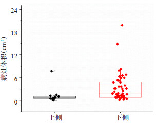
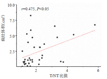
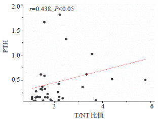
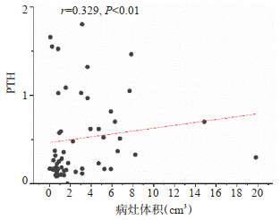
目的 99Tcm-MIBI SPECT/CT显像结合T/NT半定量分析对原发甲状旁腺腺瘤术前定位的价值。 方法 回顾性分析我院术后诊断为原发甲状旁腺腺瘤81例患者的99Tcm--MIBI SPECT/CT显像及临床资料,结合T/NT半定量分析,探讨不同部位腺瘤的检出率。 结果 99Tcm-MIBI SPECT/CT断层显像、平面显像对腺瘤检出率分别为95.7%(67/70)、90%(63/70);99Tcm-MIBI SPECT/ CT断层和平面显像腺瘤检出率差异无统计学意义( χ2=1.723,P=0.189)。根据腺瘤的位置将腺瘤分为上侧和下侧。99Tcm-MIBI SPECT/CT断层显像、平面显像对上侧腺瘤检出率分别为92.8%(13/14)、85.7%(12/14),对下侧腺瘤检出率分别为100%(53/ 53)、100%(53/53)。99Tcm-MIBI SPECT/CT断层显像上下侧腺瘤检出率差异无统计学意义( χ2=3.84,P=0.20),99Tcm-MIBI SPECT/CT平面显像下侧腺瘤检出率高于上侧( χ2=7.80,P=0.04)。下侧腺瘤体积大于上侧(Z=-3.19,P=0.001),腺瘤体积和早期相T/NT比值及血清甲状旁腺激素具有弱相关性(r=0.475、0.329,P < 0.05)。 结论 通过对原发甲状旁腺瘤患者行术前99Tcm-MIBI SPECT/CT显像,并结合T/NT半定量分析,提高了不同部位腺瘤定位的准确度,能够为手术提供有价值的参考。 -
关键词:
- 甲状旁腺腺瘤 /
- 99Tcm-MIBI SPECT/CT /
- 甲状旁腺激素 /
- T/NT比值
Abstract:Objective To investigate the value of 99Tcm-MIBI SPECT/CT imaging combined with T/NT semi-quantitative analysis in pre-operative of primary parathyroid adenoma. Methods The 99Tcm-MIBI SPECT/CT imaging and clinical data of 81 patients diagnosed with primary parathyroid adenoma were retrospectively analyze, to investigate the detection rate of parathyroid adenoma in different sites. Results The detection rate of 99Tcm-MIBI SPECT/CT tomography imaging, 99Tcm-MIBI SPECT/CT planar imaging were 95.7%(67/70) and 90%(63/70), respectively. There was no significant difference in detection rate between 99Tcm-MIBI SPECT/CT tomography imaging and planar imaging ( χ2=1.723, P=0.189). Adenomas are classified as superior and inferior according to their location, the detection rate of 99Tcm-MIBI SPECT/CT tomography imaging, 99Tcm-MIBI SPECT/CT planar imaging for superior adenomas were 92.8%(13/14)and 85.7%(12/14), and for inferior adenomas were 100%(53/53)and 100%(53/53), respectively. There was no significant difference in detection rate of adenomas between the upper and lower sides on 99Tcm-MIBI SPECT/CT tomography imaging ( χ2=3.84, P=0.20). The detection rate of inferior adenomas in 99Tcm-MIBI SPECT/CT planar imaging was higher on the lower side than on the upper side ( χ2=7.80, P=0.04). Adenoma volume on the inferior side was greater than on the superior side (Z=-3.19, P=0.001), and there was a weak correlation between adenoma volume and early phase T/NT ratio and serum parathyroid hormone (r=0.475, 0.329, P < 0.05). Conclusion By performing preoperation 99Tcm-MIBI SPECT/CT imaging in patients with primary parathyroid adenoma combined with T/NT semi-quantitative analysis, the accuracy of adenoma localization at different sites is improved and could provide a valuable reference for surgery. -
Key words:
- primary hyperparathyroidism /
- 99Tcm-MIBI SPECT/CT /
- parathyroid hormone /
- T/NT
-

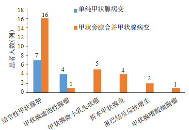
图 1 PHPT合并甲状腺病变及单纯甲状腺病变类型分布情况
Figure 1. Distribution of PHPT combined with thyroid disease and simplex thyroid disease.

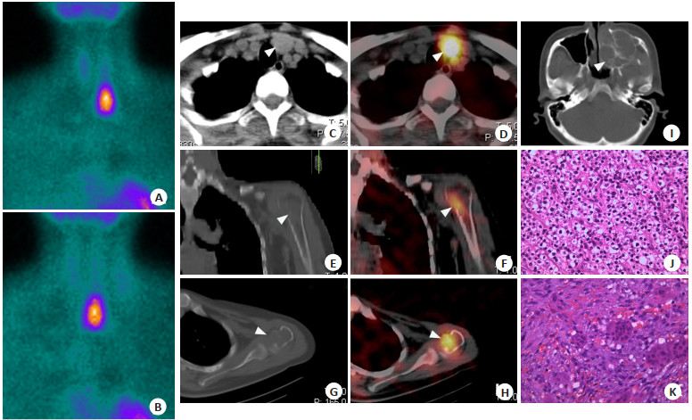
图 2 某25岁女性PHPT患者影像学表现
A~B:99Tcm-MIBI SPECT双时相显像,早期相(20 min)甲状腺左侧叶结节状放射性浓聚影,延迟相放射性浓聚影未见明显消退;C~D、J:CT及融合影像示:甲状腺左叶下极放射性浓聚影CT下定位于甲状腺左叶下极后方,大小约2.0 cm×1.0 cm×0.8 cm,术后病理结果为左侧甲状旁腺腺瘤;E~H:左侧喙突及左肱骨头囊性变,邻近骨质吸收变薄,相应部位见放射性浓聚影;I、K.左侧上颌窦占位性病变,术后病理为间叶源性梭形细胞肿瘤组织,符合甲旁亢所致的棕色瘤.
Figure 2. Imaging features of a 25-year-old female PHPT patient.

图 5 早期相T/NT值和血清PTH的相关性
Figure 5. Correlation between early phase T/NT value and serum PTH.
表 1 两种显像方法对不同部位腺瘤检出率的比较
Table 1. Comparison of detection rate of adenoma in different sites by two imaging methods
显像方法 上侧 下侧 χ2 P 左上 右上 左下 右下 SPECT/CT 6/6 7/8 22/22 31/31 3.84 0.209 SPECT 6/6 6/8 22/22 31/31 7.80 0.041 χ2 - 0.14 - - P - 1.00 - -  下载: 导出CSV
-
[1] Sun B, Guo B, Wu B, et al. Characteristics, management, and outcome of primary hyperparathyroidism at a single clinical center from 2005 to 2016[J]. Osteoporos Int, 2018, 29(3): 635-42. doi: 10.1007/s00198-017-4322-7 [2] Ozderya A, Temizkan S, Gul AE, et al. Biochemical and pathologic factors affecting technetium-99m-methoxyisobutylisonitrile imaging results in patients with primary hyperparathyroidism[J]. Ann Nucl Med, 2018, 32(4): 250-5. doi: 10.1007/s12149-018-1239-y [3] Treglia G, Trimboli P, Huellner M, et al. Imaging in primary hyperparathyroidism: focus on the evidence-based diagnostic performance of different methods[J]. Minerva Endocrinol, 2018, 43 (2): 133-43. [4] 郑玉婷, 池晓华, 齐永帅, 等. 99mTc-MIBI SPECT/CT显像结合半定量分析在甲状旁腺功能亢进症中的诊断价值及影响因素[J]. 南方医科大学学报, 2021, 41(10): 1577-82. doi: 10.12122/j.issn.1673-4254.2021.10.18 [5] 李秀梅, 李军, 王宏桥, 等. 高频超声、超声造影与99mTc-MIBI SPECT/CT在难治性甲状旁腺功能亢进术前定位中的比较[J]. 中华医学超声杂志: 电子版, 2018, 15(7): 522-9. doi: 10.3877/cma.j.issn.1672-6448.2018.07.010 [6] Mallick R, Malik J, Yip L, et al. Novel findings on SPECT-CT Tc- 99 sestamibi imaging for primary hyperparathyroidism[J]. J Surg Res, 2020, 252: 216-21. doi: 10.1016/j.jss.2020.03.014 [7] Guilmette J, Sadow PM. Parathyroid pathology[J]. Surg Pathol Clin, 2019, 12(4): 1007-19. doi: 10.1016/j.path.2019.08.006 [8] Khan AA, Hanley DA, Rizzoli R, et al. Primary hyperparathyroidism: review and recommendations on evaluation, diagnosis, and management.[J]. Osteoporos Int, 2017, 28(1): 1-19. doi: 10.1007/s00198-016-3716-2 [9] Li WB, Zhu QL, Lai XJ, et al. Value of preoperative ultrasoundguided fine-needle aspiration for localization in Tc-99m MIBInegative primary hyperparathyroidism patients[J]. Medicine, 2017, 96(49): e9051. doi: 10.1097/MD.0000000000009051 [10] Ozderya A, Temizkan S, Cetin K, et al. The results of parathyroid hormone assay in parathyroid aspirates in pre-operative localization of parathyroid adenomas for focused parathyroidectomy in patients with negative or suspicious technetium-99m-sestamibi scans[J]. Endocr Pract, 2017, 23(9): 1101-6. doi: 10.4158/EP171921.OR [11] Wojtczak B, Syrycka J, Kaliszewski K, et al. Surgical implications of recent modalities for parathyroid imaging[J]. Gland Surg, 2020, 9 (Suppl 2): S86-S94. [12] 刘岩, 王岐, 杨路路, 等. 99Tcm-MIBI双时相显像对甲状旁腺腺瘤和甲状旁腺增生的诊断价值[J]. 中国普外基础与临床杂志, 2021, 28 (11): 1457-61. https://www.cnki.com.cn/Article/CJFDTOTAL-ZPWL202111007.htm [13] 唐嘉励, 段东, 张辉. 99Tcm-MIBI SPECT/CT显像及平面显像半定量分析对原发性甲状旁腺功能亢进症的诊断价值[J]. 中华内分泌外科杂志, 2019, 13(1): 17-21. https://www.cnki.com.cn/Article/CJFDTOTAL-HAIN201223042.htm [14] 徐奇奇, 孔娜, 梁春蕊, 等. 超声、核素及CT对原发性甲状旁腺功能亢进症术前定位诊断价值的研究[J]. 中华普通外科杂志, 2021, 36(12): 922-5. doi: 10.3760/cma.j.cn113855-20210518-00304 [15] 曾鸣, 柳卫, 王宁宁, 等. 99mTc-MIBI SPECT-CT技术在甲状旁腺切除术前定位诊断中的增益价值[J]. 中华肾脏病杂志, 2017, 33(2): 86-91. doi: 10.3760/cma.j.issn.1001-7097.2017.02.002 [16] Yuan LL, Kan Y, Ma DQ, et al. Combined application of ultrasound and SPECT/CT has incremental value in detecting parathyroid tissue in SHPT patients[J]. Diagn Interv Imaging, 2016, 97(2): 219-25. doi: 10.1016/j.diii.2015.08.007 [17] 吴书婷, 朱小华, 张国鹏, 等. 甲状旁腺99Tcm-MIBI SPECT/CT显像在原发性甲状旁腺功能亢进症术前诊断中的增益价值[J]. 中华核医学与分子影像杂志, 2016, 36(5): 436-40. doi: 10.3760/cma.j.issn.2095-2848.2016.05.012 [18] Ding Y, Zou Q, Jin YT, et al. Relationship between parathyroid oxyphil cell proportion and clinical characteristics of patients with chronic kidney disease[J]. Int Urol Nephrol, 2020, 52(1): 155-9. doi: 10.1007/s11255-019-02330-y [19] Cordes M, Dworak O, Papadopoulos T, et al. MIBI scintigraphy of parathyroid adenomas: correlation with biochemical and histological markers[J]. Endocr Res, 2018, 43(3): 141-8. doi: 10.1080/07435800.2018.1437747 [20] Lee JY, Song HS, Choi JH, et al. Detecting synchronous thyroid adenoma and false-positive findings on technetium-99m MIBI single photon-emission computed tomography/computed tomography[J]. Diagnostics (Basel), 2019, 9(2): 57. doi: 10.3390/diagnostics9020057 [21] Jeong C, Kwon HI, Baek H, et al. Association of hyperparathyroidism and papillary thyroid cancer: a multicenter retrospective study[J]. Endocrinol Metab (Seoul), 2020, 35(4): 925-32. doi: 10.3803/EnM.2020.725 -

 点击查看大图
计量
- 文章访问数: 134
- HTML全文浏览量: 121
- PDF下载量: 13
- 被引次数: 0