


Diagnostic value of gray scale ultrasound, color Doppler ultrasound and shear wave elastography Young's modulus in papillary thyroid microcarcinoma
-
摘要:
目的 探讨灰阶超声、多普勒超声及剪切波杨氏模量诊断甲状腺微小乳头状癌的价值。 方法 选择2018年2月~2020年5月由我院诊治的甲状腺结节患者136例,分别进行灰阶超声、多普勒超声及剪切波杨氏模量技术及病理学检查,以病理诊断为金标准,分析灰阶超声、多普勒超声及剪切波杨氏模量和三者联合诊断甲状腺微小乳头状癌的价值。 结果 灰阶超声敏感度43.21%,特异性68.30%,准确度57.30%;多普勒超声敏感度75.32%,特异性90.74%,准确度84.32%;根据病理结果分为良性、恶性组进行ME值比较,良性组MEmean、MEmin、MEmax值明显低于恶性组(P < 0.05);MEmean、MEmin和MEmax预测微小乳头状态癌的ROC曲线最佳切值点分别为40.25、16.85、65.92 kPa,其对应的敏感度和特异性分别为85.5%、68.8%,67.5%、82.4%,58.1%、88.2%;联合检测准确度高于灰阶、多普勒超声、剪切波杨氏准确度,差异有统计学意义(P < 0.05)。 结论 灰阶超声、多普勒超声及剪切波杨氏模量三者联合诊断甲状腺微小乳头状癌可提高其诊断准确率,具有较高诊断价值。 Abstract:Objective To investigate the diagnostic value of gray scale ultrasound, Doppler ultrasound and shear wave elastography Young's modulus in papillary thyroid microcarcinoma. Methods A total of 136 patients with papillary thyroid microcarcinoma in our hospital from February 2018 to May 2020 were enrolled. Gray scale ultrasound, Doppler ultrasound, shear wave elastography Young's modulus and pathological examination were performed, respectively. Taking pathological examination results as golden standards, the value of remaining three methods in the diagnosis of papillary thyroid microcarcinoma was analyzed. Results The sensitivity, specificity and accuracy of gray-scale ultrasound were 43.21%, 68.30% and 57.30%, respectively, and those of Doppler ultrasound were 75.32%, 90.74% and 84.32%, respectively. Based on pathological examinations, patients were divided into benign group and malignant group for ME value comparison. Values of MEmean, MEmin and MEmax in benign group were significantly lower than those in malignant group (P < 0.05). The optimal ROC curve cutoff points of MEmean, MEmin and MEmax for predicting thyroid microcarcinoma were 40.25, 16.85 and 65.92 kPa, respectively, and the corresponding sensitivity and specificity were 85.5%, 68.8%, 67.5%, 82.4%, 58.1%, and 88.2%, respectively. The accuracy of combined examinations was significantly higher than that of single examination (P < 0.05). Conclusion Combined examinations of gray scale ultrasound, Doppler ultrasound and shear wave elastography Young's modulus in papillary thyroid microcarcinoma has a high diagnosis accuracy. -

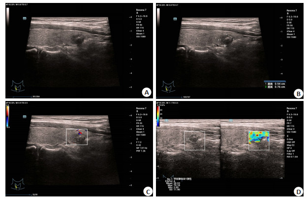
图 1 女,42岁,病理结果为甲状腺微小乳头状癌
A~B:甲状腺右侧叶下极可见一低回声结节,边界欠清,形态规则,纵横径比<1,内回声欠均匀,内可见粗大钙化,大小0.94 cm×0.76 cm;C:甲状腺右侧叶内未见明显血流信号;D:低回声结节呈蓝绿渲染,以蓝色渲染为主,评分3分.
Figure 1. Images of a 42-year-old woman with pathologically confirmed papillary thyroid microcarcinoma

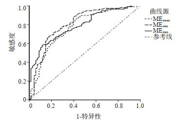
图 2 ME界值诊断甲状腺微小乳头状癌的ROC曲线
Figure 2. ROC curve of me cutoff value in diagnosis of papillary thyroid microcarcinoma
表 1 灰阶超声与病理诊断甲状腺微小乳头状癌性质准确性比较
Table 1. Accuracy comparison of gray scale ultrasound and pathology in the diagnosis of papillary thyroid microcarcinoma (n)
灰阶超声 病理 合计 良性 恶性 良性 35 46 81 恶性 33 71 104 合计 68 117 185  下载: 导出CSV
表 2 多普勒超声技术与病理诊断甲状腺微小乳头状癌性质准确性比较
Table 2. Comparison of the accuracy of Doppler ultrasound and pathology in the diagnosis of papillary thyroid microcarcinoma (n)
多普勒超声 病理 合计 良性 恶性 良性 58 19 77 恶性 10 98 108 合计 68 117 185
下载: 导出CSV
表 3 两组剪切波杨氏模量测定结果
Table 3. Measurement results of Young's modulus of two groups of shear waves (kPa, Mean±SD)
组別 MEmean MEmin MEmax 良性组
(n=68)32.12±20.87 16.68±5.76 51.55±11.43 恶性组
(n=117)51.67±10.56 27.30±8.54 68.58±17.13 t 8.451 9.115 7.303 P < 0.001 < 0.001 < 0.001
下载: 导出CSV
表 4 联合检测结果准确性比较
Table 4. Accuracy comparison of combined detection results(n)
联合 病理 合计 良性 恶性 良性 62 8 70 恶性 6 109 115 合计 68 117 185
下载: 导出CSV
-
[1] 付劲锋, 尹霞, 姜涛. 甲状腺微小乳头状癌术中冰冻病理诊断准确性分析及影响因素[J]. 河北医学, 2019, 25(6): 1041-5. doi: 10.3969/j.issn.1006-6233.2019.06.043 [2] 陈聪, 李灵敏. 甲状腺微小乳头状癌的超声表现及病理对照分析[J]. 影像研究与医学应用, 2018, 2(5): 60-1. doi: 10.3969/j.issn.2096-3807.2018.05.036 [3] Seib CD, Sosa JA. Evolving understanding of the epidemiology of thyroid cancer[J]. Endocrinol Metab Clin N Am, 2019, 48(1): 23-35. doi: 10.1016/j.ecl.2018.10.002 [4] 李婷婷, 李素梅, 许琳琳. 甲状腺癌流行病学分析及超声诊断价值[J]. 医学信息, 2020, 33(5): 85-7. https://www.cnki.com.cn/Article/CJFDTOTAL-YXXX202005025.htm [5] 曲俐. 甲状腺癌超声诊断与病理诊断效果分析[J]. 影像研究与医学应用, 2019, 3(19): 140-1. https://www.cnki.com.cn/Article/CJFDTOTAL-YXYY201919082.htm [6] 杭菁, 袁涛, 叶新华, 等. 常规超声及实时剪切波弹性成像超声表现与甲状腺乳头状癌BRAF基因突变的关系探讨[J]. 临床超声医学杂志, 2019, 21(2): 111-4. doi: 10.3969/j.issn.1008-6978.2019.02.011 [7] 牟泳霖, 曹军英, 里子彧, 等. 实时剪切波弹性成像技术对甲状腺良恶性结节鉴别研究[J]. 临床军医杂志, 2018, 46(9): 1030-2. https://www.cnki.com.cn/Article/CJFDTOTAL-JYGZ201809014.htm [8] 李婷, 林雁潮, 王涵, 等. 二维及彩色多普勒超声诊断甲状腺癌的价值[J]. 实用癌症杂志, 2020, 35(2): 205-7. doi: 10.3969/j.issn.1001-5930.2020.02.009 [9] 谢颖坤, 张巧莲, 郑晓慧, 等. 实时剪切波弹性成像在飞行员甲状腺结节诊断中的应用[J]. 中华航空航天医学杂志, 2018, 29(1): 39-43. doi: 10.3760/cma.j.issn.1007-6239.2018.01.009 [10] 中华人民共和国国家卫生健康委员会. 甲状腺癌诊疗规范(2018年版) [J]. 中华普通外科学文献: 电子版, 2019, 13(1): 1-15. doi: 10.3877/cma.j.issn.1674-0793.2019.01.001 [11] 高明. 甲状腺结节和分化型甲状腺癌诊治指南[J]. 中国肿瘤临床, 2012, 39(17): 1249-72. https://www.cnki.com.cn/Article/CJFDTOTAL-ZGWK201010018.htm [12] 冯莉莉, 杨琛, 孙乔. 2010-2014年上海市浦东新区甲状腺癌发病情况分析[J]. 上海预防医学, 2018, 30(7): 574-8. https://www.cnki.com.cn/Article/CJFDTOTAL-SHYI201807010.htm [13] 王南, 刘辉, 朱成涛, 等. 实时剪切波弹性成像杨氏模量最大值诊断甲状腺乳头状癌BRAF V600E基因突变的价值[J]. 中国医学物理学杂志, 2020, 37(8): 1040-4. doi: 10.3969/j.issn.1005-202X.2020.08.019 [14] 孙丽丽, 李玉宏, 王帅, 等. 多模态超声联合促甲状腺激素对老年甲状腺微小乳头状癌的诊断价值[J]. 中国老年学杂志, 2019, 39(16): 3928-31. doi: 10.3969/j.issn.1005-9202.2019.16.021 [15] 孟彬, 朱文军, 全丽娟, 等. 甲状腺微小乳头状癌超声图像分析[J]. 中国临床医学影像杂志, 2013, 24(1): 53-5. doi: 10.3969/j.issn.1008-1062.2013.01.015 [16] 薛丹, 罗晶, 梁凯迪, 等. 常规超声联合超声造影诊断甲状腺微小乳头状癌研究[J]. 临床军医杂志, 2018, 46(1): 25-7. https://www.cnki.com.cn/Article/CJFDTOTAL-JYGZ201801009.htm [17] 温智峰, 刘晓宇, 张文, 等. 应用高频彩超诊断甲状腺良恶性结节的价值及超声学特点分析[J]. 解放军预防医学杂志, 2019, 37(11): 78-9. https://www.cnki.com.cn/Article/CJFDTOTAL-JYYX201911080.htm [18] Du Y R, Ji C L, Wu Y, et al. Combination of Ultrasound Elastography with TI-RADS in the Diagnosis of Small Thyroid Nodules(≤10 mm): A New Method to Increase the Diagnostic Performance[J]. Eur J Radiol, 2018, 11(3): 107-9. [19] Liu J, Zhang Y, Ji Y, et al. The value of shear wave elastography in diffuse thyroid disease[J]. Clin Imaging, 2018, 8(6): 85-8. [20] Lin, Chen, Yi-Xin, et al. The values of shear wave elastography in avoiding repeat fine-needle aspiration for thyroid nodules with nondiagnostic and undetermined cytology[J]. Clin Endocrinol, 2019, 11(16): 154-6. [21] 韩蕊君, 杜晶, 陈翠, 等. 超声TI-RADS分级联合三维剪切波弹性成像对甲状腺微小癌的诊断效能[J]. 上海交通大学学报: 医学版, 2020, 040(001): 76-80. https://www.cnki.com.cn/Article/CJFDTOTAL-SHEY202001018.htm [22] 贾志莺, 武秀兰, 张银华, 等. 局灶性甲状腺炎与甲状腺乳头状癌常规超声及实时剪切波弹性成像表现[J]. 中国介入影像与治疗学, 2020, 151(11): 29-32. https://www.cnki.com.cn/Article/CJFDTOTAL-JRYX202011008.htm [23] 宋越, 谢明星, 王静, 等. 实时剪切波弹性成像技术鉴别甲状腺良恶性结节的诊断价值及影响因素分析[J]. 中国医师杂志, 2019, 12(19): 215-7. https://www.cnki.com.cn/Article/CJFDTOTAL-ZHCD201411015.htm [24] Ozturk M, Selcuk MB, Polat A V, et al. The diagnostic value of ultrasound and shear wave elastography in the differentiation of benign and malignant soft tissue tumors[J]. Skeletal Radiol, 2020, 8 (3): 320-2. -

 点击查看大图
计量
- 文章访问数: 160
- HTML全文浏览量: 119
- PDF下载量: 2
- 被引次数: 0