


A prospective study of the effects of salbutamol on cardiac structure and function by cardiac magnetic resonance imaging
-
摘要:
目的 通过心脏磁共振成像(CMR)评估沙格列汀对未存在心力衰竭的2型糖尿病患者心脏结构和功能的影响。 方法 本研究为前瞻性研究,2型糖尿病无心力衰竭患者使用沙格列汀作为常规指南指导治疗的一部分,共纳入16例患者。临床评估、CMR成像和生物标志物以盲法方式进行评估,并在6月的持续治疗后进行比较。分析治疗6月后左室射血分数的变化和左室及右室舒张末期容量、心室质量、左室整体应变和心脏生物标志物(N末端B型利钠肽前蛋白和高敏感性C反应蛋白)的变化。 结果 本研究中患者(n=16)的平均年龄为59.9岁,其中69%为男性。平均糖化血红蛋白为(8.3±0.9)%,平均治疗前左室射血分数为(57.4±3.4)%,6月后无显著变化(0.2%,95%CI:-2.5~2.1,P=0.86)。CMR分析显示,包括左心室/右心室容积、左心室质量和特征跟踪衍生的应变变化无统计学意义(P>0.05)。N末端B型利钠肽前蛋白和高敏感性C反应蛋白水平变化无统计学意义(P>0.20)。 结论 根据CMR和生物标志物的评估,在本次稳定的2型糖尿病非心衰患者中,沙格列汀治疗6月后与不良的心室重构无关。 Abstract:Objective To access the effect of salbutamol on cardiac structure and function by cardiac magnetic resonance imaging (CMR) in T2DM patients without heart failure. Methods In this prospective study, patients with T2DM without heart failure were treated with salbutamol as part of routine guideline-guided treatment. Clinical evaluation, CMR imaging and biomarkers were evaluated in a blinded manner and compared after 6 months of continuous treatment. The changes of left ventricular ejection fraction, changes of right ventricular end-diastolic volume, ventricular mass, global strain and cardiac biomarkers (NT-proBNP and hsCRP) were analyzed after 6 months of treatment. Results The average age of patients (n=16) in this study was 59.9 years old, 69% of whom were male. The average HbA1c was (8.3±0.9)%, and the mean pretreatment score was (57.4±3.4)%, with no significant change after 6 months [0.2%, 95% CI: (-2.5, 2.1), P=0.86]. CMR showed no change including left/right ventricular volumes, left ventricular mass and characteristic track-derived strain (P>0.05). Levels of N-terminal pro-B-type natriuretic peptide and high-sensitivity C-reactive protein did not change significantly (P>0.20). Conclusion According to the evaluation of CMR and biomarkers, salbutamol is not associated with adverse ventricular remodeling after 6 months of treatment in this stable type 2 diabetic non-heart failure patient. -
Key words:
- cardiac MRI /
- Salmonidine /
- cardiac structure /
- cardiac biomarkers
-

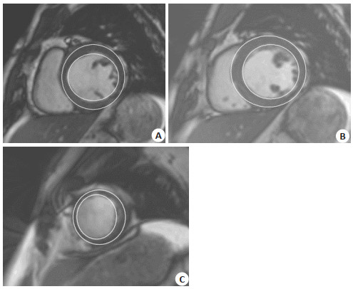
图 1 治疗前心底-心尖层面心内膜和心外膜描边图像
Figure 1. Stroke images of endocardium and epicardium at the heart- apex level before treatment.

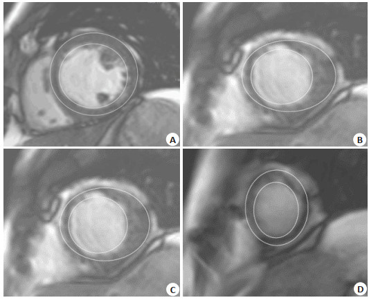
图 2 治疗后心底-心尖层面心内膜和心外膜描边图像
Figure 2. Stroke images of endocardium and epicardium at heart apex level after treatment.
表 1 患者一般资料
Table 1. General data of patients in this study
项目 值 年龄(岁, Mean±SD) 59.9±13.4 女性 5/16 (31%) BMI (kg/m2, Mean±SD) 27.5±3.7 BMI > 30 kg/m2比例 3/16 (19%) 心脏收缩压(mmHg, Mean±SD) 142±16.6 心脏舒张压(mmHg, Mean±SD) 84.6±15.6 心率(次/min, Mean±SD) 77.8±14.7 HbA1C (%, Mean±SD) 8.3±0.9 糖尿病确诊周期(年) 7 (6~10) 复发病变 高血压 9/16 (56%) 血脂异常 13/16 (81%) 冠状动脉疾病 3/16 (19%) 冠状动脉搭桥手术 2/16 (13%) 抽烟史 3/16 (19%) 房颤 1/16 (6%) 心力衰竭史 0/16 (0%) 神经病变 2/16 (13%) 肾病 1/16 (6%) 药物治疗 抗高血压 10/16 (63%) 血管紧张素转化酶抑制剂 5/16 (31%) 血管紧张素 4/16 (25%) β受体阻滞剂 3/16 (19%) 盐皮质激素受体拮抗剂 0/16 (0%) 利尿剂 2/16 (13%) 钙通道阻滞剂 2/16 (13%) 抗高血糖药物 二甲双胍 9/16 (56%) 胰岛素 4/16 (25%) 磺酰脲类 3/16 (19%) SGLT-2抑制剂 1/16 (6%) 噻唑烷二酮 0/16 (0%) 美格列奈 0/16 (0%) α葡萄糖苷抑制剂 0/16 (0%) 阿司匹林 1/16 (6%) 他汀类 16/16 (100%)  下载: 导出CSV
表 2 使用沙格列汀前和使用6月后MRI测量值比较
Table 2. Comparison of MRI measurements before and after six months of use of saxagliptin (Mean±SD)
指标 治疗前 治疗6月后 6月的平均变化(95%CI) P LVEF (%) 57.4±3.4 57.2±4.7 -0.2(-2.5, 2.1) 0.86 LVEDV (mL) 122±25.3 123±24.4 0.7(-8.7, 7.3) 0.25 LVESV (mL) 52±11.6 52±10.6 0.3(-3.6, 3.0) 0.95 LVSV (mL) 70±15.8 71±16.6 0.4(-6.0, 6.8) 0.89 左心室质量(g) 84±19.5 87±21.4 2.3(-2.6, 7.1) 0.34 LVEDV指数(mL/m2) 66±9.8 66±9.5 0.2(-4.1, 4.5) 0.92 LVESV指数(mL/m2) 28±4.8 28±4.2 0.1(-1.6, 1.8) 0.92 左心室质量指数(g/m2) 45±7.3 46±8.2 1.0(-1.3, 3.4) 0.38 RVEF (%) 55.5±4.9 55.3±4.0 -0.2(-3.4, 3.0) 0.91 RVEDV (mL) 123±27.1 125±26.6 1.4(-3.8, 6.6) 0.57 RVESV (mL) 55±14.4 56±12.4 0.5(-3.5, 4.7) 0.78 RVSV (mL) 68±15.9 69±16.3 0.9(-4.9, 6.6) 0.75 右心室质量(g) 20±4.9 20±4.3 -0.3(-1.4, 0.8) 0.59 RVEDV指数(mL/m2) 66±10.5 67±10.8 0.6(-2.4, 3.6) 0.67 RVESV指数(mL/m2) 30±6.1 30±4.9 0.3(-2.0, 2.5) 0.80 右心室质量指数(g/m2) 11±1.9 11±1.7 -0.2(-0.8, 0.5) 0.64 GRS (%) 36.9±8.7 37.3±7.8 0.5(-5.9, 6.8) 0.88 GCS (%) -17.8±3.0 -17.8±2.8 0.1(-2.1, 2.3) 0.93 GLS (%) -13.9±2.3 -13.3±2.7 0.5(-1.4, 2.5) 0.57 左心室射血分数;LVEDV:左室舒张末期体积;LVESV:左室收缩末期体积;RVEF:右心室射血分数;RVEDV:右室舒张末期体积;RVESV:右室收缩末期体积;GLS:全局纵向应变;GRS:全局径向应变;GCS:全局周向应变.
下载: 导出CSV
表 3 使用沙格列汀前和使用6月后血液生物标志物比较
Table 3. Comparison of blood biomarkers before and after 6 months of use of saxagliptin
指标 治疗前(Mean±SD) 治疗6月后(Mean±SD) 6月的平均变化(95%CI) P 肌酸酐(µmol/L) 76±21 77±20 1(- 4, 4) 0.79 NT-proBNP (ng/L) 88±183 49±40 - 39(- 142, 102) 0.42 hsCRP (mg/L) 3.9±4.3 3.1±2.9 - 0.8(- 2.2, 1.4) 0.22 HbA1c (%) 8.3±0.9 7.7±0.9 - 0.6(- 1.2, 0.6) 0.06 收缩压(mmHg) 142±16.6 130±13.3 - 11.9(- 24.2, 12.4) 0.06 舒张压(mmHg) 83±15.6 77±10.4 - 5.5(- 13.6, 8.2) 0.17 糖化血红蛋白;NT-proBNP:N末端B型利钠肽前蛋白;hsCRP:高敏感性C反应蛋白.
下载: 导出CSV
表 4 治疗前患者心脏MRI结构和功能的Spearman相关系数
Table 4. Spearman correlation coefficient of cardiac MRI structure and function before treatment
指标 LVEDV指数 LVESV指数 左心室质量指数 RVEF RVEDV指数 RVESV指数 右心室质量指数 GRS GCS GLS LVEF -0.08(P=0.76) -0.53(P=0.04) -0.03(P=0.91) 0.74(P=0.001) -0.16(P=0.56) -0.45(P=0.08) -0.36(P=0.18) 0.40(P=0.12) -0.47(P=0.06) -0.69(P=0.003) LVEDV指数 - 0.84(P < 0.001) 0.45(P=0.08) 0.24(P=0.38) 0.76(P=0.001) 0.36(P=0.17) 0.61(P=0.01) -0.24(P=0.36) 0.17(P=0.54) -0.07(P=0.80) LVESV指数 - - 0.36(P=0.17) -0.07(P=0.80) 0.66(P=0.006) 0.48(P=0.06) 0.70(P=0.003) - 0.43(P=0.10) 0.35(P=0.18) 0.26(P=0.34) 左心室质量指数 - - - 0.04(P=0.88) 0.47(P=0.07) 0.28(P=0.30) 0.65(P=0.007) 0.37(P=0.15) -0.30(P=0.25) 0.37(P=0.16) RVEF - - - - -0.16(P=0.56) -0.64(P=0.007) -0.24(P=0.36) 0.21(P=0.44) -0.32(P=0.23) -0.64(P=0.007) GRS - - - - - - - - -0.89(P < 0.001) -0.07(P=0.80) GCS - - - - - - - - - 0.17(P=0.52)
下载: 导出CSV
-
[1] 伍希, 唐露, 邓巧, 等. MRI纹理分析预测心肌淀粉样变有无延迟强化的价值初探[J]. 磁共振成像, 2021, 12(12): 6-11. doi: 10.12015/issn.1674-8034.2021.12.002 [2] 赵培君, 李浩杰, 夏黎明. 提高认识和加强心脏MR定量技术的临床应用与研究[J]. 中华放射学杂志, 2021, 55(12): 1235-40. doi: 10.3760/cma.j.cn112149-20210128-00079 [3] Petersen SE, Selvanayagam JB, Wiesmann F, et al. Left ventricular non-compaction: insights from cardiovascular magnetic resonance imaging[J]. J Am Coll Cardiol, 2005, 46(1): 101-5. doi: 10.1016/j.jacc.2005.03.045 [4] Fink EL, Wisnowski J, Clark R, et al. Brain MR imaging and spectroscopy for outcome prognostication after pediatric cardiac arrest[J]. Resuscitation, 2020, 157: 185-94. doi: 10.1016/j.resuscitation.2020.06.033 [5] 兰春伟, 刘涛, 尉娜, 等. 心脏MRI对神经系统疾病患者右向左分流的诊断价值[J]. 山东医药, 2021, 61(34): 76-9. doi: 10.3969/j.issn.1002-266X.2021.34.019 [6] Mason S, Osborn JS, Dhar R, et al. Real world MRI experience with nonconditional and conditional cardiac rhythm devices after MagnaSafe[J]. J Cardiovasc Electrophysiol, 2017, 28(12): 1468-74. doi: 10.1111/jce.13351 [7] 熊俊峰, 朱希松, 江朝根, 等. 心脏磁共振T2-mapping定量技术在急性心肌炎诊断中的应用价值[J]. 心电与循环, 2021, 40(6): 590-3. https://www.cnki.com.cn/Article/CJFDTOTAL-XDXZ202106009.htm [8] 董亚玲. 心脏MRI及超声心动图双模态对心尖室壁瘤切除术治疗后心功能评估价值[J]. 中国CT和MRI杂志, 2021, 19(12): 71-4. https://www.cnki.com.cn/Article/CJFDTOTAL-CTMR202112023.htm [9] Lupo P, Cappato R, Di Leo G, et al. An eight-year prospective controlled study about the safety and diagnostic value of cardiac and non-cardiac 1.5-T MRI in patients with a conventional pacemaker or a conventional implantable cardioverter defibrillator [J]. Eur Radiol, 2018, 28(6): 2406-16. doi: 10.1007/s00330-017-5098-z [10] 毋晓萌, 刘帅, 霍力, 等. 基于无监督深度学习的心脏PET/CT和MRI图像配准[J]. 中国医学影像学杂志, 2021, 29(11): 1158-64. doi: 10.3969/j.issn.1005-5185.2021.11.023 [11] 伍希, 唐玲玲, 胡云涛, 等. MRI影像组学在心脏疾病中的研究进展[J]. 磁共振成像, 2021, 12(11): 113-6. https://www.cnki.com.cn/Article/CJFDTOTAL-CGZC202111028.htm [12] Sandhu P, Ong JP, Garg V, et al. The effects of saxagliptin on cardiac structure and function using cardiac MRI (SCARF)[J]. Acta Diabetol, 2021, 58(5): 633-41. doi: 10.1007/s00592-020-01661-y [13] 颜龙, 胡春峰, 李强, 等. 冠状动脉磁共振血管成像和CT对可疑冠心病患者心脏事件的预测价值的比较研究[J]. 现代生物医学进展, 2021, 21(21): 4123-6, 4131. https://www.cnki.com.cn/Article/CJFDTOTAL-SWCX202121026.htm [14] 黄倩. 缺血性心肌病采用心脏磁共振检测的特征性表现及诊断价值分析[J]. 影像研究与医学应用, 2021, 5(21): 197-8. doi: 10.3969/j.issn.2096-3807.2021.21.095 [15] Cassidy S, Thoma C, Hallsworth K, et al. High intensity intermittent exercise improves cardiac structure and function and reduces liver fat in patients with type 2 diabetes: a randomised controlled trial[J]. Diabetologia, 2016, 59(1): 56-66. doi: 10.1007/s00125-015-3741-2 [16] 余鎏, 夏睿, 陶黎, 等. 扩张型心肌病患者心肌应变与左心室球形指数相关性的初步研究[J]. 中国医疗设备, 2021, 36(10): 113-6. https://www.cnki.com.cn/Article/CJFDTOTAL-YLSX202110027.htm [17] 孙学彪, 刘敏. 心脏MR评估肺动脉高压右心室结构及预后的研究进展[J]. 中华放射学杂志, 2021, 55(9): 993-6. doi: 10.3760/cma.j.cn112149-20201208-01292 [18] Li JN, He Y, Dong W, et al. Comparison of cardiac MRI with PET for assessment of myocardial viability in patients with coronary chronic total occlusion[J]. Clin Radiol, 2019, 74(5): 410. e1-410. e9. [19] 孙恒, 胡宇才, 张岚, 等. 心脏磁共振在评估急性心肌梗死后心室重构中作用的研究进展[J]. 中华实用诊断与治疗杂志, 2021, 35(9): 964-6. https://www.cnki.com.cn/Article/CJFDTOTAL-HNZD202109025.htm [20] 姜博文, 韩珂. 心肌保护策略的研究进展[J]. 分子影像学杂志, 2018, 41(2): 196-200. doi: 10.3969/j.issn.1674-4500.2018.02.15 [21] Ching CK, Chakraborty RN, Kler TS, et al. Clinical safety and performance of a MRI conditional pacing system in patients undergoing cardiac MRI[J]. Pacing Clin Electrophysiol, 2017, 40 (12): 1389-95. [22] Cowan BR, Young AA, Anderson C, et al. The cardiac MRI substudy to ongoing telmisartan alone and in combination with ramipril global endpoint trial/telmisartan randomized assessment study in ACE-intolerant subjects with cardiovascular disease: analysis protocol and baseline characteristics[J]. Clin Res Cardiol, 2009, 98(7): 421-33. [23] Nazarian S, Cantillon DJ, Woodard PK, et al. MRI safety for patients implanted with the MRI ready ICD system: MRI ready study results[J]. JACC Clin Electrophysiol, 2019, 5(8): 935-43. [24] Kapadia SR, Kodali S, Makkar R, et al. Protection against cerebral embolism during transcatheter aortic valve replacement[J]. J Am Coll Cardiol, 2017, 69(4): 367-77. -

 点击查看大图
计量
- 文章访问数: 217
- HTML全文浏览量: 228
- PDF下载量: 6
- 被引次数: 0