


The Diagnostic Value of MRI in Combination with clinical FIGO staging system in stages of cervical cancer
-
摘要:
目的 分析MRI联合临床国际妇产科协会(FIGO)分期标准在宫颈癌不同分期中的诊断价值。 方法 选取2014年1月~2020年12月于我院就诊的85例宫颈癌患者,回顾性分析患者临床资料和影像学资料等,与病理结果进行对照,分别使用临床FIGO分期标准和MRI联合临床FIGO分期标准对患者进行诊断分析,并对FIGO分期和MRI联合临床FIGO分期结果进行敏感度、特异性及一致性检验分析,比较MRI联合临床FIGO分期与临床FIGO分期在诊断宫颈癌不同分期上的准确性和一致性。 结果 FIGO分期检查Ⅰa期准确率为92.94%,Ⅰb期准确率为90.59%,Ⅱa期准确率为85.88%,Ⅱb期准确率为82.35%;Ⅲb准确率为97.65%;Ⅳ期准确率为96.47%,整体准确率为72.94%,FIGO分期检查诊断Ⅰa和Ⅰb期宫颈癌的一致性较好(Kappa > 0.60),FIGO分期检查诊断Ⅱa、Ⅱb、Ⅲb以及Ⅳ期宫颈癌的一致性较差(Kappa < 0.60);MRI联合临床FIGO分期诊断Ⅰa期准确率为92.94%,Ⅰb期准确率为90.59%,Ⅱa期准确率为94.12%,Ⅱb期准确率为96.47 %,Ⅲb期准确率为100.00%,Ⅳ期准确率为100.00%,整体准确率为88.24%,MRI联合临床FIGO分期诊断Ⅰa、Ⅰb、Ⅱa、Ⅱb、Ⅲb以及Ⅳ期宫颈癌的一致性较好(Kappa > 0.60);MRI联合临床FIGO分期在Ⅱa、Ⅱb以及整体上的准确性显著高于临床FIGO分期(P < 0.05)。 结论 MRI联合临床FIGO分期可以有效的对宫颈癌进行分期,其准确性高于单独使用临床FIGO分期。 Abstract:Objective To analyze the diagnostic value of MRI combined with Federation International of Gynecology and Obstetrics (FIGO) staging system in different stages of cervical cancer. Methods A total of 85 patients with cervical cancer who attended our hospital from January 2014 to December 2020 were selected, and the patients' clinical data and imaging data, etc. were retrospectively analyzed and compared with the pathological findings. The FIGO staging system and MRI combined with FIGO staging system were used for diagnosis. The sensitivity, specificity and consistencies tests were performed to compare, the accuracy rates and consistencies of MRI combined with clinical FIGO staging and clinical FIGO staging alone in diagnosing different stages of cervical cancer. Results The accuracy of FIGO staging was 92.94% for stage Ⅰa, 90.59% for stage Ⅰb, 85.88% for stage Ⅱa, 82.35% for stage Ⅱb, 97.65% for stage Ⅲb and 96.47% for stage Ⅳ. The overall accuracy rate was 72.94%. The consistency of FIGO staging was good in the diagnosis of cervical cancer at stages Ⅰa and Ⅰb (Kappa > 0.60), while the consistency was poor in the diagnosis of stages Ⅱa, Ⅱb and Ⅲb (Kappa < 0.60). The accuracy rate of MRI combined with clinical FIGO staging tests was 92.94% for stage Ⅰa, 90.59% for stage Ⅰb, 94.12% for stage Ⅱa, 96.47 % for stage Ⅱb, 100.00% for stage Ⅲb, 100.00% for stage Ⅳ and 88.24% for overall. The consistency of MRI combined with clinical FIGO staging in the diagnosis of cervical cancer at stages Ⅰa, Ⅰb, Ⅱa, Ⅱb, Ⅲb and Ⅳ was good (Kappa > 0.60). The accuracy rates of MRI combined with clinical FIGO staging were significantly higher than those of clinical FIGO staging alone in the diagnosis of stages Ⅱa, Ⅱb and overall (P < 0.05). Conclusion MRI combined with clinical FIGO staging can effectively staging cervical cancer, and its accuracy is higher than that of clinical FIGO staging alone. -
Key words:
- cervical cancer /
- magnetic resonance imaging /
- clinical staging
-

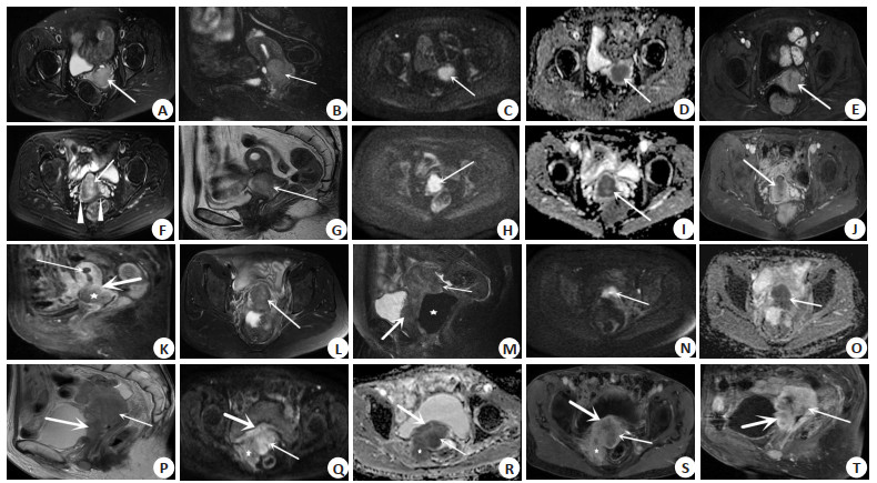
图 1 影像资料
A~E: FIGO IB期宫颈癌(A: 横轴位T2WI FSE FS; B: 矢状位T2WI FSE FS; C: DWI; D: ADC; E: 增强扫描横轴位);F~K: FIGO ⅡB期宫颈癌(F: 横轴位T2WI FSE FS; G: 矢状位T2WI FSE; H: DWI; I: ADC; J: 增强扫描横轴位; K: 增强扫描矢状位);L~O: FIGO ⅢB期宫颈癌(L: 横轴位T2WI FSE FS; M: 矢状位T2WI FSE FS; N: DWI; O: ADC; P~T: FIGO Ⅳ期宫颈癌(P: 矢状位T2WI FSE; Q: DWI; R: ADC; S: 增强扫描横轴位; T: 增强扫描矢状位).
Figure 1. Image data.
表 1 FIGO分期标准与手术病理比较
Table 1. Comparison of the diagnostic results of FIGO staging system and surgical pathological results (n)
FIGO分期 手术病理 Ⅰa Ⅰb Ⅱa Ⅱb Ⅲb Ⅳ 合计 Ⅰa 11 4 0 0 0 0 15 Ⅰb 2 34 0 0 0 0 36 Ⅱa 0 2 4 7 0 0 13 Ⅱb 0 0 3 13 2 3 21 Ⅲb 0 0 0 0 0 0 0 Ⅳ 0 0 0 0 0 0 0 合计 13 40 7 20 2 3 85  下载: 导出CSV
表 2 FIGO分期标准诊断价值分析
Table 2. Diagnostic value of FIGO staging system
FIGO分期 敏感度(%) 特异性(%) 准确率(%) 阳性预测值(%) 阴性预测值(%) Kappa值 Ⅰa 84.62 94.44 92.94 73.33 97.14 0.74 Ⅰb 85.00 95.56 90.59 94.44 87.76 0.81 Ⅱa 57.14 88.46 85.88 30.77 95.83 0.33 Ⅱb 65.00 87.69 82.35 61.9 89.06 0.52 Ⅲb 0.00 100.00 97.65 0.00 97.65 0.00 Ⅳ 0.00 100.00 96.47 0.00 96.47 0.00
下载: 导出CSV
表 3 MRI联合临床FIGO分期标准与手术病理比较
Table 3. Comparison of the results of MRI combined with clinical FIGO staging system and surgical pathological results (n)
MRI分期 手术病理 Ⅰa Ⅰb Ⅱa Ⅱb Ⅲb Ⅳ 合计 Ⅰa 9 2 0 0 0 0 11 Ⅰb 4 36 0 0 0 0 40 Ⅱa 0 2 6 1 0 0 10 Ⅱb 0 0 1 19 0 0 19 Ⅲb 0 0 0 0 2 0 2 Ⅳ 0 0 0 0 0 3 3 合计 13 40 7 20 2 3 85
下载: 导出CSV
表 4 MRI联合临床FIGO分期标准诊断价值分析
Table 4. Diagnostic value of MRI combined with clinical FIGO staging system
MRI分期 敏感度(%) 特异性(%) 准确率(%) 阳性预测值(%) 阴性预测值(%) Kappa值 Ⅰa 69.23 97.22 92.94 81.82 94.59 0.71 Ⅰb 90.00 91.11 90.59 90.00 91.11 0.81 Ⅱa 85.71 96.15 95.29 66.67 98.68 0.72 Ⅱb 95.00 98.46 97.65 95.00 98.46 0.93 Ⅲb 100.00 100.00 100.00 100.00 100.00 1.00 Ⅳ 100.00 100.00 100.00 100.00 100.00 1.00
下载: 导出CSV
表 5 两种分期方式准确性比较
Table 5. Comparison of the accuracy rates of the two staging methods
分期 FIGO分期(%) MRI联合临床FIGO分期(%) χ2 P Ⅰa 92.94 92.94 0.000 1.000 Ⅰb 90.59 90.59 0.000 1.000 Ⅱa 85.88 95.29 4.337 0.037 Ⅱb 82.35 97.65 10.835 0.001 Ⅲb 97.65 100.00 2.02 0.155 Ⅳ 96.47 100.00 3.044 0.081 整体 72.94 88.24 6.355 0.012 FIGO: 国际妇产科协会.
下载: 导出CSV
-
[1] Lin J, Chen LT, Qiu XM, et al. Traditional Chinese medicine for human papillomavirus (HPV) infections: a systematic review[J]. Biosci Trends, 2017, 11(3): 267-73. doi: 10.5582/bst.2017.01056 [2] Zou ZR, Fairley CK, Ong JJ, et al. Domestic HPV vaccine price and economic returns for cervical cancer prevention in China: a cost-effectiveness analysis[J]. Lancet Glob Health, 2020, 8(10): e1335-e1344. doi: 10.1016/S2214-109X(20)30277-1 [3] 王建东, 孔为民, 姜昊. 国际妇产科联盟2018年宫颈癌分期及有关问题[J]. 中华肿瘤杂志, 2020, 42(2): 94-8. doi: 10.3760/cma.j.issn.0253-3766.2020.02.002 [4] 张伟峰, 李朋飞, 陈春林, 等. 2004—2016年我国宫颈癌住院患者FIGO临床分期应用情况调查[J]. 中国实用妇科与产科杂志, 2018, 34 (1): 67-71. https://www.cnki.com.cn/Article/CJFDTOTAL-ZGSF201801020.htm [5] 李珂, 宋园园, 倪莉. MR I对宫颈癌分期的准确性研究[J]. 实用放射学杂志, 2019, 35(10): 1623-6. doi: 10.3969/j.issn.1002-1671.2019.10.017 [6] 王飞, 刘宗谕, 陈军, 等. 磁共振在宫颈癌诊断中的应用[J]. 中华医学杂志, 2020, 100(14): 1081-3. doi: 10.3760/cma.j.cn112137-20191203-02633 [7] 彭传勇, 徐启兰, 周瑾, 等. 多参数MRI在宫颈癌早期诊断及临床分期中的应用价值[J]. 实用放射学杂志, 2021, 37(5): 826-9. doi: 10.3969/j.issn.1002-1671.2021.05.031 [8] 马勇, 杨子权, 李瑞梅, 等. 高清MRI联合DWI在宫颈癌分期诊断中的应用观察[J]. 中国妇幼保健, 2019, 34(21): 5056-8. https://www.cnki.com.cn/Article/CJFDTOTAL-ZFYB201921079.htm [9] Hu Z, Ma D. The precision prevention and therapy of HPV-related cervical cancer: new concepts and clinical implications[J]. Cancer Med, 2018, 7(10): 5217-36. doi: 10.1002/cam4.1501 [10] Pecorelli S. Revised FIGO staging for carcinoma of the vulva, cervix, and endometrium[J]. Int J Gynaecol Obstet, 2009, 105(2): 103-4. doi: 10.1016/j.ijgo.2009.02.012 [11] 马丽华, 娄阁. MRI指导子宫颈癌分期的临床意义[J]. 中国妇幼保健, 2009, 24(28): 4021-3. https://www.cnki.com.cn/Article/CJFDTOTAL-ZFYB200928053.htm [12] Merz J, Bossart M, Bamberg F, et al. Revised FIGO Staging for Cervical Cancer-A New Role for MRI. Staging des Zervixkarzinoms-die neue Rolle der MRT-Bildgebung[J]. Rofo, 2020, 192(10): 937-44. doi: 10.1055/a-1198-5729 [13] Salvo G, Odetto D, Pareja R, et al. Revised 2018 International Federation of Gynecology and Obstetrics (FIGO) cervical cancer staging: a review of gaps and questions that remain[J]. Int J Gynecol Cancer, 2020, 30(6): 873-8. doi: 10.1136/ijgc-2020-001257 [14] 宋凯林, 边芳, 刘玉玲, 等. 多模态磁共振成像在评估IB期宫颈癌大小及分期中的应用价值[J]. 暨南大学学报: 自然科学与医学版, 2021, 42(2): 203-9. https://www.cnki.com.cn/Article/CJFDTOTAL-JNDX202102012.htm [15] Balcacer P, Shergill A, Litkouhi B. MRI of cervical cancer with a surgical perspective: staging, prognostic implications and pitfalls[J]. Abdom Radiol (NY), 2019, 44(7): 2557-71. doi: 10.1007/s00261-019-01984-7 [16] Matsuo K, Machida H, Mandelbaum RS, et al. Validation of the 2018 FIGO cervical cancer staging system[J]. Gynecol Oncol, 2019, 152 (1): 87-93. doi: 10.1016/j.ygyno.2018.10.026 [17] Soneji ND, Bharwani N, Ferri A, et al. Pre-operative MRI staging of endometrial cancer in a multicentre cancer network: can we match single centre study results?[J]. Eur Radiol, 2018, 28(11): 4725-34. doi: 10.1007/s00330-018-5465-4 [18] 彭澎, 向阳. 体现实践成果, 引领未来方向: 国际妇产科联盟2018年宫颈癌分期解读[J]. 协和医学杂志, 2020, 11(1): 12-5. https://www.cnki.com.cn/Article/CJFDTOTAL-XHYX202001004.htm [19] Saleh M, Virarkar M, Javadi S, et al. Cervical cancer: 2018 revised international federation of gynecology and obstetrics staging system and the role of imaging[J]. AJR Am J Roentgenol, 2020, 214(5): 1182-95. doi: 10.2214/AJR.19.21819 [20] Fournier LS, Bats AS, Durdux C. Diffusion MRI: Technical principles and application to uterine cervical cancer[J]. Cancer/Radiothérapie, 2020, 24(5): 368-73. doi: 10.1016/j.canrad.2020.02.008 [21] Devine C, Viswanathan C, Faria S, et al. Imaging and staging of cervical cancer[J]. Semin Ultrasound CT MRI, 2019, 40(4): 280-6. doi: 10.1053/j.sult.2019.03.001 [22] Luo L, Luo Q, Tang L. Diagnostic value and clinical significance of MRI and CT in detecting lymph node metastasis of early cervical cancer[J]. Oncol Lett, 2020, 19(1): 700-6. [23] 邱书珺, 陆晓兰, 蒋小平, 等. MSCT和MRI对浸润性宫颈癌术前分期的价值对比[J]. 放射学实践, 2012, 27(1): 77-80. https://www.cnki.com.cn/Article/CJFDTOTAL-FSXS201201027.htm [24] 秦凤英, 马锦涛, 赵明丽, 等. 多参数MRI联合临床分期预测ⅢCr期宫颈癌同步放化疗后复发[J]. 放射学实践, 2021, 36(6): 767-72. https://www.cnki.com.cn/Article/CJFDTOTAL-FSXS202106022.htm [25] Yu L, Zhang HF, Jiang DW, et al. Comparison of imaging features and diagnostic values of MRI, CT and contrast-enhanced ultrasonography in the diagnosis of cervical carcinoma staging[J]. Eur Rev Med Pharmacol Sci, 2018, 22(15): 4784-91. [26] Moro F, Gui B, Arciuolo D, et al. Fusion imaging of ultrasound and MRI in the assessment of locally advanced cervical cancer: a prospective study[J]. Int J Gynecol Cancer, 2020, 30(4): 456-65. doi: 10.1136/ijgc-2019-000902 [27] 谢宗源, 李伟兰, 谭志斌, 等. 术前动态对比增强磁共振成像、磁共振扩散加权成像在宫颈癌病理分期评估中的应用[J]. 山东医药, 2019, 59(9): 71-3. https://www.cnki.com.cn/Article/CJFDTOTAL-SDYY201909023.htm -

 点击查看大图
计量
- 文章访问数: 209
- HTML全文浏览量: 117
- PDF下载量: 8
- 被引次数: 0