


Application of dual-phase CT enhancement combined with three-dimensional reconstruction technique in the qualitative diagnosis of thyroid tumors
-
摘要:
目的 观察双期CT增强联合三维重建技术在甲状腺肿瘤定性诊断中的应用价值。 方法 回顾性分析2017年1月~2021年5月于我院收治的98例甲状腺肿瘤患者临床资料,患者均行双期CT增强联合三维重建技术检查,以手术病理检查作为金标准。探究双期CT增强、双期CT增强联合三维重建技术对甲状腺肿瘤的诊断价值,分析双期CT增强联合三维重建技术对甲状腺肿瘤病理特征的诊断价值。 结果 双期CT增强对甲状腺肿瘤的诊断敏感度为84.91%,特异性为82.22%,准确率为83.67%;双期CT增强联合三维重建技术对甲状腺肿瘤的诊断敏感度为92.45%,特异性为80.00%,准确率为86.73%。双期CT增强联合三维重建技术对患者单侧或双侧淋巴结肿大、粗钙化、微钙化、病灶数目的诊断准确率为94.23%、72.73%、89.74%、87.12%。双期CT增强联合三维重建技术检查显示,甲状腺癌患者的单侧或双侧淋巴结肿大占检出总数的71.43%,甲状腺腺瘤的单侧或双侧淋巴结肿大占检出总数的28.57%;甲状腺癌患者的70个病灶中钙化占比为52.86%,甲状腺腺瘤患者的45个病灶中钙化占比为31.11%。 结论 双期CT增强联合三维重建技术在甲状腺肿瘤定性诊断中的应用价值较高,颈部淋巴结肿大、钙化具备辅助诊断的价值。 Abstract:Objective To observe the application value of dual-phase CT enhancement combined with three-dimensional reconstruction in the qualitative diagnosis of thyroid tumors. Methods The clinical data of 98 patients with thyroid tumors admitted to the hospital from January 2017 and May 2021 were retrospectively analyzed. All patients underwent dual-phase CT enhancement combined with three-dimensional reconstruction, and surgical pathological examination was used as the gold standard. The diagnostic value of dual-phase CT enhancement and dual-phase CT enhancement combined with threedimensional reconstruction for thyroid tumors was explored, and the diagnostic value of dual-phase CT enhancement combined with three-dimensional reconstruction for pathological features of thyroid tumors was analyzed. Results The diagnostic sensitivity of dual-phase CT enhancement for thyroid tumors was 84.91%, the specificity was 82.22% and the accuracy rate was 83.67%, the diagnostic sensitivity of duplex CT enhancement combined with 3D reconstruction technique for thyroid tumors was 92.45%, the specificity was 80.00% and the accuracy was 86.73%. The diagnostic accuracy rates of dualphase CT enhancement combined with three-dimensional reconstruction for unilateral or bilateral lymph node enlargement, coarse calcification, microcalcification and the number of lesions were 94.23%, 72.73%, 89.74% and 87.12% respectively. Dualphase CT enhancement combined with three-dimensional reconstruction showed that the unilateral or bilateral lymph node enlargement in patients with thyroid cancer accounted for 71.43% of the total detection, and the unilateral or bilateral lymph node enlargement in thyroid adenoma accounted for 28.57% of the total detection, and calcification accounted for 52.86% of the 70 lesions in patients with thyroid cancer and 31.11% of the 45 lesions in patients with thyroid adenoma. Conclusion Dualphase CT enhancement combined with three-dimensional reconstruction are of high value in the qualitative diagnosis of thyroid tumors, and cervical lymph node enlargement and calcification are of value as an aid to diagnosis. -
Key words:
- CT enhancement /
- three-dimensional reconstruction /
- thyroid tumors /
- qualitative diagnosis /
- diagnosis
-

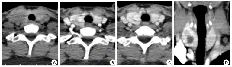
图 1 患者女,30岁,确诊为甲状腺乳头状癌(T1bN1aM0期)
A: CT平扫,使用512×512高分辨率平扫, 可见病灶微小钙化; B: CT平扫图; C~D: CT双期扫描图, 显示病灶呈渐进性强化, 包膜不完整呈“咬饼样”改变; E: 放大后的CT平扫图, 气管前(Ⅵ区)可见转移性淋巴结; F~G: CT双期扫描图,动脉期扫描可见转移性淋巴结与周围组织对比欠佳, 而静脉期明显强化.
Figure 1. A30-year-old female patient, diagnosed with papillary thyroid carcinoma (T1bN1aM0 stage).

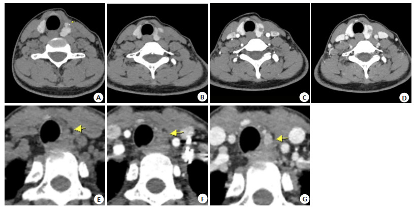
图 2 患者女,27岁,右侧滤泡性腺瘤
A: CT平扫,甲状腺右侧叶密度减低,病变边界不清; B~C: CT双期扫描图病变呈渐进性明显强化, 内见囊变; D: 三维重建图直观显示病变全貌且病变包膜完整.
Figure 2. A27-year-old female patient with a right follicular adenoma.
表 1 双期CT增强、三维重建技术及其联合诊断与病理诊断甲状腺肿瘤的比较
Table 1. Comparison of dual-phase CT enhancement, three-dimensional reconstruction and their combined diagnosis with pathological diagnosis of thyroid tumors (n)
检查方法 手术病理结果 合计 甲状腺癌 甲状腺腺瘤 双期CT增强 甲状腺癌 45 8 53 甲状腺腺瘤 8 37 45 合计 53 45 98 双期CT增强联合三维重建技术 甲状腺癌 49 9 58 甲状腺腺瘤 4 36 40 合计 53 45 98  下载: 导出CSV
表 2 CT增强、三维重建技术及其联合诊断的敏感度、特异性及准确率比较
Table 2. Comparison of sensitivity, specificity and accuracy rate of CT enhancement, three-dimensional reconstruction and their combined diagnosis
检查方法 敏感度 特异性 准确率 双期CT增强 45/53(84.91%) 37/45(82.22%) 82/98(83.67%) 双期CT增强联合三维重建技术 49/53(92.45%) 36/45(80.00%) 85/98(86.73%) χ2 1.504 0.073 0.364 P 0.220 0.788 0.546
下载: 导出CSV
表 3 双期CT增强联合三维重建技术对病理特征的诊断效能
Table 3. Diagnostic efficiency of dual-phase CT enhancement combined with three-dimensional reconstruction for pathological characteristics (n)
病理特征 双期CT增强联合三维重建技术(甲状腺癌/甲状腺腺瘤) 手术病理结果(甲状腺癌/甲状腺腺瘤) 准确率(%) 单侧或双侧淋巴结肿大 49(35/14) 52(37/15) 94.23 钙化 51(37/14) 61(42/19) 83.61 粗钙化 16(4/12) 22(6/16) 72.73 微钙化 35(33/2) 39(36/3) 89.74 病灶数目 115(70/45) 132(83/49) 87.12
下载: 导出CSV
-
[1] Pool C, Walter V, Bann D, et al. Molecular characterization of tumors meeting diagnostic criteria for the non-invasive follicular thyroid neoplasm with papillary-like nuclear features (NIFTP)[J]. Virchows Arch, 2019, 474(3): 341-51. doi: 10.1007/s00428-018-02512-6 [2] Cameselle-Teijeiro JM, Eloy C, Sobrinho-Simões M. Pitfalls in challenging thyroid tumors: emphasis on differential diagnosis and ancillary biomarkers[J]. Endocr Pathol, 2020, 31(3): 197-217. doi: 10.1007/s12022-020-09638-x [3] Tang PZ, Ren CY, Shen LJ, et al. Development and validation of a diagnostic nomogram for the preoperative differentiation between follicular thyroid carcinoma and follicular thyroid adenomas[J]. J ComputAssist Tomogr, 2021, 45(1): 128-34. doi: 10.1097/RCT.0000000000001078 [4] 许立龙, 李世岩, 朱江, 等. 高频超声联合超声引导下粗针穿刺活检在诊断原发性甲状腺淋巴瘤中的应用价值[J]. 中华耳鼻咽喉头颈外科杂志, 2021, 56(8): 858-62. [5] Leimbach RD, Hoang TD, Shakir MKM. Diagnostic challenges of medullary thyroid carcinoma[J]. Oncology, 2021, 99(7): 422-32. doi: 10.1159/000515373 [6] Caresio C, Caballo M, Deandrea M, et al. Quantitative analysis of thyroid tumors vascularity: a comparison between 3-D contrast-enhanced ultrasound and 3-D Power Doppler on benign and malignant thyroid nodules[J]. Med Phys, 2018, 45(7): 3173-84. doi: 10.1002/mp.12971 [7] 许梦苗, 郭小燕, 王鼎, 等. CT平扫图像三维纹理分析对甲状腺良恶性结节的鉴别诊断价值[J]. 现代肿瘤医学, 2020, 28(18): 3226-30. doi: 10.3969/j.issn.1672-4992.2020.18.029 [8] 何慕真, 马明平, 林阳, 等. 双能量CT成像在诊断甲状腺乳头状癌颈部淋巴结转移中的临床应用价值[J]. 中国癌症杂志, 2018, 28(7): 497-504. https://www.cnki.com.cn/Article/CJFDTOTAL-ZGAZ201807005.htm [9] Ramírez-Moya J, Baker AR, Slack FJ, et al. ADAR1-mediated RNA editing is a novel oncogenic process in thyroid cancer and regulates miR-200 activity[J]. Oncogene, 2020, 39(18): 3738-53. doi: 10.1038/s41388-020-1248-x [10] Wang Q, Shen YL, Ye B, et al. Gene expression differences between thyroid carcinoma, thyroid adenoma and normal thyroid tissue[J]. Oncol Rep, 2018, 40(6): 3359-69. [11] Qiu ZL, Shen CT, Sun ZK, et al. Distant metastases from pathologically proven benign follicular nodules of the thyroid: clinicopathological features and predictors of long-term survival[J]. Endocrine, 2020, 69(1): 113-25. doi: 10.1007/s12020-020-02237-0 [12] Jayakody S, Reagh J, Bullock M, et al. Medullary thyroid carcinoma: survival analysis and evaluation of mutation-specific immuno-histochemistry in detection of sporadic disease[J]. World J Surg, 2018, 42(5): 1432-9. doi: 10.1007/s00268-018-4551-8 [13] Prete A, Lo AS, Sadow PM, et al. Pericytes elicit resistance to vemurafenib and sorafenib therapy in thyroid carcinoma via the TSP-1/TGFβ1 axis[J]. Clin Cancer Res, 2018, 24(23): 6078-97. doi: 10.1158/1078-0432.CCR-18-0693 [14] 魏培英, 蒋念东, 韩志江, 等. CT强化程度对甲状腺乳头状癌颈部淋巴结转移的诊断价值[J]. 中华内分泌外科杂志, 2020, 14(2): 144-8. [15] 袁辉, 段永利, 陈孝柏, 等. MSCT双期增强扫描对甲状旁腺腺瘤的诊断价值[J]. CT理论与应用研究, 2019, 28(3): 377-84. https://www.cnki.com.cn/Article/CJFDTOTAL-CTLL201903014.htm [16] Cai HF, Wang R, Li Y, et al. Role of 3D reconstruction in the evaluation of patients with lower segment oesophageal cancer[J]. J Thorac Dis, 2018, 10(7): 3940-7. doi: 10.21037/jtd.2018.06.119 [17] 刘天, 黄品同, 张超. 甲状腺结节内粗大钙化位置与甲状腺癌的关系[J]. 中国超声医学杂志, 2021, 37(7): 721-4. doi: 10.3969/j.issn.1002-0101.2021.07.001 [18] 周健, 赖旭峰, 韩志江, 等. 多种CT征象对甲状腺良、恶性结节的预测价值[J]. 中华全科医师杂志, 2018, 17(1): 44-9. [19] 唐世龙, 刘先凡, 程卓, 等. 迭代重组算法iDose4结合剂量指数在学龄前儿童胸部低剂量CT扫描中的应用[J]. 临床放射学杂志, 2019, 38 (8): 1536-41. https://www.cnki.com.cn/Article/CJFDTOTAL-LCFS201908044.htm [20] 张昆, 黄华. CT三维成像对甲状腺外科手术指导及随访预后评估的价值研究[J]. 中国CT和MRI杂志, 2021, 19(4): 21-3. https://www.cnki.com.cn/Article/CJFDTOTAL-CTMR202104007.htm -

 点击查看大图
计量
- 文章访问数: 322
- HTML全文浏览量: 167
- PDF下载量: 6
- 被引次数: 0