


Clinical value of dynamic contrast enhancement magnetic resonance imaging for the semiquantitative differential diagnosis of benign and malignant pulmonary occupying lesions
-
摘要:
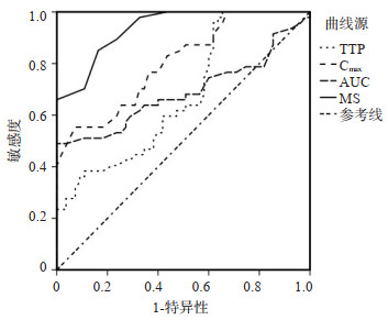
目的 探讨动态对比增强磁共振成像(DCE-MRI)半定量鉴别诊断良恶性肺部占位性病变的临床价值。 方法 回顾性分析2020年3月~2021年3月在我院就诊的102例肺部占位性病变患者病例资料。根据手术切除后的病理结果,将肺癌患者作为恶性组(n=47),将良性肺部占位病变患者作为良性组(n=55)。所有患者均行DCE、MRI扫描,获得半定量参数,包括达峰时间、最大浓度、对比浓度-时间曲线下面积、最大线性斜率。比较良恶性肺部占位性病变组半定量参数的差异,并用ROC曲线评价上述各参数鉴别价值。 结果 恶性组病例中病灶以囊实性、不均匀强化、不规则形态、边界不清晰为主,良性组病例病变以囊性、均匀强化、规则形态、边界清晰或欠清晰为主。两组常规MRI特征差异无统计学意义(P > 0.05);恶性组达峰时间参数水平低于良性组(P < 0.05),恶性组最大浓度、曲线下面积、最大线性斜率参数水平高于良性组(P < 0.05);以病理结果为标准,绘制半定量参数ROC曲线,最大线性斜率曲线下面积最大为0.937,诊断效能较其余参数高,最大线性斜率准确度最高84.4%,最大浓度特异度最高为92.7%,达峰时间敏感度最高为97.9%。 结论 DCE-MRI半定量参数可鉴别诊断良恶性肺部占位性病变。 -
关键词:
- 增强磁共振成像 /
- 半定量 /
- 良恶性肺部占位性病变 /
- 临床价值
Abstract:Objective To investigate the clinical dynamic contrast enhancement magnetic resonance imaging (DCE-MRI) for the semi-quantitative differential diagnosis of benign and malignant pulmonary occupying lesions. Methods Clinical data of 102 patients with pulmonary occupying lesions in our hospital from March 2020 to March 2021 were retrospectively analyzed. Based on the pathological findings, patients with pulmonary cancer were considered as the malignant group (n=47) and patients with benign pulmonary occupying lesions were considered as the benign group (n=55). All patients received DCE and MRI scans to obtain semi-quantitative parameters including time to peak, maximum concentration, AUC of time-concentration curve, maximum slope were obtained. The differences in semi-quantitative parameters between benign and malignant groups were compared, and ROC curve was used to evaluate the discriminatory value of the each of these parameters. Results In the malignant group, the lesions were mainly cystic solidity, heterogeneous enhancement, irregular morphology and unclear borders, while those in benign group, the lesions were mainly cystic, homogeneous enhancement, regular morphology and clear or poorly defined borders. The lesion features of two groups showed no significant difference in conventional MRI (P > 0.05). the levels of the parameters of time to peak in malignant group were lower than those in benign group (P < 0.05), and the levels of the parameters of maximum concentration, AUC and maximum slope were higher than those in benign group (P < 0.05); Based on pathological results, ROC curve of semi-quantitative parameters showed that maximum slope curve, and the maximum area under the curve of maximum linear slope was 0.937. The diagnostic efficacy was higher than the rest of the parameters, with a maximum linear slope accuracy of 84.4%, a maximum concentration specificity of 92.7%, and a peak attainment time sensitivity of 97.9%. Conclusion Semi-quantitative parameters of DCE-MRI are effective in the differential diagnosis of benign and malignant pulmonary occupying lesions. -

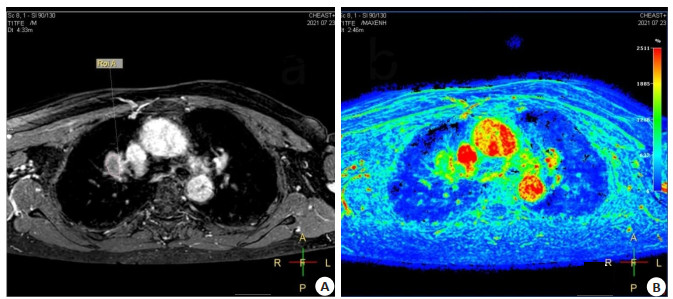
图 1 患者女,42岁,病理结果:肺腺癌
A: 增强扫描病灶实性、囊性成分征象: 右上肺门区见类圆形等T1长T2信号肿块, 边缘毛糙, 弥散受限,增强扫描明显强化; B: TIP、Cmax、AUC、MS伪彩图示病灶实性部分分别呈青色、绿色色信号, 对应值分别为TTP=165.52 s、Cmax=2140 μg/mL、AUC=318 613 μg·h/mL、MS 18.38%.
Figure 1. Female patient, 42 years old, pathological results: lung adenocarcinoma.

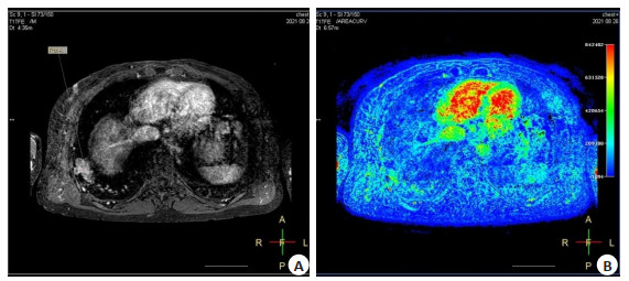
图 2 男,47岁,病理结果:肺部炎性病灶
A: 增强扫描病灶实性、囊性成分征象: 右肺下叶见类圆形稍短T1稍长T2信号肿块,边缘尚清,弥散受限,增强扫描明显强化; B: TIP、Cmax、AUC、MS伪彩图示病灶实性部分分别呈绿色、蓝色色信号,对应值分别为TTP=274.57 s、Cmax=2150 μg/mL、AUC=151 207 μg·h/mL.
Figure 2. Male patient, 47 years old, pathological results: pulmonary inflammatory lesions.
表 1 常规MRI特征
Table 1. Conventional MRI features
特征 恶性组(n=47) 良性组(n=55) χ2 P 肿块类型 2.322 0.313 囊性为主 15 24 实性为主 15 11 囊实性 17 20 强化特点 1.632 0.442 均匀 13 17 不均匀 21 18 无强化 13 20 形态 0.006 0.938 不规则 26 30 规则 21 25 边界 2.279 0.320 清晰 4 9 不清晰 22 19 欠清晰 21 27  下载: 导出CSV
表 2 半定量参数的组间、组内一致性
Table 2. Inter group and intra group consistency of semi quantitative parameters
参数 组间 组内 TTP 0.965 0.964 Cmax 0.974 0.981 AUC 0.978 0.986 MS 0.980 0.980 TTP: 达峰时间; Cmax: 最大浓度; AUC: 浓度-时间曲线下面积; MS: 最大斜度.
下载: 导出CSV
表 3 DCE-MRI半定量参数比较
Table 3. Comparison of semi quantitative parameters of DCE-MRI
参数 恶性组(n=47) 良性组(n=55) t P TTP (s) 3.03±0.88 3.51±0.74 2.993 0.004 Cmax (μg/mL) 0.46±0.27 0.23±0.18 5.126 < 0.001 AUC (μg·h/mL) 0.52±0.32 0.37±0.16 3.059 0.003 MS (%) 0.31±0.08 0.22±0.05 6.914 < 0.001
下载: 导出CSV
表 4 DCE-MRI半定量参数鉴别诊断效能
Table 4. Differential diagnostic efficacy of DCE-MRI semi quantitative parameters
参数 最佳阈值 ROC面积 准确度(%) 特异性(%) 敏感度(%) TTP 2.77 0.675 72.1 46.3 97.9 Cmax 0.38 0.804 64.0 92.7 55.3 AUC 0.53 0.689 72.7 96.4 48.9 MS 0.26 0.937 84.4 83.6 85.1
下载: 导出CSV
-
[1] 刘磊, 庞新路, 尚文俊, 等. DCE-MRI鉴别诊断前列腺良恶性病变的临床价值[J]. 中国CT和MRI杂志, 2018, 16(4): 120-2. doi: 10.3969/j.issn.1672-5131.2018.04.037 [2] Zhang Q, Peng YS, Liu W, et al. Radiomics based on multimodal MRI for the differential diagnosis of benign and malignant breast lesions [J]. J Magn Reson Imaging, 2020, 52(2): 596-607. doi: 10.1002/jmri.27098 [3] 卢海涛, 邢伟, 张艳文, 等. 动态对比增强磁共振成像预测高级别胶质瘤IDH基因突变的价值[J]. 中华医学杂志, 2019, 99(39): 3105-9. doi: 10.3760/cma.j.issn.0376-2491.2019.39.013 [4] 刘雅怡, 岳斌, 孙玲玲, 等. 动态对比增强MRI定量参数评价软组织肿瘤生物学行为的价值[J]. 中华放射学杂志, 2020, 54(10): 980-5. doi: 10.3760/cma.j.cn112149-20200224-00238 [5] Digumarthy SR, Padole AM, Lo Gullo R, et al. CT texture analysis of histologically proven benign and malignant lung lesions[J]. Medicine, 2018, 97(26): e11172. doi: 10.1097/MD.0000000000011172 [6] 解礼冰, 田兴仓, 马丽, 等. DCE-MRI诊断肺癌和肺部不同类型良性肿块的价值[J]. 磁共振成像, 2018, 9(3): 192-6. https://www.cnki.com.cn/Article/CJFDTOTAL-CGZC201803008.htm [7] 刘宏, 张凤翔, 张芳. DCE-MRI半定量及定量分析在鉴别颈部淋巴结良恶性中的研究现状[J]. 磁共振成像, 2021, 12(1): 103-5. https://www.cnki.com.cn/Article/CJFDTOTAL-CGZC202101026.htm [8] 焦志云, 胡春洪, 杜芳, 等. 磁共振动态增强联合多b值扩散加权成像鉴别诊断肺部良恶性病变的价值[J]. 临床放射学杂志, 2019, 38(2): 239-43. https://www.cnki.com.cn/Article/CJFDTOTAL-LCFS201902013.htm [9] 左金, 闫海龙, 韩东明. DCE-MRI在卵巢交界性肿瘤与上皮性卵巢癌中的鉴别诊断价值[J]. 医学影像学杂志, 2020, 30(5): 798-802. https://www.cnki.com.cn/Article/CJFDTOTAL-XYXZ202005021.htm [10] 孔维丹, 岳秀慧, 陶晓峰. 动态对比增强MRI在甲状腺良恶性结节鉴别诊断中的应用价值[J]. 实用放射学杂志, 2020, 36(1): 21-4. doi: 10.3969/j.issn.1002-1671.2020.01.006 [11] 周意明, 徐筑津, 华彬, 等. 比较乳腺动态对比增强MRI定量和半定量参数预测乳腺癌新辅助化疗疗效的价值[J]. 实用放射学杂志, 2020, 36(3): 396-400. doi: 10.3969/j.issn.1002-1671.2020.03.015 [12] 杜永浩, 梁挺, 杨健, 等. DCE-MRI Extended tofts模型和Reference Region模型在肺部结节/肿块良恶性诊断中的应用价值[J]. 西安交通大学学报: 医学版, 2019, 40(3): 417-20, 450. https://www.cnki.com.cn/Article/CJFDTOTAL-XAYX201903015.htm [13] Choi YJ, Lee IS, Song YS, et al. Diagnostic performance of diffusion-weighted (DWI) and dynamic contrast-enhanced (DCE) MRI for the differentiation of benign from malignant soft-tissue tumors[J]. J Magn Reson Imaging, 2019, 50(3): 798-809. doi: 10.1002/jmri.26607 [14] 周文华, 徐大伟, 王立军, 等. 多层螺旋CT及动态增强磁共振成像血流参数在肝良恶性结节鉴别诊断中应用价值探究[J]. 中华生物医学工程杂志, 2019, 25(5): 518-22. doi: 10.3760/cma.j.issn.1674-1927.2019.05.002 [15] Lu JP. Microvessel density of malignant and benign hepatic lesions and MRI evaluation[J]. World J Gastroenterol, 2004, 10(12): 1730. doi: 10.3748/wjg.v10.i12.1730 [16] 孟思, 周超, 李小华, 等. 肺癌患者DCE-MRI表现特点与其临床病理类型、疾病严重程度的相关性分析[J]. 哈尔滨医科大学学报, 2020, 54(2): 160-3. doi: 10.3969/j.issn.1000-1905.2020.02.012 [17] 杜永浩, 梁挺, 张硕, 等. 3D非刚性图像配准在DCE-MRI诊断肺结节/肿块良恶性中的应用价值[J]. 现代肿瘤医学, 2020, 28(14): 2478-81. doi: 10.3969/j.issn.1672-4992.2020.14.024 -

 点击查看大图
计量
- 文章访问数: 155
- HTML全文浏览量: 181
- PDF下载量: 5
- 被引次数: 0