


Diagnostic value of whole-body bone imaging and SPECT/CT in bone destruction caused by Talaromyces marneffei
-
摘要:
目的 探讨全身骨显像与SPECT/CT在马尔尼菲篮状菌感染骨破坏中的诊断价值及显像特征分析。 方法 回顾性分析我院2016年6月~2021年6月于核医学科行全身骨显像及SEPCT/CT断层融合显像的马尔尼菲篮状菌患者47例,其中男25例,女22例,年龄17~72岁,中位年龄51岁。比较全身骨显像、SPECT/CT对马尔尼菲篮状菌骨破坏的诊断效能,并探讨马尔尼菲篮状菌骨破坏的显像特征。 结果 全身骨显像,SPECT/CT断层融合显像对马尔尼菲篮状菌骨破坏诊断符合率比较,差异有统计学意义(P < 0.05)。全身骨平面显像对骨破坏的诊断敏感度为84.4%,特异性为100%,诊断符合率85.1%;SPECT/CT对TM骨破坏的诊断敏感度为97.8%,特异性为100%,诊断符合率98.9%。马尔尼菲篮状菌骨破坏主要侵犯中轴骨,并伴有全身多个位置受累,四肢骨及颅骨为其相对特异性的侵犯位置。 结论 全身骨显像可以一次性观察全身病灶,在病变的早期即可检出骨质代谢的异常活跃,SPECT/CT诊断符合率较高,二者联合对马尔尼菲篮状菌骨破坏具有很好的应用价值。 Abstract:Objective To explore the diagnostic value and imaging characteristics of whole-body bone imaging and SPECT/CT in bone destruction caused by Talaromyces marneffei. Methods We retrospectively analyzed 47 patients with Talaromyces marneffei who underwent whole-body bone imaging and SPECT/CT tomographic fusion imaging in the Department of Nuclear Medicine in our hospital from June 2016 to June 2021, including 25 males and 22 females, aged from 17 to 72 years old, with a median age of 51 years. We compared the diagnostic efficacy of whole-body bone imaging and SPECT/CT in bone destruction, and discussed the imaging characteristics of bone destruction. Results There was a significant difference in the coincidence rate of whole-body bone imaging and SPECT/CT tomographic fusion imaging in the diagnosis of bone destruction caused by Talaromyces marneffei (P < 0.05). The sensitivity of whole-body bone imaging in the diagnosis of bone destruction was 84.4%, the specificity was 100%, and the diagnostic coincidence rate was 85.1%. The diagnostic sensitivity of SPECT/CT for TM bone destruction was 97.8%, the specificity was 100%, and the diagnostic compliance rate was 98.9%. Bone destruction of Talaromyces marneffei mainly affects the axial bones and is associated with the involvement of several locations throughout the body, with the bones of the extremities and the skull being the relatively specific sites of invasion. Conclusion Whole-body bone imaging can observe the whole-body focus at one time, and the abnormal activity of bone metabolism can be detected in the early stage of the lesion. The coincidence rate of SPECT/CT diagnosis is high. The combination of SPECT/CT has a high diagnostic yield and is of good use in the case of bone destruction caused by Talaromyces marneffei. -
Key words:
- whole-body bone imaging /
- SPECT/CT /
- Talaromyces marneffei /
- bone destruction
-

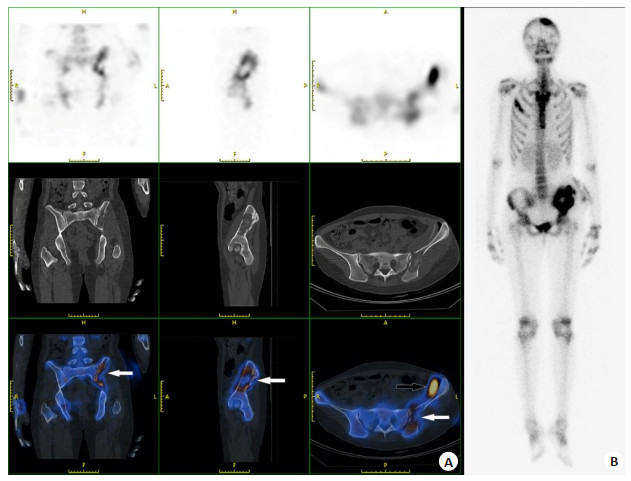
图 1 一例24岁女性患者的SPECT/CT断层融合显像及全身骨显像
A: SPECT/CT断层融合显像示左侧髂骨见的小片状显像剂分布异常浓聚影, 其靠近骶髂关节处见显像剂分布稀疏, 呈现中间缺损周边浓聚的特点. 融合CT示左侧髂骨明显骨质破坏(白色箭头), 其上方见条索状软组织影,密度不均(黑色箭头); B: 全身骨显像示左侧顶骨、右侧第3前肋、左侧髂骨见片状显像剂异常浓聚影.
Figure 1. SPECT/CT tomographic fusion imaging and whole-body bone imaging of a 24-year-old female patient
表 1 全身骨显像与SPECT/CT断层融合显像对TM感染骨破坏的诊断结果对比
Table 1. Comparison of diagnostic results between whole-body bone imaging and SPECT/CT tomographic fusion imaging in TM infected bone destruction[n (%)]
全身骨显像 SPECT/CT断层融合显像 合计 骨破坏 无骨破坏 骨破坏 38 (80.9) 0 (0.0) 38 (80.9) 无骨破坏 6 (12.8) 3 (6.4) 9 (19.1) 合计 44 (93.6) 3 (6.4) 47 (100.0) χ2=4.167, P=0.041.  下载: 导出CSV
表 2 全身骨显像与SPECT/CT断层融合显像对TM感染骨破坏的诊断效能对比
Table 2. Diagnostic efficiency of whole-body bone scintigraphy compared with SPECT/CT tomographic fusion imaging in TM infected bone destruction (%)
检测方法 敏感度 特异性 诊断符合率 全身骨显像 84.4 100 85.1 SPECT/CT断层融合显像 97.8 100 98.9
下载: 导出CSV
表 3 TM感染骨破坏病灶分布特点
Table 3. Characteristics of distribution of bone destruction lesions in TM infection
骨破坏部位 例数(n) 病灶数(个) 肋骨 35 285 脊柱 29 88 颅骨 32 69 股骨 28 50 骨盆 25 48 胸骨 15 34 肩关节 17 32 肱骨 15 25 膝关节 10 21 胫骨 10 20 合计 - 672
下载: 导出CSV
-
[1] Vanittanakom N, Cooper CR Jr, Fisher MC, et al. Penicillium marneffei infection and recent advances in the epidemiology and molecular biology aspects[J]. Clin Microbiol Rev, 2006, 19(1): 95-110. doi: 10.1128/CMR.19.1.95-110.2006 [2] 夏露, 蓝秀万, 温波. 巨噬细胞对马尔尼菲篮状菌免疫作用机制研究进展[J]. 中国真菌学杂志, 2021, 16(1): 60-4, 68. doi: 10.3969/j.issn.1673-3827.2021.01.015 [3] 王浩迪, 唐小平, 李凌华. HIV阴性人群马尔尼菲篮状菌病研究进展[J]. 国际流行病学传染病学杂志, 2018, 45(1): 45-7. doi: 10.3760/cma.j.issn.1673-4149.2018.01.011 [4] Browne SK, Burbelo PD, Chetchotisakd P, et al. Adult-onset immunodeficiency in Thailand and Taiwan[J]. N Engl J Med, 2012, 367(8): 725-34. doi: 10.1056/NEJMoa1111160 [5] Liu GN, Huang JS, Zhong XN, et al. Penicillium marneffei infection within an osteolytic lesion in an HIV-negative patient[J]. Int J Infect Dis, 2014, 23: 1-3. doi: 10.1016/j.ijid.2013.12.019 [6] Qiu Y, Zhang JQ, Liu GN, et al. Retrospective analysis of 14 cases of disseminated Penicillium marneffei infection with osteolytic lesions [J]. BMC Infect Dis, 2015, 15: 47. doi: 10.1186/s12879-015-0782-6 [7] 农云洁, 农恒荣, 黄小桂, 等. 艾滋病相关浅表淋巴结马尔尼菲蓝状菌病超声影像分析[J]. 中国超声医学杂志, 2021, 37(4): 435-8. doi: 10.3969/j.issn.1002-0101.2021.04.023 [8] 史晓天, 顾玉婷, 李凯, 等. 马尔尼菲蓝状菌骨感染病变的影像学分析[J]. 实用放射学杂志, 2021, 37(6): 985-8. doi: 10.3969/j.issn.1002-1671.2021.06.027 [9] 胡文清, 陈俐, 包盈莹, 等. 非HIV感染的马尔尼菲青霉病的影像学诊断及临床分析[J]. 实用放射学杂志, 2017, 33(7): 1002-5. doi: 10.3969/j.issn.1002-1671.2017.07.005 [10] 张峰, 焦举, 谢良骏. 99mTc-MDP SPECT/CT全身骨显像诊断前列腺癌骨转移的临床价值[J]. 实用医学杂志, 2017, 33(11): 1774-7. doi: 10.3969/j.issn.1006-5725.2017.11.014 [11] Bai Y, Xi D, Chen Q, et al. Characteristics of Talaromyces marneffei with bone destruction in Guangxi Province, China: a retrospective study[J]. Am J Transl Res, 2021, 13(10): 11491-500. [12] Wei HY, Liang WJ, Li B, et al. Clinical characteristics and risk factors of Talaromyces marneffei infection in human immunodeficiency virus-negative patients: a retrospective observational study[J]. World J Emerg Med, 2021, 12(4): 281-6. doi: 10.5847/wjem.j.1920-8642.2021.04.005 [13] 邓卓霖. 马尔尼菲青霉病研究新发现: 溶骨病变及关节炎[J]. 广西科学, 1994, 1(1): 53-8. https://www.cnki.com.cn/Article/CJFDTOTAL-GXKK401.014.htm [14] 邓卓霖, 马韵, 李山. 马尔尼菲青霉所致的骨与关节病的临床与病理研究[J]. 广西医学院学报, 1993, 10(3): 262-7. https://www.cnki.com.cn/Article/CJFDTOTAL-GXYD199303001.htm [15] Deng Z, Ribas JL, Gibson DW, et al. Infections caused by Penicillium marneffei in China and Southeast Asia: review of eighteen published cases and report of four more Chinese cases[J]. Rev Infect Dis, 1988, 10(3): 640-52. doi: 10.1093/clinids/10.3.640 [16] 何清, 史育红, 王朝点. SEPCT/CT在分化型甲状腺癌诊治的增益价值及高剂量131I的治疗疗效[J]. 分子影像学杂志, 2021, 44(4): 664-7. doi: 10.12122/j.issn.1674-4500.2021.04.17 [17] 邓群力, 莫逸, 刘康龙. 99mTc-亚甲基二膦酸盐骨显像评价乳腺癌骨转移的特征[J]. 分子影像学杂志, 2018, 41(3): 316-9. doi: 10.3969/j.issn.1674-4500.2018.03.07 [18] 黄谋清, 章梦芝, 曾小建. 马尔尼菲青霉菌感染骨损害99mTc-MDP骨显像1例[J]. 中国临床医学影像杂志, 2020, 31(2): 148-50. https://www.cnki.com.cn/Article/CJFDTOTAL-LYYX202002026.htm [19] 梁锐烘, 刘艳雯, 曾庆思. 免疫功能正常者马尔尼菲蓝状菌病的胸部CT及PET-CT表现[J]. 放射学实践, 2019, 34(12): 1313-7. https://www.cnki.com.cn/Article/CJFDTOTAL-FSXS201912010.htm [20] 张建全, 柳广南, 杨美玲, 等. 马尔尼菲青霉病并发溶骨性破坏八例临床分析[J]. 中华临床医师杂志: 电子版, 2011, 5(13): 3912-5. doi: 10.3877/cma.j.issn.1674-0785.2011.13.052 [21] 卢朝辉, 刘鸿瑞, 谢秀丽, 等. 马尔尼菲青霉菌感染[J]. 中华病理学杂志, 2004, 33(6): 536-40. doi: 10.3760/j.issn:0529-5807.2004.06.009 [22] Chan YF, Woo KC. Penicillium marneffei osteomyelitis[J]. J Bone Jo Surg Br Vol, 1990, 72-B(3): 500-3. doi: 10.1302/0301-620X.72B3.2341456 [23] Zheng JD, Gui XE, Cao Q, et al. A clinical study of acquired immunodeficiency syndrome associated Penicillium marneffei infection from a non-endemic area in China[J]. PLoS One, 2015, 10 (6): e0130376. doi: 10.1371/journal.pone.0130376 -

 点击查看大图
计量
- 文章访问数: 204
- HTML全文浏览量: 125
- PDF下载量: 7
- 被引次数: 0