


Value of P- selection combined with right echocardiography in the diagnosis of patients with migraine with unclosed foramen ovale
-
摘要:
目的 分析P选择素联合右心声学造影(cTTE)诊断偏头痛合并卵圆孔未闭患者的价值。 方法 选取2019年4月~2021年4月我院收治的64例偏头痛患者,根据是否并发卵圆孔未闭分为发生组47例,未发生组17例,比较所有患者血清中的P选择素水平,并进行右心声学造影检查,比较不同检查方法的诊断准确率。 结果 并发卵圆孔未闭患者P选择素水平明显高于未发卵圆孔未闭患者,差异有统计学意义(P < 0.05);以术中封堵为金标准,P选择素诊断偏头痛合并卵圆孔未闭的敏感度为68.09%、特异性为82.35%、阳性预测值为91.43%、阴性预测值为48.28%、诊断符合率为71.88%,一致性为0.412;以术中封堵为金标准,P选择素联合cTTE诊断偏头痛合并卵圆孔未闭的敏感度为82.98%、特异性为88.24%、阳性预测值为95.12%、阴性预测值为65.22%、诊断符合率为84.38%,一致性为0.640。 结论 P选择素联合右心声学造影有利于提高偏头痛合并卵圆孔未闭患者的诊断准确率,在临床上有一定的应用价值。 Abstract:Objective To analyze the value of P-selection combined with right echocardiography (cTTE) diagnosing patients with migraine combined with unclosed foramen ovale. Methods A total of 64 migraine patients admitted to our hospital from April 2019 to April 2021 were selected and divided into the occurrence group (n=47) and the non-occurrence group (n=17) according to the complication of patent foramina ovale. The serum P-selection levels of all patients was compared, and right echocardiography was performed to compare the diagnostic accuracy of different examination methods. Results The levels of P selectin were significant higher in patients with patent foramen ovale than in those without patent foramen ovale, with a statistically significant difference (P < 0.05). Using intraoperative closure as the gold standard, the sensitivity of P-selectin for the diagnosis of migraine combined with oval foramen nonocclusion was 68.09%, specificity was 82.35%, positive predictive value was 91.43%, negative predictive value was 48.28%, and diagnostic compliance rate was 71.88% and concordance 0.412. Using intraoperative closure as the gold standard, the sensitivity of P-selectin combined with cTTE for the diagnosis of migraine combined with patent foramen ovale was 82.98%, specificity was 88.24%, positive predictive value was 95.12%, negative predictive value was 65.22%, diagnostic compliance was 84.38%, with a consistency of 0.640. Conclusion P-selection combined with right aspiration imaging can improve the diagnostic accuracy in patients with migraine combined with unclosed foramen ovale. -
Key words:
- P-selection /
- radiography of right aspiration /
- migraine /
- patent foramen ovale /
- diagnostic value
-

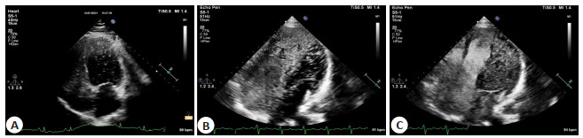
图 1 Vasalva动作下偏头痛合并卵圆孔未闭患者右心声学造影图像
Figure 1. Right echocardiography images of migraine patients with patent foramen ovale under Vasalva motion.
表 1 P选素与金标准对偏头痛合并卵圆孔未闭的诊断结果比较
Table 1. Comparison of the diagnostic results of migraine with patent foramen ovale between P-selective and gold standard (n)
P选择素 金标准 合计 阳性 阴性 阳性 32 3 35 阴性 15 14 29 合计 47 17 64  下载: 导出CSV
表 2 P选择素联合cTTE与金标准对偏头痛合并卵圆孔未闭的诊断结果比较
Table 2. Comparison of P-selection combined with cTTE and gold standard in the diagnosis of migraine combined with patent foramen ovale (n)
P选择素联合cTTE 金标准 合计 阳性 阴性 阳性 39 2 41 阴性 8 15 23 合计 47 17 64 cTTE: 右心声学造影.
下载: 导出CSV
-
[1] 刘文娟, 张玉顺, 成革胜, 等. 两种不同封堵器对不明原因脑卒中合并大量右向左分流患者卵圆孔未闭的疗效对比[J]. 中国循环杂志, 2018, 33(4): 385-9. doi: 10.3969/j.issn.1000-3614.2018.04.017 [2] Zhao EF, Wei YJ, Zhang YF, et al. A comparison of transthroracic echocardiograpy and transcranial Doppler with contrast agent for detection of patent foramen ovale with or without the Valsalva maneuver[J]. Medicine, 2015, 94(43): e1937. doi: 10.1097/MD.0000000000001937 [3] 翁纽周, 赖志雄. 超声心动图结合右心声学造影诊断卵圆孔未闭的临床价值探讨[J]. 黑龙江医学, 2018, 42(10): 1021-2. [4] Zhao EF, Wei YJ, Zhang YF, et al. A comparison of transthroracic echocardiograpy and transcranial Doppler with contrast agent for detection of patent foramen ovale with or without the Valsalva maneuver[J]. Medicine, 2015, 94(43): e1937. doi: 10.1097/MD.0000000000001937 [5] Yue L, Zhai YN, Wei LQ. Which technique is better for detection of right-to-left shunt in patients with patent foramen ovale: comparing contrast transthoracic echocardiography with contrast transesophageal echocardiography[J]. Echocardiography, 2014, 31(9): 1050-5. doi: 10.1111/echo.12523 [6] 匡永芳, 朱海锋, 郭远峰, 等. 经胸超声心动图与经食道超声心动图联合右心声学造影在卵圆孔未闭封堵术中的应用对比[J]. 影像技术, 2021, 33(4): 44-8. https://www.cnki.com.cn/Article/CJFDTOTAL-YIXI202104011.htm [7] 陆燕飞, 苏海庆, 宋海国, 等. 实时三维超声心动图联合右心声学造影诊断成人卵圆孔未闭的临床分析[J]. 医学影像学杂志, 2018, 28(11): 1936-9. https://www.cnki.com.cn/Article/CJFDTOTAL-XYXZ201811049.htm [8] Mongodi S, Via G, Riccardi M, et al. Patent foramen ovale diagnosis: the importance of provocative maneuvers[J]. J Clin Ultrasound, 2017, 45(1): 58-61. doi: 10.1002/jcu.22383 [9] 张秀玲. 超声不同方法联合应用对提高卵圆孔未闭检出率的应用价值[J]. 广州医药, 2020, 51(5): 76-8, 83. https://www.cnki.com.cn/Article/CJFDTOTAL-GZYY202005017.htm [10] Yue L, Zhai YN, Wei LQ. Which technique is better for detection of right-to-left shunt in patients with patent foramen ovale: comparing contrast transthoracic echocardiography with contrast transesophageal echocardiography[J]. Echocardiography, 2014, 31(9): 1050-5. doi: 10.1111/echo.12523 [11] Kong XJ, Chai QF, Guo K, et al. Analysis of contrast echocardiography for detecting right to left shunt in adults with patent foramen ovale[J]. Zhonghua Yi Xue Za Zhi, 2017, 97(43): 3380-3. [12] 岳庆雄, 刘佳, 周瑜, 等. TEE右心声学造影在微小卵圆孔未闭右向左分流检测中的应用[C]//海峡两岸医药卫生交流协会, 厦门大学: 2018. [13] 刘峰, 叶钜亨, 贺俊波, 等. 偏头痛患者采用TCD发泡试验筛查卵圆孔未闭的价值及封堵PFO后的疗效分析[J]. 中国实用医药, 2020, 15 (14): 51-3. https://www.cnki.com.cn/Article/CJFDTOTAL-ZSSA202014020.htm [14] Zhao EF, Wei YJ, Zhang YF, et al. A comparison of transthroracic echocardiograpy and transcranial Doppler with contrast agent for detection of patent foramen ovale with or without the Valsalva maneuver[J]. Medicine, 2015, 94(43): e1937. doi: 10.1097/MD.0000000000001937 [15] 孙立华, 匡瑞娟, 孙伟华. 中青年隐源性脑梗死合并卵圆孔未闭的临床及MRI特点分析[J]. 国际医药卫生导报, 2019, 25(15): 2496-7. doi: 10.3760/cma.j.issn.1007-1245.2019.15.029 [16] Wilmshurst P, Nightingale S. The role of cardiac and pulmonary pathology in migraine: a hypothesis[J]. Headache, 2006, 46(3): 429-34. [17] Rigatelli G, Dell'avvocata F, Cardaioli P, et al. Improving migraine by means of primary transcatheter patent foramen ovale closure: long-term follow-up[J]. Am J Cardiovasc Dis, 2012, 2(2): 89-95. [18] Bender E. In the clinic-patent foramen ovale closure and stroke: PFO closure associated with fewer recurrent ischemic strokes compared to medical therapy[J]. Neurol Today, 2017, 17(21): 8-11. [19] 鞠冠毅, 刘福秀, 李兴梅, 等. 右心声学超声造影对卵圆孔未闭的探讨[J]. 影像研究与医学应用, 2020, 4(5): 56-8. https://www.cnki.com.cn/Article/CJFDTOTAL-YXYY202005033.htm [20] 王文婷, 黄海韵, 柳强维, 等. 经胸超声心动图结合右心声学造影在判断卵圆孔未闭右向左分流中的临床价值[J]. 第三军医大学学报, 2017, 39(16): 1648-53. https://www.cnki.com.cn/Article/CJFDTOTAL-DSDX201716011.htm -

 点击查看大图
计量
- 文章访问数: 136
- HTML全文浏览量: 76
- PDF下载量: 2
- 被引次数: 0