Exploring the changes of brain functional connectivity in patients with acute ischemic stroke in non-dominant cerebral hemisphere: based on rs-fMRI
-
摘要:
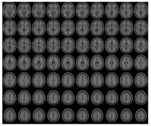
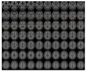
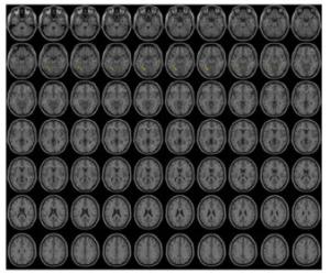
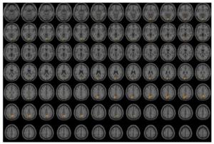
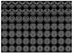
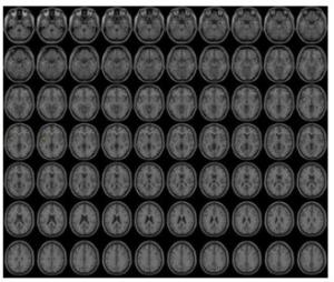
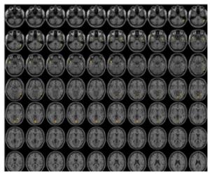
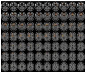
目的运用静息态脑功能成像探究非优势大脑半球缺血性脑卒中患者在急性期的脑功能连接的变化。 方法以15例健康受试者作为正常对照组,15例非优势大脑半球(右半球)中动脉急性梗塞(发病 < 72 h)的患者作为卒中组,采用西门子3.0 T超导MRI采集两组患者相关数据,以功能连接为结局指标,并基于Matlab 2012a平台使用DPABI工具包对两组数据进行预处理和统计学分析。 结果最终10例患者纳入试验,与健康受试者对比,差异有统计学意义(P < 0.005)的感兴趣区(ROI)有11个。以右背外侧额上回为ROI,与左后扣带回(T=6.5173)等脑区功能连接增强;以右眶部额上回为ROI,与左BA18(T=6.0674)等脑区功能连接增强;以左中央沟盖为ROI,与左距状裂周围皮层(T=5.7831)等脑区功能连接增强;以右眶内额上回为ROI,与左梭状回(T=5.7361)等脑区功能连接增强;以左岛叶为ROI,与左梭状回(T=4.5436)等脑区功能连接增强;以右岛叶为ROI,与右小脑后叶(T=4.9352)等脑区功能连接增强;以左楔叶为ROI,与左岛叶(T=5.6924)等脑区功能连接增强;与右梭状回为ROI,与左眶回(T=8.4505)等脑区功能连接增强;以左楔前叶为ROI,与左豆状壳核(T=5.1894)等脑区功能连接增强;以左中央旁小叶为ROI,与左豆状壳核(T=7.9109)等脑区功能连接增强;以左颞横回为ROI,与左舌回(T=5.9146)等脑区功能连接增强。 结论非优势大脑半球缺血性脑卒中患者在急性期存在与视觉、听觉、高级认知及运动相关的脑区之间功能连接增强的脑功能变化。 -
关键词:
- 静息态功能磁共振成像 /
- 脑功能 /
- 急性缺血性脑卒中 /
- 非优势大脑半球
Abstract:ObjectiveTo explore the functional network changes in patients with acute ischemic stroke in non-dominant cerebral hemisphere. MethodsIn this trial, 15 healthy subjects (HSs) were recruited as controls, 15 subjects with middle cerebral artery acute infarction in non-dominant cerebral hemisphere were recruited into stroke group. Rs-fMRI data of subjects were obtained by Siemens 3.0 T superconducting MRI. It was based on Matlab 2012a platform, using DPABI software for data analysis and mapping with functional connectivity as an outcome indicator. ResultsFinally, the changes of functional connectivity changes (P < 0.005) in ten subjects were as follows: Centered to the seed region of the dorsolateral superior frontal gyrus, FC increased at the left posterior cingulate cortex(T=6.5173). Centered to the seed region of the right orbital superior frontal gyrus, FC increased at the left BA18(T=6.0674). Centered to the seed region of the Rolandic_Oper_L(ALL), FC increased at the cortex around the left fissura calcarina(T=5.7831). Centered to the seed region of the right intraorbital superior frontal gyrus, FC increased at the cortex around the left lobule fusiform(T=5.7361). Centered to the seed region of the left insular lobe, FC increased at the cortex around the left lobule fusiform(T=4.5436). Centered to the seed region of the right insular lobe, FC increased at the cortex around the right posterior cerbellar lobe(T=4.9352). Centered to the seed region of the left cuneus, FC increased at the cortex around the left insular lobe(T=5.6924). Centered to the seed region of the right lobule fusiform, FC increased at the cortex around the left gyri orbitales(T=8.4505). Centered to the seed region of the left precuneus, FC increased at the cortex around the left lenticular putamen(T=5.1894). Centered to the seed region of the left paracentral lobule, FC increased at the cortex around the left lenticular putamen(T=7.9109). Centered to the seed region of the left gyri temporales transversi, FC increased at the cortex around the left gyrus lingualis(T=5.9146). ConclusionSpecific functional network changes present in patients with acute ischemic stroke in non-dominant cerebral hemisphere, and function connection enhancement among the brain regions related to visual, uditory, higher cognition and motor. -
表 1 正常组和卒中组的一般资料
Table 1. General information of normal group ans stroke patients group
分组 性别(n) 年龄(岁, Mean±SD) 男性 女性 正常组(n=12) 4 8 56.17±3.83 卒中组(n=10) 8 2 59.40±7.65 F/χ2 5.728 4.437 P 0.057 0.021 表 2 与ROI4种子点的功能连接出现特异性变化的脑区
Table 2. Brain regions with enhanced functional connectivity to ROI4
种子点 脑区效应 脑区 MiNi坐标 激活强度(T) X Y Z ROI4 增强 左后扣带回(BA30),左BA18、BA19、梭状回、距状沟周围皮层 -24 -69 3 6.5173 增强 左BA18 -30 -99 -12 4.3214 增强 左BA18、BA19 -18 -93 21 4.1986 增强 左楔叶(BA18) -12 -75 24 3.9032 增强 左楔叶(BA19) -12 -96 30 4.528 增强 左楔前叶 -15 -57 42 4.9078 X、Y、Z代表脑空间定位轴,分别表示在MiNi标准化空间坐标系中左右、前后和上下方位的坐标位置. 表 3 与ROI6种子点的功能连接出现特异性变化的脑区
Table 3. Brain regions with enhanced functional connectivity to ROI6
种子点 脑区效应 脑区 MiNi坐标 激活强度(T) X Y Z ROI6 增强 左BA18 -48 -78 -6 6.0674 增强 左颞上回(BA42) -63 -18 6 7.2853 增强 右楔前叶、楔叶 21 -72 30 4.0274 增强 右楔前叶(BA7) 21 -78 39 4.4489 增强 左楔叶,左楔前叶 -12 -78 27 5.1324 增强 右楔前叶 9 -75 42 5.6006 X、Y、Z代表脑空间定位轴,分别表示在MiNi标准化空间坐标系中左右、前后和上下方位的坐标位置. 表 4 与ROI17种子点的功能连接出现特异性变化的脑区
Table 4. Brain regions with enhanced functional connectivity to ROI17
种子点 脑区效应 脑区 MiNi坐标 激活强度(T) X Y Z ROI17 增强 左距状裂周围皮层,左舌回 0 -87 -6 5.7831 增强 左楔叶(BA19) -6 -93 24 7.7859 X、Y、Z代表脑空间定位轴,分别表示在MiNi标准化空间坐标系中左右、前后和上下方位的坐标位置. 表 5 与ROI26种子点的功能连接出现特异性变化的脑区
Table 5. Brain regions with enhanced functional connectivity to ROI26
种子点 脑区效应 脑区 MiNi坐标 激活强度(T) X Y Z ROI26 增强 左梭状回(BA37) -36 -54 -18 5.7361 增强 左小脑后叶(山坡)、梭状回 -24 -60 -15 6.3606 增强 右梭状回 33 -54 -18 5.4181 增强 右后扣带回(BA30)、距状裂周围皮层 21 -69 6 6.6042 表 6 与ROI29种子点的功能连接出现特异性变化的脑区
Table 6. Brain regions with enhanced functional connectivity to ROI29
种子点 脑区效应 脑区 MiNi坐标 激活强度(T) X Y Z ROI29 增强 左梭状回、舌回(BA19) -21 -69 -15 4.5436 增强 左舌回、距状裂周围皮层 0 -90 -6 5.8961 增强 左后扣带回(BA30)、距状裂周围皮层 -18 -66 9 4.9249 增强 右楔前叶(BA7),左楔叶、BA7 6 -57 45 5.8495 X、Y、Z代表脑空间定位轴,分别表示在MiNi标准化空间坐标系中左右、前后和上下方位的坐标位置. 表 7 与ROI30种子点的功能连接出现特异性变化的脑区
Table 7. Brain regions with enhanced functional connectivity to ROI30
种子点 脑区效应 脑区 MiNi坐标 激活强度(T) X Y Z ROI30 增强 右小脑后叶(蚓结节) 51 -57 -36 4.9352 增强 右颞中回(BA21) 48 3 -27 5.1089 增强 左颞上回(BA38)、颞中回(BA21) -51 9 -24 6.3894 增强 右梭状回(BA37)、颞下回(BA20) 51 -63 -18 4.8646 增强 右侧舌回、距状裂周围皮层 3 -84 -3 4.8811 增强 左侧舌回(BA19) -21 -63 -9 5.1046 增强 右枕下回(BA19) 42 -75 -6 4.5885 增强 右侧舌回 27 -57 -6 5.0313 X、Y、Z代表脑空间定位轴,分别表示在MiNi标准化空间坐标系中左右、前后和上下方位的坐标位置. 表 8 与ROI69种子点的功能连接出现特异性变化的脑区
Table 8. Brain regions with enhanced functional connectivity to ROI69
种子点 脑区效应 脑区 MiNi坐标 激活强度(T) X Y Z ROI69 增强 左侧豆状壳核,右侧BA11、左侧岛叶(BA13)、右侧额内侧回、左侧直回 -27 -3 -3 7.9109 增强 左侧额上回 -9 54 -24 4.2597 增强 左侧额上回(BA10) -15 48 -9 3.9595 增强 左侧BA10 -21 63 12 5.2083 增强 右侧尾状核头,右侧豆状壳核 12 9 0 6.808 增强 左侧额下回三角部 -42 18 18 4.2427 X、Y、Z代表脑空间定位轴,分别表示在MiNi标准化空间坐标系中左右、前后和上下方位的坐标位置. -
[1] Wang D, Liu J, Liu M, et al. Patterns of stroke between university hospitals and nonuniversity hospitals in mainland China: prospective multicenter hospital-based registry study[J]. World Neurosurg, 2017, 98: 258-65. doi: 10.1016/j.wneu.2016.11.006 [2] Huang YN, Wang JG, Wei JW, et al. Age and gender variations in the management of ischaemic stroke in China[J]. Int J Stroke, 2010, 5 (5): 351-9. doi: 10.1111/j.1747-4949.2010.00460.x [3] 郝子龙, 谈颂, 阳清伟, 等. 成都卒中登记方法及3123例患者基本特征和功能结局[J]. 中华神经科杂志, 2011, 44(12): 826-31. doi: 10.3760/cma.j.issn.1006-7876.2011.12.006 [4] Wang Z, Li J, Wang C, et al. Gender differences in 1-year clinical characteristics and outcomes after stroke: results from the China National Stroke Registry[J]. PLoS One, 2013, 8(2): e56459. doi: 10.1371/journal.pone.0056459 [5] 曹京波, 张彤, 马越涛, 等. 大脑半球的分工与协同[J]. 中国临床康复, 2006, 10(38): 143-5. https://www.cnki.com.cn/Article/CJFDTOTAL-XDKF200638047.htm [6] 曾珍. 静息态功能磁共振成像在神经精神疾病的应用研究进展[J]. 功能与分子医学影像学杂志: 电子版, 2020, 9(2): 1856-9. doi: 10.3969/j.issn.2095-2252.2020.02.009 [7] Yoon BW, Morillo CA, Cechetto DF, et al. Cerebral hemispheric lateralization in cardiac autonomic control[J]. Arch Neurol, 1997, 54(6): 741-4. doi: 10.1001/archneur.1997.00550180055012 [8] Guo Q, Thabane L, Hall G, et al. A systematic review of the reporting of sample size calculations and corresponding data components in observational functional magnetic resonance imaging studies[J]. NeuroImage, 2014, 86: 172-81. doi: 10.1016/j.neuroimage.2013.08.012 [9] Desmond JE, Glover GH. Estimating sample size in functional MRI (fMRI) neuroimaging studies: statistical power analyses[J]. J Neurosci Methods, 2002, 118(2): 115-28. doi: 10.1016/S0165-0270(02)00121-8 [10] 张玉梅, 王拥军, 马锐华, 等. 利手与语言优势半球关系的临床研究[J]. 中国康复医学杂志, 2005, 20(4): 281-2. doi: 10.3969/j.issn.1001-1242.2005.04.014 [11] 刘鸣, 贺茂林. 中国急性缺血性脑卒中诊治指南2014[J]. 中华神经科杂志, 2015, 48(4): 246-57. https://www.cnki.com.cn/Article/CJFDTOTAL-YXZL201606005.htm [12] 关莹, 朱路文, 王丰, 等. 不同rs-fMRI数据处理方法在血管性认知障碍中的应用[J]. 康复学报, 2019, 29(1): 70-4. https://www.cnki.com.cn/Article/CJFDTOTAL-FYXB201901011.htm [13] Cauda F, D'Agata F, Sacco K, et al. Functional connectivity of the Insula in the resting brain[J]. Neuroimage, 2011, 55(1): 8-23. doi: 10.1016/j.neuroimage.2010.11.049 [14] Sutoko S, Atsumori H, Obata A, et al. Lesions in the right Rolandic operculum are associated with self-rating affective and apathetic depressive symptoms for post-stroke patients[J]. Sci Rep, 2020, 10 (1): 20264. doi: 10.1038/s41598-020-77136-5 [15] Carter AR, Astafiev SV, Lang CE, et al. Resting interhemispheric functional magnetic resonance imaging connectivity predicts performance after stroke[J]. Ann Neurol, 2010, 67(3): 365-75. http://psycnet.apa.org/psycinfo/2010-06948-012 [16] Lin LY, Ramsey L, Metcalf NV, et al. Stronger prediction of motor recovery and outcome post-stroke by cortico-spinal tract integrity than functional connectivity[J]. PLoS One, 2018, 13(8): e0202504. http://www.researchgate.net/publication/327195087_Stronger_prediction_of_motor_recovery_and_outcome_post-stroke_by_cortico-spinal_tract_integrity_than_functional_connectivity/download [17] Lu Q, Huang G, Chen L, et al. Structural and functional reorganization following unilateral internal capsule infarction contribute to neurological function recovery[J]. Neuroradiology, 2019, 61(10): 1181-90. doi: 10.1007/s00234-019-02278-x [18] Wu CT, Weissman DH, Roberts KC, et al. The neural circuitry underlying the executive control of auditory spatial attention[J]. Brain Res, 2007, 1134(1): 187-98. http://europepmc.org/abstract/MED/17204249 [19] Fransson P, Marrelec G. The precuneus/posterior cingulate cortex plays a pivotal role in the default mode network: Evidence from a partial correlation network analysis[J]. NeuroImage, 2008, 42(3): 1178-84. http://www.ncbi.nlm.nih.gov/pubmed/18598773 [20] Le AD, Vesia M, Yan XG, et al. Parietal area BA7 integrates motor programs for reaching, grasping, and bimanual coordination[J]. J Neurophysiol, 2017, 117(2): 624-36. http://www.ncbi.nlm.nih.gov/pubmed/27832593 [21] Chen H, Shi M, Zhang H, et al. Different patterns of functional connectivity alterations within the default-mode network and sensorimotor network in basal Ganglia and pontine stroke[J]. Med Sci Monit, 2019, 25: 9585-93. http://www.ncbi.nlm.nih.gov/pubmed/31838483 [22] Ueda K, Fujimoto G, Ubukata S, et al. Brodmann areas 11, 46, and 47: emotion, memory, and empathy[J]. Brain Nerve, 2017, 69(4): 367-74. http://www.ncbi.nlm.nih.gov/pubmed/28424391 [23] Peng K, Steele SC, Becerra L, et al. Brodmann area 10: Collating, integrating and high level processing of nociception and pain[J]. Prog Neurobiol, 2018, 161: 1-22. http://smartsearch.nstl.gov.cn/paper_detail.html?id=cebaa9c60dad8d3b408f422b3fccf8b0 [24] 耿海洋, 王菲, 汤艳清, 等. 首发未用药青少年重性抑郁障碍患者前额叶-海马静息态下功能连接和解剖连接的多模态磁共振研究[C]//中华医学会第十三次全国精神医学学术会议论文集. 济南, 2015: 33-4. -


 下载:
下载:
