


Application of high-resolution magnetic resonance imaging in the rupture risk assessment of unruptured intracranial aneurysms
-
摘要:
目的探究高分辨磁共振(HR-MRI)成像在颅内未破裂动脉瘤破裂风险评估中的应用效果。 方法回顾性分析2017年1月~2020年12月我院收治经过三维数字减影血管造影技术诊断为颅内动脉瘤的88例患者(101个动脉瘤)的临床资料,根据动脉是否破裂分为破裂组(n=30,35个动脉瘤)、未破裂组(n=58,66个动脉瘤),并将未破裂组分为强化亚组(n=22,26个动脉瘤)和未强化亚组(n=36,40个动脉瘤),所有患者均行HR-MRI成像,观察各组动脉瘤特征。 结果HR-MRI成像检查与数字减影血管造影诊断结果相比,准确率为79.55%。破裂组强化率明显高于未破裂组,差异有统计学意义(P < 0.05)。破裂组与未破裂组动脉瘤位置、动脉瘤大小、是否为宽颈动脉瘤、瘤体体率比较差异无统计学意义(P>0.05);破裂组有子囊、体颈比≥2的比例明显高于未破裂组,差异有统计学意义(P < 0.05)。强化组与未强化组动脉瘤位置、动脉瘤大小、是否为宽颈动脉瘤比较差异无统计学意义(P>0.05);强化组有子囊、体颈比≥2、瘤体体率≥2的比例明显高于未强化组,差异有统计学意义(P < 0.05)。 结论HR-MRI成像在颅内未破裂动脉瘤破裂风险评估中有良好应用效果,瘤体破裂风险与瘤壁强化有一定关系,影响瘤壁强化的因素有子囊情况及体颈比、瘤体体率等动脉瘤形态学特征。 Abstract:ObjectiveTo explore the application effect of high-resolution magnetic resonance imaging (HR-MRI) in the rupture risk assessment of unruptured intracranial aneurysms. MethodsThe clinical data of 88 patients (101 aneurysms) diagnosed as intracranial aneurysms by three-dimensional digital subtraction angiography in the hospital were retrospectively analyzed between January 2017 and December 2020. According to whether the artery was ruptured, the patients were divided into ruptured group (n=30, 35 aneurysms) and unruptured group (n=58, 66 aneurysms), and the patients in the unruptured group were classified as enhanced subgroup (n=22, 26 aneurysms) and unenhanced subgroup (n=36, 40 aneurysms). All patients underwent HR-MRI imaging to observe the characteristics of aneurysms in each group. ResultsCompared with the diagnostic result of digital subtraction angiography, the accuracy rate of HR-MRI imaging examination was 79.55%. The enhancement rate of ruptured group was significantly higher than that of unruptured group (P < 0.05). There was no statistical significance in aneurysm location, aneurysm size, presence or absence of wide-necked aneurysm or size ratio between ruptured group and unruptured group (P>0.05). The ratios of ascus and aspect ratio≥2 in ruptured group were significantly higher than those in unruptured group (P < 0.05). The differences in the location of aneurysm, the size of aneurysm, and presence or absence wide-necked aneurysm were not significant between enhanced group and unenhanced group (P>0.05). The ratios of ascuss, aspect ratio≥2, and size ratio≥2 were significantly higher in enhanced group than those in unenhanced group (P < 0.05). ConclusionHR-MRI imaging has a good application effect in the rupture risk assessment of unruptured intracranial aneurysms. The risk of tumor rupture has a certain relationship with the enhancement of the tumor wall. And the factors affecting the enhancement of the tumor wall are ascus status and morphological characteristics of aneurysm such as aspect ratio and size ratio. -
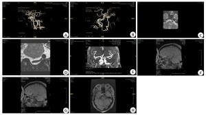
图 1 HR-MRI图像
A:3D磁共振血管造影,V4动脉瘤左侧后前位;B:3D磁共振血管造影,V4动脉瘤右侧后前位;C:T2WI呈低信号血管流空;D:T1WI+C示瘤壁强化均匀;E:3D磁共振血管造影,右侧C4段动脉瘤;F:T1WI,虚线内为等信号血栓影;G:T1WI,虚线内为低信号新月形流空影;H:T1WI+C示动脉瘤壁明显强化.
Figure 1. HR-MRI images.
表 1 颅内动脉瘤瘤体破裂与瘤壁强化的关系
Table 1. Relationship between tumor rupture and tumor wall enhancement of intracranial aneurysms (n)
组别 瘤体数 强化 未强化 强化率(%) 破裂组 35 35 0 100.00 未破裂组 66 26 40 39.39 χ2 35.122 P < 0.001  下载: 导出CSV
表 2 破裂组与未破裂组动脉瘤形态学特征比较
Table 2. Comparison of morphological characteristics of aneurysms between ruptured group and unruptured group
病变特征 破裂组(35个动脉瘤) 未破裂组(66个动脉瘤) χ2 P 动脉瘤位置 0.011 0.917 前循环 20 37 后循环 15 29 动脉瘤大小(mm) 0.008 0.929 < 5 14 27 ≥5 21 39 宽颈动脉瘤 1.987 0.159 是 28 44 否 7 22 子囊 30.676 < 0.001 有 26 12 无 9 54 AR 6.576 0.010 < 2 19 52 ≥2 16 14 SR 0.788 0.375 < 2 18 40 ≥2 17 26 AR:体颈比;SR:瘤体体率.
下载: 导出CSV
表 3 强化组与未强化组动脉瘤形态学特征比较
Table 3. Comparison of morphological characteristics of aneurysms between enhanced group and unenhanced group
病变特征 强化组(26个动脉瘤) 未强化(40个动脉瘤) χ2 P 动脉瘤位置 0.523 0.470 前循环 16 21 后循环 10 19 动脉瘤大小(mm) 0.703 0.402 < 5 9 18 ≥5 17 22 宽颈动脉瘤 0.127 0.722 是 18 26 否 8 14 子囊 22.564 <0.001 有 12 0 无 14 40 AR 15.967 <0.001 < 2 14 38 ≥2 12 2 SR 6.016 0.014 < 2 11 29 ≥2 15 11
下载: 导出CSV
-
[1] Can A, Castro VM, Yu S, et al. Antihyperglycemic agents are inversely associated with intracranial aneurysm rupture[J]. Stroke, 2018, 49(1): 34-9. doi: 10.1161/STROKEAHA.117.019249 [2] Ollikainen E, Tulamo R, Kaitainen S, et al. Macrophage infiltration in the saccular intracranial aneurysm wall as a response to locally lysed erythrocytes that promote degeneration[J]. J Neuropathol Exp Neurol, 2018, 77(10): 890-903. doi: 10.1093/jnen/nly068 [3] Chen Y, Fan H, He X, et al. China Intracranial Aneurysm Project (CIAP): protocol for a prospective cohort study of interventional treatment and craniotomy for unruptured aneurysms[J]. BMJ Open, 2018, 8(5): e019333. http://www.ncbi.nlm.nih.gov/pubmed/29794089 [4] 贾璐琼, 刘鹏, 吕明. 应用动态对比增强磁共振成像预警颅内动脉瘤破裂风险的研究进展[J]. 中华神经外科杂志, 2021, 37(1): 100-2. doi: 10.3760/cma.j.cn112050-20200224-00075 [5] 张翠兰, 郭书丽, 王延岗, 等. CTA与MRA在颅内不同部位动脉瘤诊断及破裂出血风险预测的价值研究[J]. 中国医学装备, 2019, 16(7): 84-7. doi: 10.3969/J.ISSN.1672-8270.2019.07.019 [6] 胡瑞婷, 韦武鹏, 李雪花, 等. 高分辨率磁共振成像与CT血管造影评价大脑中动脉狭窄和斑块性质的效果对比[J]. 广西医学, 2017, 39 (7): 994-7. https://www.cnki.com.cn/Article/CJFDTOTAL-GYYX201707019.htm [7] Feng X, Blemker SS, Inouye J, et al. Assessment of velopharyngeal function with dual-planar high-resolution real-time spiral dynamic MRI[J]. Magn Reson Med, 2018, 80(4): 1467-74. doi: 10.1002/mrm.27139 [8] 梁汉祥. 高分辨磁共振管壁成像技术在颅内动脉瘤破裂风险评估中的可行性[J]. 检验医学与临床, 2017, 14(3): 331-2, 335. doi: 10.3969/j.issn.1672-9455.2017.03.008 [9] 饶明俐. 《中国脑血管病防治指南》摘要(三) [J]. 中风与神经疾病杂志, 2006, 23(1): 4-8. doi: 10.3969/j.issn.1003-2754.2006.01.001 [10] Pawlowska E, Szczepanska J, Wisniewski K, et al. NF-κB-Mediated Inflammation in the Pathogenesis of Intracranial Aneurysm and Subarachnoid Hemorrhage. Does Autophagy Play a Role?[J]. Int J Mol Sci, 2018, 19(4): 1245. doi: 10.3390/ijms19041245 [11] Zhou G, Zhu Y, Yin Y, et al. Author Correction: Association of wall shear stress with intracranial aneurysm rupture: systematic review and meta-analysis[J]. Sci Rep, 2018, 8(1): 5244. doi: 10.1038/s41598-018-23445-9 [12] 吴俊, 刘清源, 王诺川, 等. 颅内动脉瘤形态学和血流动力学特点对夹闭术中动脉瘤破裂的预测作用[J]. 中华神经外科杂志, 2019, 35(3): 288-92. doi: 10.3760/cma.j.issn.1001-2346.2019.03.012 [13] Kirsch V, Ertl-Wagner B, Berman A, et al. High-resolution MRI of the inner ear enables syndrome differentiation and specific treatment of cerebellar downbeat nystagmus and secondary endolymphatic Hydrops in a postoperative ELST patient[J]. J Neurol, 2018, 265(1): 48-50. doi: 10.1007/s00415-018-8858-z [14] Lu X, Li C, Qu C, et al. A high resolution MRI study of the relationship between plaque enhancement and perforator stroke after stenting for symptomatic vertebrobasilar artery Stenosis[J]. J Stroke Cerebrovasc Dis, 2021, 30(3): 105558. doi: 10.1016/j.jstrokecerebrovasdis.2020.105558 [15] 付其昌, 程敬亮, 管生, 等. 高分辨率MRI在颅内动脉瘤评估中的应用进展[J]. 中华放射学杂志, 2020, 54(4): 372-5. doi: 10.3760/cma.j.cn112149-20190414-00158 [16] Freitag MT, Kesch C, Cardinale J, et al. Simultaneous whole-body 18F-PSMA-1007-PET/MRI with integrated high-resolution multiparametric imaging of the prostatic Fossa for comprehensive oncological staging of patients with prostate cancer: a pilot study [J]. Eur J Nucl Med Mol Imaging, 2018, 45(3): 340-7. doi: 10.1007/s00259-017-3854-6 [17] 张雪凤, 李帅, 史张, 等. 高分辨率MRI在大脑中动脉粥样硬化斑块药物治疗随访中的应用[J]. 中华放射学杂志, 2020, 54(4) : 318-24. doi: 10.3760/cma.j.cn112149-20190415-00288 [18] 李力, 段国礼, 赵瑞, 等. 颅内未破裂动脉瘤介入治疗术后神经系统并发症的危险因素分析[J]. 第二军医大学学报, 2018, 39(3): 238-44. https://www.cnki.com.cn/Article/CJFDTOTAL-DEJD201803003.htm [19] 王晖, 康凯. 直径≤7 mm颅内动脉瘤破裂的危险因素分析和预测模型构建[J]. 中国脑血管病杂志, 2018, 15(10): 523-9. doi: 10.3969/j.issn.1672-5921.2018.10.004 [20] 顾艳, 张永刚, 骆孟, 等. 四维CT血管成像对颅内小动脉瘤破裂风险因素的分析研究[J]. 中华放射学杂志, 2019, 53(6): 480-4. [21] 姜梦达, 刘玉, 陶晓峰, 等. 高分辨率CT、常规及功能MRI对颅底低级别软骨肉瘤的诊断价值[J]. 分子影像学杂志, 2021, 44(2): 213-8. doi: 10.12122/j.issn.1674-4500.2021.02.01 [22] 张梁, 周志斌. 高分辨率磁共振血管壁成像在颅内动脉粥样硬化性疾病中的应用[J]. 分子影像学杂志, 2020, 43(1): 45-8. doi: 10.12122/j.issn.1674-4500.2020.01.10 -

 点击查看大图

计量
- 文章访问数: 442
- HTML全文浏览量: 166
- PDF下载量: 5
- 被引次数: 0