


Correlation of MRI findings with vasculogenic mimicry and reticular fibers in diffuse large B-cell lymphoma
-
摘要:
目的分析颅内弥漫大B细胞淋巴瘤(DLBCL)MRI征象与血管生成拟态(VM)、网状纤维的相关性。 方法收集2017年3月~2020年4月经我院确诊并做MRI检查的颅内DLBCL患者93例。分别记录所有患者MRI征象,并对肿瘤标本进行染色、分析,不同性质VM及网状纤维的强化指数,VM、网状纤维与强化指数的关系,MRI表现与血管生成拟态、网状纤维的关系。 结果肿瘤内微血管密度42.42±18.78 mm2,病灶成熟血管数目9.65±4.02 mm2。所有颅内患者中单发62例,多发31例,共149个病灶;93例MRI结果中53个T1WI呈等信号、40个稍低信号;44个T2WI呈等信号、49个稍高信号;所有病灶DWI呈高信号;62个分叶征病灶,47个脐凹征病灶,31个尖角征病灶,26个握拳征病灶;18个脑膜浸润病灶;所有病灶均有PAS染色阳性索状或网状结构,其中53个强阳性,40个弱阳性;网状纤维染色:49个病灶(强阳性)内分布较多网状纤维;44个病灶(弱阳性)中散布网状纤维;所有患者均有强化,强化指数为0.996±0.368。患者微血管密度与强化指数呈正相关(P < 0.05),强化指数与成熟血管数量正相关(P < 0.05)。VM弱阳性组强化指数低于强阳性组(P < 0.05);网状纤维弱阳性组强化指数低于强阳性组(P < 0.05)。VM、网状纤维与强化指数呈明确的正相关(P < 0.05)。坏死MRI表现与VM呈负相关(P < 0.05)。 结论DLBCL MRI表现坏死与VM呈负相关,DLBCL在MRI表现中有一定特点,可按其表现进行诊断。 -
关键词:
- 颅内弥漫大B细胞淋巴瘤 /
- MRI征象 /
- 血管生成拟态 /
- 网状纤维
Abstract:ObjectiveTo analyze the correlation between MRI findings of intracranial diffuse large B-cell lymphoma (DLBCL) and vasculogenic mimicry(VM), reticular fiber. MethodsNinety-three patients with intracranial DLBCL who were diagnosed and underwent MRI examination in our hospital from March 2017 to April 2020 were collected. The MRI signs of all patients were recorded, and the tumor specimens were stained and analyzed. We analyzed the enhancement index of VM and reticular fiber with different properties, the relationship between VM, reticular fiber and enhancement index, and the relationship between MRI findings and vasculogenic mimicry, reticular fiber. ResultsThe density of microvessels in the tumor was 42.42± 18.78 mm2, and the number of mature vessels in the lesion was 9.65±4.02 mm2. Among all intracranial patients, there were 62 single cases and 31 multiple cases, with a total of 149 lesions. Among the 93 MRI results, 53 T1WI showed isointensity and 40 slightly hypointensity. 44 T2WI showed equal signal and 49 showed slightly higher signal. All lesions showed high DWI signal. There were 62 lobulation sign lesions, 47 umbilical concave sign lesions, 31 sharp Angle sign lesions and 26 clenched fist sign lesions.18 meningeal infiltrating lesions. PAS staining was positive in all lesions, including 53 strong positive lesions and 40 weak positive lesions. Reticular fiber staining: 49 lesions (strongly positive) had more reticular fibers. Reticular fibers were scattered in 44 lesions (weakly positive). All patients had enhancement, enhancement index 0.996 ± 0.368. The microvessel density of patients was positively correlated with the enhancement index(P < 0.05), and the enhancement index of patients was positively correlated with the number of mature vessels (P < 0.05). The enhancement index of weak positive VM group was lower than that of strong positive VM group (P < 0.05). The strengthening index of weak positive reticular fiber group was lower than that of strong positive group (P < 0.05). VM, reticular fiber and enhancement index were positively correlated(P < 0.05). MRI findings of necrosis were negatively correlated with VM (P < 0.05). ConclusionThe MRI manifestation necrosis of DLBCL is negatively correlated with VM. DLBCL has certain characteristics in MRI manifestation and can be diagnosed according to its manifestation. -
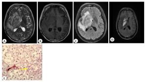
图 1 患者女,66岁,MRI图像及病理图片
A~D: MRI图像中右侧基底节区见片状T2WI高-低混杂信号,右侧侧脑室受压变窄,脑中线略左偏,增强扫描呈明显强化,大小约5.2 cm×2.8 cm×3.6 cm,向上至右侧侧脑室顶部,向下累及右侧海马,中脑;E: PAS+CD34染色(×400)示红染网状结构为PAS阳性结构,呈“背靠背”样环形排列,为图案基质型血管生成拟态.
Figure 1. MRI and pathological images of a 66-year-old female patient

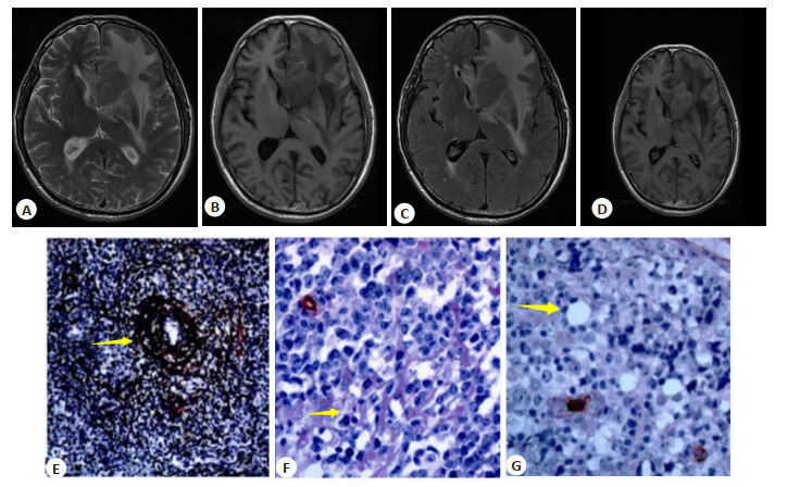
图 2 患者男,71岁,MRI图像及病理图片
A~D: MRI像中左额叶示不规则团块状异常信号,侵犯胼胝体膝部左侧,T2WI呈稍高信号,局部脑实质及侧脑室受压,中线结构稍右移;增强扫描:左侧额叶明显强化,大小约3.6 cm×3.0 cm×3.0 cm; E: Gordon-Sweet染色(×200),肿瘤内网状纤维丰富,肿瘤细胞“套袖样”浸润于血管周围,肿瘤间内见呈环形、放射状排列的网状纤维(白色箭头);F: PAS+CD34染色(×400),肿瘤内呈索状PAS阳性结构,相互连接呈网状,为图案基质型血管生成拟态(黑色箭头);G: 肿瘤细胞围成的小圆形结构,内衬薄层PAS红染基膜(×300),为管道型为图案基质型血管生成拟态.
Figure 2. MRI and pathological images of a 71-year-old male patient
表 1 不同性质VM及网状纤维的强化指数比较
Table 1. Comparison of enhancement index of VM and reticular fiber with different properties(Mean±SD)
指标 强化指数 t P 血管生成拟态 6.163 < 0.05 弱阳性(n=53) 0.75±0.37 强阳性(n=40) 1.16±0.23 网状纤维 5.158 < 0.05 弱阳性(n=49) 0.81±0.34 强阳性(n=44) 1.15±0.29 VM:血管生成拟态.  下载: 导出CSV
表 2 MRI表现与血管生成拟态、网状纤维的关系
Table 2. Correlation of MRI findings with vasculogenic mimicry and reticular fibers
MRI表现 血管生成拟态 网状纤维 r P r P 分叶征 0.000 0.532 0.133 0.545 握拳征 0.126 0.351 -0.040 0.879 尖角征 -0.079 0.182 -0.031 0.894 脐凹征 0.126 0.704 0.043 0.844 坏死 -0.174 0.013 -0.115 0.614 脑膜浸润 -0.068 0.051 -0.021 0.923
下载: 导出CSV
-
[1] 于宝华, 薛田, 张岩, 等. 弥漫性大B细胞淋巴瘤中MYD88基因突变及其临床病理相关性分析[J]. 中国癌症杂志, 2018, 28(9): 679-85. https://www.cnki.com.cn/Article/CJFDTOTAL-ZGAZ201809009.htm [2] Hasturk AE, Eyupoglu EE, Gel G, et al. P14.14 Scalp invasion of diffuse large b-cell lymphoma without systemic involvement[J]. Neuro-Oncology, 2019, 21(Supplement_3): 69. http://www.researchgate.net/publication/335673270_P1414_Scalp_invasion_of_diffuse_large_b-cell_lymphoma_without_systemic_involvement [3] 张婷婷, 王先火, 孟斌, 等. A2aR在弥漫大B细胞淋巴瘤组织和CD8+T细胞上的表达和预后价值的研究[J]. 中国肿瘤临床, 2018, 45 (13): 673-7. doi: 10.3969/j.issn.1000-8179.2018.13.365 [4] 刘巧遇. 肿瘤血管生成拟态的分子病理和影像学研究进展[J]. 放射学实践, 2019, 34(5): 565-8. https://www.cnki.com.cn/Article/CJFDTOTAL-FSXS201905018.htm [5] 饶慕圣, 曹胜华, 徐兴东, 等. 肿瘤细胞主导的血管生成拟态与肿瘤侵袭转移[J]. 国际外科学杂志, 2019(1): 60-3, 封4. doi: 10.3760/cma.j.issn.1673-4203.2019.01.016 [6] 殷仁斌, 刘玉霞, 任伟丹, 等. 经典型霍奇金淋巴瘤累及骨髓的临床病理分析[J]. 诊断病理学杂志, 2020, 27(10): 702-5. doi: 10.3969/j.issn.1007-8096.2020.10.004 [7] 李明, 谭诗云. 肿瘤相关成纤维细胞对大肠癌LoVo细胞增殖和侵袭能力的影响[J]. 胃肠病学和肝病学杂志, 2019, 28(5): 514-8. doi: 10.3969/j.issn.1006-5709.2019.05.007 [8] Ma S, Sen SS, Jug R, et al. Adrenal relapse of primary central nervous system diffuse large B-cell lymphoma: a case report[J]. Medicine: Baltimore, 2018, 97(38): e12482. doi: 10.1097/MD.0000000000012482 [9] Folberg R, Hendrix MJ, Maniotis AJ. Vasculogenic mimicry and tumor angiogenesis[J]. Am J Pathol, 2000, 156(2): 361-81. doi: 10.1016/S0002-9440(10)64739-6 [10] Xu T, Jia Q, Wang Y, et al. Rare cases of primary central nervous system anaplastic variant of diffuse large B-cell lymphoma[J]. Diagn Pathol, 2019, 14(1): 45. doi: 10.1186/s13000-019-0826-0 [11] 林巧贤, 杨阿碰, 曾志勇, 等. 原发中枢神经系统弥漫大B细胞淋巴瘤的临床特征及预后因素分析[J]. 福建医科大学学报, 2019, 53(4): 229-34. https://www.cnki.com.cn/Article/CJFDTOTAL-FJYD201904005.htm [12] 严金海, 罗东兰, 张芬, 等. 心房黏液瘤伴纤维素相关弥漫性大B细胞淋巴瘤六例临床病理学特征[J]. 中华病理学杂志, 2020, 49(10): 1027-30. doi: 10.3760/cma.j.cn112151-20200302-00160 [13] Lin FH, Zhang XH, Zhang J, et al. Fluorescein sodium-guided biopsy or resection in primary central nervous system lymphomas with contrast-enhancing lesion in MRI[J]. J Neuro Oncol, 2018, 139 (3): 757-65. doi: 10.1007/s11060-018-2924-3 [14] Santiago R, Ortiz Jimenez J, Forghani R, et al. Prediction of highrisk group of primary refractory diffuse large B-cell lymphoma (DLBCL) patients using a CT-based radiomics model with machine learning[J]. Blood, 2019, 134(Supplement_1): 4136. doi: 10.1182/blood-2019-126745 [15] 秦英. 弥漫大B细胞淋巴瘤中HK-Ⅱ、TNFAIP3异常表达与肿瘤细胞恶性特征的相关性[J]. 海南医学院学报, 2018, 24(22): 2015-8, 2022. https://www.cnki.com.cn/Article/CJFDTOTAL-HNYY201822018.htm [16] 樊静, 许国庆, 王蓓, 等. 乳腺癌超声造影特征与血管生成拟态的相关性分析[J]. 临床超声医学杂志, 2020, 22(2): 125-8. doi: 10.3969/j.issn.1008-6978.2020.02.017 [17] 陶明珠, 胡书娅, 郭庆喜, 等. 肝细胞癌组织中肿瘤相关成纤维细胞与血管生成拟态的相关性及临床意义[J]. 现代肿瘤医学, 2019, 27(23): 4234-7. doi: 10.3969/j.issn.1672-4992.2019.23.025 [18] Meling TR, Latysheva A, Da Broi M, et al. Is deep brain involvement in intracranial primary central nervous system lymphoma of importance for penetration of chemotherapeutic agents?[J]. Neuroradiology, 2018, 60(7): 703-13. doi: 10.1007/s00234-018-2038-9 [19] Shen G, Kou Y, Liu B, et al. Multiple subcutaneous nodules as initial presentation of diffuse large B-cell non-Hodgkin lymphoma (DLBCL) detected by PET/CT[J]. Clin Nucl Med, 2018, 43(10): 759-61. doi: 10.1097/RLU.0000000000002221 [20] 王雪晴, 王国庆, 许娇娇, 等. 良恶性子宫平滑肌肿瘤坏死组织中网状纤维及masson染色评分差异研究[J]. 中国妇产科临床杂志, 2019, 20 (4): 354-5. [21] Muly S, Liu S, Lee R, et al. MRI of intracranial intraventricular lesions[J]. Clin Imaging, 2018, 52: 226-39. doi: 10.1016/j.clinimag.2018.07.021 [22] 张金彩, 时传迎, 李明利, 等. 原发性中枢神经系统血管内淋巴瘤的影像表现: 附4例报道并文献复习[J]. 磁共振成像, 2020, 11(10): 900-3. doi: 10.12015/issn.1674-8034.2020.10.013 [23] Chen Y, Wang X, Li L, et al. Correction to: Differential diagnosis of sinonasal extranodal NK/T cell lymphoma and diffuse large B cell lymphoma on MRI[J]. Neuroradiology, 2020, 62(9): 1201. doi: 10.1007/s00234-020-02488-8 [24] Choi EJ, Jin GY, Chung MJ. Serial chest CT findings of intravascular large B-cell lymphoma of the lungs[J]. J Thorac Dis, 2018, 10(3): E218-20. DOI: 10.21037/jtd.2018.02.44. [25] 张玉琴, 邓艳, 何茂远, 等. 原发性中枢神经系统淋巴瘤的多模态磁共振成像诊断价值[J]. 中国医师进修杂志, 2019, 42(11): 1013-6. doi: 10.3760/cma.j.issn.1673-4904.2019.11.012 [26] 邹少洲, 胡国栋, 蔡迎迎, 等. 以呼吸道病变首诊的弥漫大B细胞淋巴瘤的诊治[J]. 分子影像学杂志, 2019, 42(4): 457-60. doi: 10.12122/j.issn.1674-4500.2019.04.08 -

 点击查看大图
计量
- 文章访问数: 685
- HTML全文浏览量: 361
- PDF下载量: 7
- 被引次数: 0