


Comparation of MRI, multi-slice spiral CT and digital subtraction angiography in the clinical diagnosis of acute cerebral infarction
-
摘要:
目的探讨MRI、多层螺旋CT(MSCT)与数字减影血管造影(DSA)在急性脑梗死临床诊断中的应用。 方法对本院2019年3月~2020年12月100例疑似急性脑梗死患者的临床资料进行回顾性分析,所有患者均接受MRI、MSCT及DSA检查,以DSA结果为金标准,分析MRI、MSCT诊断结果与DSA结果的一致性,计算MRI、MSCT诊断急性脑梗死的准确度、特异性、敏感度、阳性预测值、阴性预测值。 结果DSA诊断结果显示,100例疑似急性脑梗死患者中,急性脑梗死患者79例,非急性脑梗死患者21例;MRI诊断结果显示,急性脑梗死患者77例,非急性脑梗死患者23例,与DSA诊断结果一致性分析,Kappa值为0.637;MSCT诊断结果显示,急性脑梗死患者71例,非急性脑梗死患者29例,与DSA诊断结果一致性分析,Kappa值为0.524。MRI对急性脑梗死的诊断准确度、敏感度分别为92.00%、93.67%均高于MSCT的82.00%、83.54%(P < 0.05)。MRI对发病24 h内、发病72 h内检出率分别为89.29%、87.50%,高于MSCT的64.29%、70.83%(P < 0.05)。 结论MRI、MSCT对急性脑梗死的诊断结果与DSA具有较好的一致性,但MRI诊断优势更加明显,可为临床尽早拟定治疗方案提供参考。 Abstract:ObjectiveTo explore the comparative analysis of magnetic resonance imaging (MRI), multi-slice spiral CT (MSCT) and digital subtraction angiography (DSA) in the clinical diagnosis of acute cerebral infarction (ACI). MethodsThe clinical data of 100 patients with suspected ACI in the hospital from March 2019 to December 2020 were retrospectively analyzed. All patients underwent MRI, MSCT and DSA. Taking DSA results as the golden standard, consistency of diagnosis results between MRI, MSCT and DSA were analyzed. The accuracy, specificity and sensitivity of MRI and MSCT in the diagnosis of ACI were calculated. ResultsThe diagnosis results of DSA showed that among the 100 patients with suspected ACI, 79 cases with ACI and 21 cases without. The diagnosis results of MRI showed that 77 cases with ACI and 23 cases without. Consistency analysis between DSA and MRI showed that Kappa value was 0.637, indicating good consistency. The diagnosis results of MSCT showed that 71 cases with ACI and 29 cases without. Consistency analysis between DSA and MSCT showed that Kappa value was 0.524, indicating good consistency. The accuracy and sensitivity of MRI in the diagnosis of ACI were higher than those in control group (P < 0.05). The detection rates of onset within 24 h and 72 h by MRI were higher than those by MSCT(P < 0.05). ConclusionThe consistency of diagnosis results between MRI, MSCT and DSA is good for ACI. However, diagnosis advantages of MRI are more significant, which can provide reference for developing clinical treatment regimens as early as possible. -
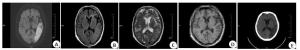
图 1 急性脑梗死患者MRI及MSCT检查
A:MRI显示左侧颞枕叶见片状等长T1长T2异常信号影;B:MRI显示自由水抑制像呈高信号;C:DWI像左侧颞枕叶见大片状高信号;D:MSCT显示左侧颞枕叶见片状低密度影;E:MSCT显示边缘模糊.
Figure 1. MRI and MSCT examination for patients with acute cerebral infarction.
表 1 MRI、MSCT诊断结果与DSA诊断结果的一致性分析
Table 1. Comparison of diagnosis results between MRI, MSCT and DSA(n)
诊断方法 类型 DSA Kappa值 阳性 阴性 MRI 0.637 阳性 74 3 阴性 5 18 MSCT 0.524 阳性 66 5 阴性 13 16 MSCT: 多层螺旋CT; DSA: 数字减影血管造影.  下载: 导出CSV
表 2 MRI、MSCT对急性脑梗死的诊断效能
Table 2. Diagnostic efficiency of MRI and MSCT for acute cerebral infarction(%)
诊断方法 准确度 敏感度 特异性 阳性预测值 阴性预测值 MRI 92.00(92/100) 93.67(74/79) 85.71(18/21) 96.10(74/77) 78.26(18/23) MSCT 82.00(82/100) 83.54(66/79) 76.19(16/21) 92.96(66/71) 55.17(16/29) χ2 4.421 4.013 0.618 0.715 3.021 P 0.036 0.045 0.432 0.398 0.082
下载: 导出CSV
表 3 MRI、MSCT对不同发病时间的诊断情况比较
Table 3. Comparison of diagnostic conditions between MRI and MSCT at different onset time[n(%)]
诊断方法 发病时间 24 h内(n=28) 72 h内(n=48) 3~5 d(n=19) >5 d(n=5) MRI 25(89.29) 42(87.50) 5(26.32) 2(40.00) MSCT 18(64.29) 34(70.83) 10(52.63) 4(80.00) χ2 4.139 4.042 2.754 1.667 P 0.042 0.044 0.097 0.197
下载: 导出CSV
-
[1] Song TJ, Chang Y, Chun MY, et al. High dietary glycemic load is associated with poor functional outcome in patients with acute cerebral infarction[J]. J Clin Neurol, 2018, 14(2): 165-73. doi: 10.3988/jcn.2018.14.2.165 [2] Liang Y, Wu J, Liu JQ, et al. The clinical implications of thrombelastography in the diagnosis of acute cerebral infarction[J]. Clin Lab, 2018, 64(1): 147-52. http://europepmc.org/abstract/MED/29479891 [3] Zeng JQ, Wang F, Feng HS, et al. Influencing factors of recanalization after intravenous thrombolysis with urokinase in acute cerebral infarction patients[J]. Eur Neurol, 2020, 83(2): 162-6. doi: 10.1159/000507288 [4] 吴雅蔚, 叶靖, 征锦, 等. 动态对比增强磁共振成像联合NLR对急性缺血性脑梗死预后的预测价值[J]. 实用放射学杂志, 2020, 36(7): 1027-30. doi: 10.3969/j.issn.1002-1671.2020.07.004 [5] Yu YJ, Xiong W. Tirofiban combined with rt- PA intraarterial thrombolysis improves the recanalization rate of acute middle cerebral artery occlusion in rabbits[J]. Eur Rev Med Pharmacol Sci, 2018, 22(9): 2888-95. http://europepmc.org/abstract/MED/29771445 [6] 张海峰. CT与MRI在老年出血性脑梗死患者中的诊断价值[J]. 中国实用医刊, 2017, 44(7): 81-3. doi: 10.3760/cma.j.issn.1674-4756.2017.07.026 [7] 高安生. DWI与MSCT在急性脑梗死患者诊断中的比较分析[J]. 医药论坛杂志, 2018, 39(9): 39-40, 44. https://www.cnki.com.cn/Article/CJFDTOTAL-HYYX201809013.htm [8] 林靖复. MRI诊断急性脑梗死的价值及梗死病灶ADC值的变化[J]. 中国医学物理学杂志, 2019, 36(6): 693-6. doi: 10.3969/j.issn.1005-202X.2019.06.014 [9] 中国脑血管病防治指南编写委员会. 中国脑血管病防治指南: 试行版[M]. 北京: 人民卫生出版社, 2007: 125-127. [10] Liu X, Rao S, Wang J. Intravenous thrombolysis in combination with mild hypothermia therapy in the treatment of acute cerebral infarction[J]. Pak J Med Sci, 2019, 35(4): 1161-6. [11] Lv G, Wang GQ, Xia ZX, et al. Influences of blood lipids on the occurrence and prognosis of hemorrhagic transformation after acute cerebral infarction: a case-control study of 732 patients[J]. Mil Med Res, 2019, 6(1): 2. http://www.cnki.com.cn/Article/CJFDTotal-JYDX201903003.htm [12] Wang T, Gong Y, Shi YB, et al. Feasibility of dual- low scheme combined with iterative reconstruction technique in acute cerebral infarction volume CT whole brain perfusion imaging[J]. Exp Ther Med, 2017, 14(1): 163-8. doi: 10.3892/etm.2017.4451 [13] 冯旭霞, 景赟航, 李转霞, 等. 多层螺旋CT与磁共振成像对原发性颅脑肿瘤的诊断对比[J]. 山西医药杂志, 2020, 49(13): 1662-4. https://www.cnki.com.cn/Article/CJFDTOTAL-SXYY202013010.htm [14] Shinohara Y, Kato A, Kuya K, et al. Perfusion MR imaging using a 3D pulsed continuous arterial spin- labeling method for acute cerebral infarction classified as branch atheromatous disease involving the lenticulostriate artery territory[J]. AJNR Am J Neuroradiol, 2017, 38(8): 1550-4. doi: 10.3174/ajnr.A5247 [15] Carré A, Klausner G, Edjlali M, et al. Standardization of brain MR images across machines and protocols: bridging the gap for MRIbased radiomics[J]. Sci Rep, 2020, 10(1): 12340. doi: 10.1038/s41598-020-69298-z [16] 郭华峰, 王晓男, 杨璐. 多层螺旋CT联合MRI检查在脑梗死合并脑出血诊断中的应用[J]. 实用临床医药杂志, 2020, 24(23): 8-10, 14. [17] 许涛, 郭静, 由秀, 等. 磁共振成像和CT对短暂性脑缺血发作患者的急性脑梗死诊断价值研究[J]. 中国医学装备, 2021, 18(1): 54-8. doi: 10.3969/J.ISSN.1672-8270.2021.01.014 [18] Nakajo Y, Zhao Q, Enmi JI, et al. Early detection of cerebral infarction after focal ischemia using a new MRI indicator[J]. Mol Neurobiol, 2019, 56(1): 658-70. doi: 10.1007/s12035-018-1073-1 [19] 刘美, 周凌燕. CT联合MRI对老年多发性急性期脑梗死患者的临床诊断价值[J]. 中国CT和MRI杂志, 2021, 19(2): 29-31. doi: 10.3969/j.issn.1672-5131.2021.02.008 [20] 赵仕懂, 丘武应, 朱志铿, 等. MSCT与MRI检查在脑梗死患者临床诊断价值[J]. 现代医用影像学, 2017, 26(5): 1210-2. https://www.cnki.com.cn/Article/CJFDTOTAL-XDYY201705021.htm [21] Hori M, Irie R, Suzuki M, et al. Teaching neuroimages: obscured cerebral infarction on MRI[J]. Clin Neuroradiol, 2017, 27(4): 519- 20. doi: 10.1007/s00062-017-0576-x [22] Zhang WB, Cheng JL, Zhang Y, et al. Analysis of CT and MRI combined examination for the diagnosis of acute cerebral infarction [J]. J Coll Physicians Surg Pak, 2019, 29(9): 898-9. doi: 10.29271/jcpsp.2019.09.898 [23] 张梁, 周志斌. 高分辨率磁共振血管壁成像在颅内动脉粥样硬化性疾病中的应用[J]. 分子影像学杂志, 2020, 43(1): 45-8. doi: 10.12122/j.issn.1674-4500.2020.01.10 [24] 刘国昌. MRI与CT在急性脑梗死早期诊断价值探讨[J]. 影像研究与医学应用, 2020, 4(9): 237-8. https://www.cnki.com.cn/Article/CJFDTOTAL-YXYY202009149.htm -

 点击查看大图
计量
- 文章访问数: 442
- HTML全文浏览量: 159
- PDF下载量: 1
- 被引次数: 0