


Establishment of peripheral nerve acute crush injury animal model and b value optimization of diffusion kurtosis imaging
-
摘要:
目的制作适合MRI监测的兔坐骨神经挤压伤模型,并对扩散峰度成像(DKI)b值的选择进行探讨。 方法选用健康新西兰大白兔27只,使用宽度约8 mm的自制扁嘴钳建立坐骨神经损伤模型,选取右后肢为手术侧,左侧为假手术侧,于术前及1 d、3 d、1周、2周、4周及8周各时间点行DKI扫描,b值分别为0、750、1500 s/mm2及0、1000、2000 s/mm2。于各时间点随机取2只兔子进行病理检查。 结果DKI1500与DKI2000各参数值具有相似的变化趋势。FA1500与FA2000均在第1天降至最低,之后在3 d~8周持续回升,各时间点组间差异均有统计学意义(P < 0.05);在第1天均明显下降,此后开始缓慢、曲折上升的趋势,双侧MK1500值在术后第2周(P=0.022)、第4周(P=0.018)、第6周(P=0.016)及第8周(P=0.016)差异有统计学意义。而MK2000仅在第4周组间差异有统计学意义(P=0.002)。RK值及AK值在绝大多数时间点差异无统计学意义。 结论使用扁嘴钳钳夹兔坐骨神经中段制作损伤与修复模型,可方便使用MRI对损伤段神经进行直接监测与定量测量;DKI周围神经成像最大b值取1500 s/mm2可能较2000 s/mm2更为合适。 Abstract:ObjectiveTo establish a rabbit sciatic nerve crush injury model suitable for MRI monitoring, and discuss the selection of b value of diffusion kurtosis imaging (DKI). MethodsTwenty-seven healthy New Zealand white rabbits were selected. The sciatic nerve injury model was established with self-made flat forceps with a width of about 8mm. The right posterior limb was selected as the injury side and the left was the sham surgical side. DKI was performed before and 1 d, 3 d, 1 week, 2 weeks, 4 weeks and 8 weeks after injury, with b values of 0, 750, 1500 s/mm2 and 0, 1000, 2000 s/mm2, respectively. Two rabbits were randomly selected at each time point for pathological examination. ResultsThe parameters of DKI1500 and DKI2000 showed similar trends. Both FA1500 and FA2000 decreased to the lowest level on the first day and then continued to increase at 3-8 weeks, with statistically significant differences among groups at each time point (P < 0.05). The MK1500 of both sides was significantly decreased on the first day, and then slowly increased. The differences in the MK1500 of both sides at the second week (P=0.022), the fourth week (P=0.018), the sixth week (P=0.016) and the eighth week (P=0.016) after injury were significant. The difference of MK2000 between groups was significant only at the fourth week (P=0.002). There was no significant difference between Rk and AK at most time points. ConclusionUsing flat forceps to clamp the middle segment of the rabbit sciatic nerve to make the injury model can facilitate the direct monitoring and quantitative measurement of the injured nerve by MRI. A maximum b value of 1500 s/mm2 for DKI imaging may be more suitable than 2000 s/mm2. -
Key words:
- peripheral nerve /
- animal model /
- magnetic resonance imaging /
- diffusion kurtosis imaging
-
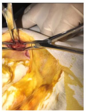
图 1 兔坐骨神经急性挤压伤模型制作
扁嘴钳钳夹30 s后兔损伤段坐骨神经,局部神经变薄变扁,呈半透明状,长度约8 mm;两端夹痕边界清楚,神经表面血管破裂出血.
Figure 1. Establishment of rabbit model of acute sciatic nerve crush injury.
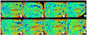
图 2 损伤前后兔坐骨神经DKI2000序列FA图
A: 损伤前,FA图清晰显示双侧坐骨神经平行走行;B: 术后第1天,坐骨神经损伤段(箭头)FA值明显降低,形态较前模糊,假手术侧坐骨神经FA值亦不均匀减低;C: 术后第3天,损伤段坐骨神经FA值较第1天稍升高,假手术侧FA值较前均匀,信号升高; D: 术后第1周,损伤段坐骨神经FA值升高,假手术侧FA值较前升高,信号基本均匀; E~H: 术后第2、4、6、8周,双侧坐骨神经FA值逐渐恢复.
Figure 2. Rabbit sciatic nerve on FA map of DKI2000 before and after crush injury.
表 1 DKI2000与DKI1500序列坐骨神经FA值比较
Table 1. Comparison of FA values between DKI2000 and DKI1500(Mean±SD)
术后时间 FA2000 FA1500 损伤侧(n=11) 假手术侧(n=11) 损伤侧(n=11) 假手术侧(n=11) 0 d 0.635±0.060 0.635±0.043 0.642±0.042 0.651±0.056 1 d 0.254±0.044* 0.343±0.062 0.297±0.050* 0.375±0.045 3 d 0.323±0.054* 0.425±0.069 0.328±0.071* 0.432±0.054 1周 0.344±0.065* 0.459±0.050 0.360±0.078* 0.452±0.054 2周 0.429±0.047* 0.535±0.048 0.419±0.049* 0.552±0.087 4周 0.481±0.031* 0.597±0.046 0.487±0.034* 0.595±0.033 6周 0.520±0.049* 0.639±0.039 0.540±0.082* 0.641±0.040 8周 0.538±0.066* 0.655±0.054 0.534±0.073* 0.635±0.027 *P < 0.05 vs假手术侧.  下载: 导出CSV
表 2 DKI2000与DKI1500序列坐骨神经MK值比较
Table 2. Comparison of MK values between DKI2000 and DKI1500(Mean±SD)
术后时间 MK2000 MK1500 损伤侧(n=11) 假手术侧(n=11) 损伤侧(n=11) 假手术侧(n=11) 0 d 1.326±0.377 1.180±0.415 1.516±0.429 1.747±0.394 1 d 0.733±0.190 0.831±0.178 0.829±0.352 0.818±0.242 3 d 0.768±0.209 0.734±0.159 0.935±0.369 1.153±0.370 1周 0.757±0.334 0.945±0.281 0.842±0.383 1.141±0.536 2周 0.932±0.448 1.377±0.574 0.836±0.345* 1.229±0.395 4周 0.795±0.274* 1.391±0.501 0.998±0.317* 1.467±0.514 6周 0.982±0.431 1.309±0.320 1.122±0.384* 1.611±0.480 8周 1.047±0.483 1.338±0.404 1.070±0.283* 1.399±0.301 *P < 0.05 vs假手术侧.
下载: 导出CSV
表 3 DKI2000与DKI1500序列坐骨神经RK值比较
Table 3. Comparison of RK values between DKI2000 and DKI1500(Mean±SD)
术后时间 RK2000 RK1500 损伤侧(n=11) 假手术侧(n=11) 损伤侧(n=11) 假手术侧(n=11) 0 d 1.398±0.427 1.422±0.541 1.781±0.519 1.912±0.520 1 d 0.834±0.322 1.087±0.311 1.095±0.495 1.060±0.341 3 d 1.127±0.476 1.062±0.410 1.292±0.586 1.648±0.395 1周 1.043±0.618 1.185±0.458 1.204±0.383 1.424±0.748 2周 1.199±0.493 1.631±0.739 1.198±0.287 1.505±0.516 4周 1.018±0.423 1.461±0.632 1.228±0.516 1.600±0.514 6周 1.025±0.448* 1.454±0.322 1.330±0.491* 1.806±0.539 8周 1.216±0.636 1.317±0.485 1.269±0.389 1.456±0.415 *P < 0.05 vs假手术侧.
下载: 导出CSV
表 4 DKI2000与DKI1500序列坐骨神经AK值比较
Table 4. Comparison of AK values between DKI2000 and DKI1500(Mean±SD)
术后时间 AK2000 AK1500 损伤侧(n=11) 假手术侧(n=11) 损伤侧(n=11) 假手术侧(n=11) 0 d 1.361±0.319 1.373±0.522 1.355±0.644 1.150±0.644 1 d 1.366±0.754 1.223±0.454 1.278±0.621 1.087±0.637 3 d 1.220±0.570 1.253±0.817 1.272±0.741 1.141±0.362 1周 1.208±0.669 1.162±0.445 1.017±0.547 1.316±0.535 2周 1.315±0.486 1.005±0.536 1.372±0.397 1.220±0.472 4周 1.375±0.483 1.352±0.620 1.469±0.559 1.263±0.677 6周 1.519±0.586 1.371±0.309 1.254±0.510 1.276±0.548 8周 1.273±0.620 1.787±0.746 1.310±0.411 1.546±0.551
下载: 导出CSV
-
[1] Kouyoumdjian J, Graç C, Ferreira VM. Peripheral nerve injuries: a retrospective survey of 1124 cases[J]. Neurol India, 2017, 65(3): 551. doi: 10.4103/neuroindia.NI_987_16 [2] 王艳, 王茜. 周围神经损伤动物模型的研究进展[J]. 中国康复理论与实践, 2014, 20(6): 537-9. doi: 10.3969/j.issn.1006-9771.2014.06.008 [3] Sun C, Hou Z, Hong G, et al. In vivo evaluation of sciatic nerve crush injury using diffusion tensor imaging: correlation with nerve function and histology[J]. J Comput Assist Tomogr, 2014, 38(5): 790-6. doi: 10.1097/RCT.0000000000000118 [4] Andersson G, Orädd G, Sultan F, et al. In vivo diffusion tensor imaging, diffusion kurtosis imaging, and tractography of a sciatic nerve injury model in rat at 9.4T[J]. Sci Rep, 2018, 8(1): 12911. doi: 10.1038/s41598-018-30961-1 [5] 李俊, 岳茜, 肖如辉, 等. MRI在周围神经病变中的应用进展[J]. 中国医学影像技术, 2019, 35(3): 455-8. https://www.cnki.com.cn/Article/CJFDTOTAL-ZYXX201903051.htm [6] Rosenkrantz AB, Padhani AR, Chenevert TL, et al. Body diffusion kurtosis imaging: Basic principles, applications, and considerations for clinical practice[J]. J Magn Reson Imaging, 2015, 42(5): 1190-202. doi: 10.1002/jmri.24985 [7] Wan Q, Zeng Q, Li X, et al. Development of a rabbit model of radiation-induced sciatic nerve injury: in vivo evaluation using T2 relaxation time measurements[J]. J Comput Assist Tomogr, 2015, 39 (4): 613-8. doi: 10.1097/RCT.0000000000000241 [8] Li X, Shen J, Chen J, et al. Magnetic resonance imaging evaluation of acute crush injury of rabbit sciatic nerve: correlation with histology[J]. Can Assoc Radiol J, 2008, 59(3): 123-30. http://europepmc.org/abstract/med/18697718 [9] Morisaki S, Kawai Y, Umeda M, et al. In vivo assessment of peripheral nerve regeneration by diffusion tensor imaging[J]. J Magn Reson Imaging, 2011, 33(3): 535-42. doi: 10.1002/jmri.22442 [10] Luís AL, Amado S, Geuna S, et al. Long-term functional and morphological assessment of a standardized rat sciatic nerve crush injury with a non-serrated clamp[J]. J Neurosci Methods, 2007, 163 (1): 92-104. doi: 10.1016/j.jneumeth.2007.02.017 [11] 李登科. 电针对坐骨神经损伤家兔的组织形态学及原癌基因C-FOS表达的影响[D]. 咸阳: 陕西中医药大学, 2007. [12] Takagi T, Nakamura M, Yamada M, et al. Visualization of peripheral nerve degeneration and regeneration: Monitoring with diffusion tensor tractography[J]. NeuroImage, 2009, 44(3): 884-92. doi: 10.1016/j.neuroimage.2008.09.022 [13] 伍鹏欢, 黄成燕, 史本超. 磁共振DTI技术在周围神经系统疾病诊断中的进展[J]. 分子影像学杂志, 2019, 42(3): 294-6. doi: 10.12122/j.issn.1674-4500.2019.03.03 [14] 方可薇, 王艺蓉, 张莉, 等. DKI技术在中枢非肿瘤及周围神经病变中的应用进展[J]. 解放军医药杂志, 2019, 31(11): 113-6. doi: 10.3969/j.issn.2095-140X.2019.11.027 [15] Chen YY, Zhang X, Lin XF, et al. DTI metrics can be used as biomarkers to determine the therapeutic effect of stem cells in acute peripheral nerve injury[J]. J Magn Reson Imaging, 2017, 45(3): 855-62. doi: 10.1002/jmri.25395 [16] 万齐, 余煜栋, 李新春, 等. 评估兔坐骨神经急性挤压伤扩散张量成像与肌电图对比研究[J]. 临床放射学杂志, 2020, 39(6): 1220-4. https://www.cnki.com.cn/Article/CJFDTOTAL-LCFS202006042.htm [17] Glenn GR, Tabesh A, Jensen JH. A simple noise correction scheme for diffusional kurtosis imaging[J]. Magn Reson Imaging, 2015, 33 (1): 124-33. doi: 10.1016/j.mri.2014.08.028 -

 点击查看大图

计量
- 文章访问数: 471
- HTML全文浏览量: 199
- PDF下载量: 2
- 被引次数: 0